Abstract
Here, we report a rare case of bilateral medial rectus palsy following closed head injury. An adult male had an accidental fall which rendered him unconscious. This was followed by diplopia and restricted ocular motility. He received supportive medical therapy. He was examined for systemic medical and ophthalmic findings. Routine laboratory tests and imaging techniques were employed as per the symptoms. Diffusion-weighted imaging on magnetic resonance imaging proved it to be a rare presentation of small bilateral midbrain infarct. He recovered fully after 8 months.
Keywords: Diffusion-weighted imaging in medial rectus palsy, infarct, medial rectus palsy
Here, we report a rare case of bilateral medial rectus (MR) palsy following closed head injury as a result of midbrain infarction. A similar case has been reported in the past without magnetic resonance imaging (MRI) reports.[1] Unilateral isolated MR palsies have been reported, but bilateral palsy is rare and has not been reported in the recent past.
Case Report
A 35-year-old male patient was hit by a bike from behind. He fell down and lost consciousness for 3 h. He noticed diplopia, unsteady gait, and speech difficulties for 2 days after, for which he visited the hospital. The patient had diplopia in the initial stage which disappeared because of suppression.
The patient was admitted to medical ward and received supportive treatment in the form of glucose saline and Vitamin B complex.
He did not have any medical history of diabetes mellitus, hypertension, or coronary heart disease. He did not have any other systemic problem.
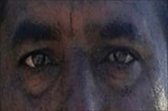
The patient was referred to the ophthalmology department for deviation of eyes and restricted ocular motility. He did not have diplopia at that time. His vision in both eyes was unaffected (6/9) and pupils were of normal size (3 mm) and reactive to light. Both eyes showed outward deviation, left eye being more divergent than the right. There was complete absence of adduction of both eyes and convergence was not possible. His fundi were normal. He had some difficulty in speech and was walking with the support of his relatives as he had giddiness [Fig. 1].
Figure 1.

Nine gaze positions of the patient revealing divergence and both eyes adduction restriction
Patient's hemogram, erythrocyte sedimentation rate, total and differential leukocyte count as well as lipid profile were normal. Computed tomographic (CT) angiography showed normal vascular pattern of brain, but there were small hypointense foci on both sides of aqueduct of Sylvius. Diffusion-weighted MRI revealed foci of restricted diffusion on both sides of aqueduct of Sylvius in midbrain, suggestive of acute midbrain infarct. Frontoparietal lobe on the right side showed infarct that could be reason for speech and gait problems, but these could not be correlated with MRI findings and were relieved very early [Fig. 2].
Figure 2.

Diffusion-weighted imaging (magnetic resonance imaging) and computed tomographic scan showing (a) infarct in frontoparietal region, (b) computed tomographic scan showing multiple infarct involving midline area, (c) and (d) foci of restricted diffusion on both sides of aqueduct of Sylvius in midbrain suggestive of acute midbrain infarct involving medial rectus nuclei
At 1-month follow-up, the patient fully recovered from his gait and speech problems, but ocular deviation and motility restrictions persisted. He did not have any diplopia. He visited the hospital again after 15 days (50 days after injury) without any recovery of ocular motility.
Eight months after injury, the patient regained his ocular movements fully with complete alignment of eyes [Fig. 3].
Figure 3.

Complete recovery with orthotropia after 8 months
Discussion
Ocular motor palsies are known to occur in cerebral concussion injuries, but selective nuclear lesion with paralysis of a particular muscle is very unusual. Isolated unilateral extraocular muscle palsy (EOMP) is usually thought to be caused by orbital lesions or muscular diseases.[1] Bilateral MR palsy due to nuclear involvement in closed head injury is reported only once in 1976 without documented CT or MRI images.[1] Recently isolated unilateral MR palsy due to midbrain lesion is reported in few cases with MRI documentation.[2,3,4] One important differential diagnosis that is internuclear ophthalmoplegia can be ruled out here because of absent convergence, and there was no abduction nystagmus. The absence of dissociated abducting nystagmus also rules out wall-eyed bilateral internuclear ophthalmoplegia from midbrain infarction which involves the bilateral medial longitudinal fasciculus which are usually supplied by the anteromedial perforators of the posterior cerebral artery.
The mechanism of nuclear EOMP in cerebral concussion injury is explained on the basis of the wave of the fluid in the third ventricle resulting from the blow. This wave with its greatest force around anterior end of aqueduct of Sylvius where the nuclei lie causes edema and petechial hemorrhages.[1,5] Small foci of midbrain infarct are the result of obstruction of blood flow in vena nervorum. Recovery may or may not be there in such lesions.
Conclusion
This is a case of nuclear oculomotor nerve palsy with an unusual presentation due to bilateral infarct in the midbrain involving the MR subnuclei which are situated most ventrally and can be diagnosed with diffusion-weighted imaging (DWI).[6] With use of DWI and other multimodality MRI, the probability of picking up midbrain infarcts causing isolated oculomotor palsies has increased.[7]
Declaration of patient consent
The authors certify that they have obtained all appropriate patient consent forms. In the form the patient(s) has/have given his/her/their consent for his/her/their images and other clinical information to be reported in the journal. The patients understand that their names and initials will not be published and due efforts will be made to conceal their identity, but anonymity cannot be guaranteed.
Financial support and sponsorship
Nil.
Conflicts of interest
There are no conflicts of interest.
References
- 1.Tehra KG, Ishwarchandra K, Kamble M. Bilateral medial rectus palsy due to nuclear involvement in closed head injury (a case report) Indian J Ophthalmol. 1976;24:25–6. [PubMed] [Google Scholar]
- 2.Sofiani MA, Lee Kwen P. Isolated medial rectus nuclear palsy as a rare presentation of midbrain infarction. Am J Case Rep. 2015;16:715–8. doi: 10.12659/AJCR.893875. [DOI] [PMC free article] [PubMed] [Google Scholar]
- 3.Rabadi MH, Beltmann MA. Midbrain infarction presenting isolated medial rectus nuclear palsy. Am J Med. 2005;118:836–7. doi: 10.1016/j.amjmed.2005.01.050. [DOI] [PubMed] [Google Scholar]
- 4.Lee HS, Yang TI, Choi KD, Kim JS. Teaching video neuroimage: Isolated medial rectus palsy in midbrain infarction. Neurology. 2008;71:e64. doi: 10.1212/01.wnl.0000335267.63332.2c. [DOI] [PubMed] [Google Scholar]
- 5.Lyle TK. Certain problems in the treatment of traumatic ocular palsy. Trans Ophthalmol Soc U K. 1959;79:519–31. [PubMed] [Google Scholar]
- 6.Kwon JH, Kwon SU, Ahn HS, Sung KB, Kim JS. Isolated superior rectus palsy due to contralateral midbrain infarction. Arch Neurol. 2003;60:1633–5. doi: 10.1001/archneur.60.11.1633. [DOI] [PubMed] [Google Scholar]
- 7.Bal S, Lal V, Khurana D, Prabhakar S. Midbrain infarct presenting as isolated medial rectus palsy. Neurol India. 2009;57:499–501. doi: 10.4103/0028-3886.55579. [DOI] [PubMed] [Google Scholar]


