Abstract
Dry eye is a common eye disease, and suitable animal models are indispensable for investigating the pathogenesis and developing treatments for dry eye. The present study was conducted to develop an androgen deficiency dry eye model induced by finasteride, and to evaluate ocular surface status and inflammatory cytokine gene expression in the lacrimal gland using a cytokine antibody array system. The results revealed that the antiandrogenic drug finasteride induced significant tear deficiency, and the histopathology results revealed significant inflammatory cell infiltration in the lacrimal gland. The cytokine antibody array system identified increased B7-2 (also known as cluster of differentiation 86), interleukin (IL)-1β, IL-4, IL-6, IL-10, matrix metalloproteinase-8, Fas ligand, tumor necrosis factor (TNF)-α and metalloproteinase inhibitor 1 levels in the lacrimal gland of the dry eye model. These cytokines were validated as candidate markers through the use of western blot analysis and reverse transcription-quantitative polymerase chain reaction. Both analyses confirmed a significant increase in proinflammatory cytokines, including IL-1β, IL-6 and TNF-α, and anti-inflammatory cytokines, including IL-4 and IL-10. The aforementioned data suggested that inflammation in antiandrogenic models resulted from a balance between inflammatory and anti-inflammatory responses. Thus, direct finasteride administration may produce an applicable model for dry eye mediated by androgen deficiency. In addition, there may be a correlation between sex, steroid deficiency and the inflammatory response. The findings of the present study have provided useful information for the pathogenesis and diagnosis of dry eye mediated by androgen deficiency.
Keywords: androgen deficiency dry eye, finasteride, cytokine antibody, inflammatory, lacrimal gland
Introduction
Dry eye is a common ocular surface disease associated with symptoms of ocular discomfort, tear film instability, and ocular surface and lacrimal gland inflammation (1–3). Dry eye not only affects the patient's daily life, i.e., reading, computer work, and driving, but also increases the risk of ocular infections and visual disturbance (4). Dry eye affects approximately 10–20% of adults (5). Epidemiological studies have indicated that among those >50 years of age, approximately 7% of women and 4% of men in the US have reported symptoms of dry eye (6). A decline in androgen levels is a major cause of dry eye given that androgens play an important role in the regulation of lacrimal gland secretion. Due to the decrease in sex hormones, the incidence of dry eye is significantly increased in postmenopausal women compared with men (7). If untreated, postmenopausal dry eye will significantly affect the quality of later life. It is prudent to explore the pathogenesis and treatment of sex steroid-deficient dry eye. To achieve this goal, an appropriate animal model for this condition is very important.
Gene expression profiling technologies have allowed large panels of genes to be analyzed at one time, which can provide important information about diseases (8). Immunostaining procedures can be used to explore the expression of proteins, but the process lacks quantification and is inefficient because only one or several proteins can be detected simultaneously. However, antibody array technology has allowed for simultaneous measurement of a large panel of genes. Such antibody arrays could provide information on the role of cytokines in the pathogenesis of dry eye.
The aim of this study was to develop a rat model of androgen deficiency dry eye and to assess changes in clinical outcomes, lacrimal gland histopathology, and inflammatory cytokine levels. Cytokine antibody assays were used to determine whether inflammatory-related cytokine levels in the lacrimal gland of the dry eye model were increased or decreased. An overall analysis of cytokine protein expression in the lacrimal gland could provide critical information for the pathogenesis and diagnosis of androgen deficiency dry eye.
Materials and methods
Animals
A total of 18 7-week-old female Wistar rats, which were obtained from the Animal Experimental Center of Nanjing Medical University (Nanjing, China), were used in accordance with the ARVO Statement for the Use of Animals in Ophthalmic and Vision Research. The experimental protocol was approved by the Animal Care and Use Committee of Nanjing University of Traditional Chinese Medicine (Nanjing, China).
Animal model
Six rats were used as controls without any treatment, and another six rats received saline gastric perfusion. The remaining six rats received finasteride gastric perfusion (MSD International GMBH Co., Ltd., Arecibo, Puerto Rico). Saline (5 ml/kg/day) or finasteride (1.16 mg/kg/day) was orally administered to the rats once a day for 4 weeks.
Phenol red thread test, tear film break up time (BUT) test and fluorescein staining
To confirm the presence of dry eye, tear flow, BUT and corneal fluorescein staining were evaluated. Measurements were made at 0, 7, 14, 21 and 28 days. Tear secretion was measured using phenol red impregnated cotton threads (Tianjin Jingming New Technological Development Co., Ltd., Tianjin, China). The threads were held with jeweler forceps and placed between the lower lid and the globe for 60 sec. Wetting of the thread was measured with a ruler with 0.5-mm precision (9). The BUT results and corneal staining were examined under a slit lamp microscope with a cobalt blue filter (Topcon SL-D7; Topcon Medical Systems, Inc., Tokyo, Japan). Staining indicates corneal epithelium damage. We used a scoring system ranging from 0 to 3 for corneal staining (2). The cornea was divided into five areas, each of which was scored as follows: No staining was given a score of 0; superficial stippling and micropunctate staining was given a score of 1; macropunctate staining with some coalescent areas was given a score of 2; numerous coalescent macropunctate areas and/or patches was given a score of 3. Then, the scores were added, and the totals ranged between 0 and 15.
Cytokine antibody array analysis
To investigate the changes in inflammatory cytokine expression levels in different groups and to select the relevant candidate genes for further study, gene microarray methods were used to analyze the expression of inflammatory cytokines in the lacrimal gland. Raybio® Rat Cytokine Antibody Arrays, G Series 2, were purchased from RayBiotech, Inc. (Norcross, GA, USA) and used according to the manufacturer's instruction. Lacrimal glands were removed and cut into small lobules (2 mm in diameter). The protein concentration was determined for each sample using the BCA method. Rat cytokine antibody arrays were used to detect the expression of 35 cytokines in the lacrimal glands of the three groups. The relative expression levels of cytokines were calculated by comparing the signal intensities. The expression of the corresponding cytokines was increased or decreased compared with the control group. A ratio >1.3 was considered to indicate high expression, whereas a ratio <0.77 was considered to represent low expression (Table I).
Table I.
Position of 35 rat inflammatory factors in the antibody-based protein microarray.
| A | B | C | D | E | F | G | H | I | J | K | L | |
|---|---|---|---|---|---|---|---|---|---|---|---|---|
| 1 | POS | POS | NEG | NEG | Activin A | Agrin | B7-2/Cd86 | β-NGF | CINC-1 | CINC-2α | CINC-3 | CNTF |
| 2 | POS | POS | NEG | NEG | Activin A | Agrin | B7-2/Cd86 | β-NGF | CINC-1 | CINC-2α | CINC-3 | CNTF |
| 3 | Fas ligand | Fractalkine | GM-CSF | ICAM-1 | IFN-γ | IL-1α | IL-1β | IL-1 R6 | IL-2 | IL-4 | IL-6 | IL-10 |
| 4 | Fas ligand | Fractalkine | GM-CSF | ICAM-1 | IFN-γ | IL-1α | IL-1β | IL-1 R6 | IL-2 | IL-4 | IL-6 | IL-10 |
| 5 | IL-13 | Leptin | LIX | L-Selectin | MCP-1 | MIP-3α | MMP-8 | PDGF-AA | Prolactin R | RAGE | Thymus Chemokine-1 | TIMP-1 |
| 6 | IL-13 | Leptin | LIX | L-Selectin | MCP-1 | MIP-3α | MMP-8 | PDGF-AA | Prolactin R | RAGE | Thymus Chemokine-1 | TIMP-1 |
| 7 | TNF-α | VEGF | BLANK | BLANK | BLANK | BLANK | BLANK | BLANK | BLANK | BLANK | BLANK | POS |
| 8 | TNF-α | VEGF | BLANK | BLANK | BLANK | BLANK | BLANK | BLANK | BLANK | BLANK | BLANK | POS |
POS, positive; NEG, negative; β-NGF, β nerve growth factor; CINC, cytokine-induced neutrophil chemoattractant; CNTF, ciliary neurotrophic factor; GM-CSF, granulocyte-macrophage colony stimulating factor; ICAM-1, intercellular adhesion molecule-1; IFN-γ, interferon-γ; IL, interleukin; MCP, monocyte chemoattractant protein; MIP, macrophage inflammatory protein; MMP, matrix metalloproteinase; PDGF, platelet-derived growth factor; RAGE, receptor for advanced glycation end products; TIMP, tissue-specific inhibitor of metalloproteinase; TNF, tumor necrosis factor; VEGF, vascular endothelial growth factor.
Western blot analysis
Based on the protein microarray results, only highly expressed cytokines were selected for further investigation using western blots. Lacrimal glands were removed from two groups of rats. Tissues samples were homogenized in isolation buffer (10 mM Tris, pH 8.0, 1 mM EDTA) and centrifuged at 2,000 × g for 20 min. The supernatants were denatured in SDS-PAGE sample buffer for 20 min at 60°C and resolved on a 4–20% gradient SDS-PAGE gel. All samples and molecular weight standards were boiled for 5 min prior to loading onto 4–15% polyacrylamide gradient gels (Bio-Rad Laboratories, Inc., Hercules, CA, USA). The gels were run for 30 min at 80 V, washed in transfer buffer for 90 min and electroblotted onto nitrocellulose membranes for 2 h at 100 mA. The blots were blocked overnight at 4°C with Tris-buffered saline containing 3% milk and incubated for 2 h at room temperature with the corresponding antibody. Following three rinses, the blots were incubated with horseradish peroxidase (HRP)-conjugated rabbit anti-goat immunoglobulin G (1:2,000) for 1 h, and the hybridized bands were detected with an enhanced chemiluminescence kit (PerkinElmer Life Sciences, Boston, MA, USA).
Quantitative real-time PCR analysis
Briefly, total RNA was extracted from lacrimal glands with TRIzol (Invitrogen, Carlsbad, CA, USA). First-strand complementary DNA (cDNA) was synthesized from 2 µg of total RNA using oligo (dT) primers and M-MLV reverse transcriptase (Invitrogen). The PCR reaction was performed in a 50-µl volume with 5 U polymerase (Takara Bio, Inc., Otsu, Japan) and cDNA samples equivalent to 1 ng RNA. SYBR-Green (1:20,000 dilution) was included in each reaction to allow relative quantification of RNA levels using the ABI StepOne Plus detection system (Applied Biosystems, Foster City, CA, USA). The primers for detecting genes were designed using Primer Express (Table II). Glyceraldehyde-3-phosphate dehydrogenase (GAPDH) was used as an endogenous reference to normalize cDNA loading. The relative expression level of each target gene among different samples and cell lines was calculated accordingly (ABI PRISM 7300 Detection system; Applied Biosystems). For the cell lines, the relative expression level (defined as ‘fold-change’) of the target gene was determined by 2−ΔCt (ΔCt=ΔtTtarget-ΔCtβ-actin) and normalized to the fold-change detected in the corresponding control cells, which was defined as 1.0 (Table II).
Table II.
Primer sequence for the PCR in this experiment.
| Gene name | Sequence | Length (bp) |
|---|---|---|
| MMP8 | F: CGTGGCTGCTCATGAATTTG | 102 |
| R: TAGGTGCTGGGTTCTCTGTA | ||
| CNTF | F: TTGGAGATGGTGGTCTCTTTG | 100 |
| R: ATGACACGAAGGTCATGGATAG | ||
| FasL | F: GGTGCTAATGGAGGAGAAGAAG | 106 |
| R: TAAATGGTCAGCAACGGTAAGA | ||
| IL-1β | F: TCCCTGAACTCAACTGTGAAATA | 103 |
| R: GGCTTGGAAGCAATCCTTAATC | ||
| IL-4 | F: GTCACCCTGTTCTGCTTTCT | 96 |
| R: GACCTGGTTCAAAGTGTTGATG | ||
| IL-6 | F: GAAGTTAGAGTCACAGAAGGAGTG | 105 |
| R: GTTTGCCGAGTAGACCTCATAG | ||
| IL-10 | F: AGTGGAGCAGGTGAAGAATG | 109 |
| R: GAGTGTCACGTAGGCTTCTATG | ||
| GAPDH | F: GGGAAACCCATCACCATCTT | 72 |
| R: ATACTCAGCACCAGCATCAC |
MMP, matrix metalloproteinase; CNTF, ciliary neurotrophic factor; FasL, Fas ligand; IL, interleukin; GAPDH, glyceraldehyde-3-phosphate dehydrogenase.
Lacrimal gland histopathology
The lacrimal glands from sacrificed animals were fixed in 4% formalin for 24 h. After incubation in 30% sucrose overnight, the specimens were embedded in paraffin, cross-sectioned, and stained with hematoxylin and eosin (H&E).
Statistical analysis
The data are presented as means ± SD (min-max). Statistical analysis was performed using SAS (version 9.3; SAS Institute Inc., Cary, NC, USA). Non-parametric (Mann-Whitney U or Wilcoxon) or appropriate parametric (t-test) statistical tests were performed for comparisons between the 2 groups. Differences were considered as statistically significant at P-value <0.05.
Results
Effect of finasteride on tear production
Compared with the Sal group, administration of finasteride, a type II and type III 5α-reductase inhibitor [5α-reductase convert stestosterone to dihydrotestosterone (DHT)], significantly reduced tear production (Fig. 1A and B). No difference in tear production was noted in the Con group compared with the Sal group. Additionally, corneal punctate staining in the Fin group was disrupted as noted by a significant increase in staining grade (Fig. 1C).
Figure 1.
Effect of finasteride administration on tear production. (A) Tear secretion changes in the three groups at different time points (unit: mm); (B) BUT changes in the three groups at different time points (unit: sec); (C) changes in corneal epithelial fluorescein staining in the three groups at different time points *P<0.05/**P<0.01 vs. baseline within the same group. ▲P<0.05/▲▲P<0.01 within the Con group at the same time point. BUT, break up time.
Cytokine assay results
The levels of 22 cytokines in the lacrimal gland were significantly altered in the Fin group compared with the Con group (Table III; Fig. 2). Most of these cytokines were inflammatory factors and chemokines. The expression levels of 15 cytokines, including B7-2/Cd86, ciliary neurotrophic factor (CNTF), Fas igand (FasL), granulocyte-macrophage colony stimulating factor (GM-CSF), intercellular adhesion molecule (ICAM)-1, interferon (IFN)-γ, interleukin (IL)-1β, IL-4, IL-6, IL-10, Leptin, L-Selectin, matrix metalloproteinase (MMP)-8, tissue-specific inhibitor of metalloproteinase (TIMP)-1, and tumor necrosis factor (TNF)-α, were significantly increased. Among the highly expressed cytokines, IL-1β, IL-6, GM-CSF, and TNF-α are proinflammatory cytokines, and IL-4 and IL-10 are anti-inflammatory genes (Table III).
Table III.
Cytokine assay results in lacrimal gland of three groups.
| Name of cytokines | Folds of group Sal to Con lacrimal gland | Folds of group Fin to Con lacrimal gland |
|---|---|---|
| Inflammatory cytokines | ||
| IL-1α | 1.191 | 1.220 |
| IL-1β | 0.990 | 1.533↑ |
| IL-2 | 1.004 | 1.283 |
| IL-6 | 0.951 | 1.357↑ |
| IFN-γ | 1.125 | 1.827↑ |
| Fas ligand | 0.772 | 3.096↑ |
| GM-CSF | 0.867 | 1.952↑ |
| TNF-α | 0.721 | 1.541↑ |
| Anti-inflammatory cytokines | ||
| IL-1 R6 | 0.949 | 1.334↑ |
| IL-4 | 0.857 | 1.716↑ |
| IL-10 | 0.816 | 1.593↑ |
| IL-13 | 0.904 | 1.435↑ |
| Chemokines | ||
| MCP-1 | 0.926 | 1.469↑ |
| MIP-3α | 1.306↑ | 1.375↑ |
| Fractalkine | 1.198 | 1.194 |
| CINC-1 | 1.114 | 1.136 |
| CINC-2α | 1.253 | 1.216 |
| CINC-3 | 1.216 | 1.229 |
| Thymus chemokine-1 | 1.125 | 1.085 |
| Growth factors | ||
| β-NGF | 1.441↑ | 1.448↑ |
| PDGF-AA | 1.323↑ | 1.330↑ |
| VEGF | 1.286 | 1.291 |
| CNTF | 0.872 | 1.809↑ |
| Activin A | 1.261 | 1.343↑ |
| Other cytokines | ||
| B7-2/Cd86 | 0.940 | 1.992↑ |
| Agrin | 1.034 | 1.054 |
| Leptin | 0.848 | 2.315↑ |
| MMP-8 | 0.640 | 3.304↑ |
| TIMP-1 | 1.045 | 1.528↑ |
| LIX | 1.246 | 1.295 |
| L-Selectin | 0.926 | 1.664↑ |
| Prolactin R | 1.240 | 1.232 |
| RAGE | 1.111 | 1.116 |
| ICAM-1 | 0.982 | 1.862↑ |
IL, interleukin; IFN-γ, interferon-γ; GM-CSF, granulocyte-macrophage colony stimulating factor; TNF, tumor necrosis factor; MCP, monocyte chemoattractant protein; MIP, macrophage inflammatory protein; CINC, cytokine-induced neutrophil chemoattractant; β-NGF, β nerve growth factor; PDGF, platelet-derived growth factor; VEGF, vascular endothelial growth factor; CNTF, ciliary neurotrophic factor; MMP, matrix metalloproteinase; TIMP, tissue-specific inhibitor of metalloproteinase; RAGE, receptor for advanced glycation end products; ICAM-1, intercellular adhesion molecule-1.
Figure 2.
Microarray analysis of secreted inflammatory factors from lacrimal glands. (A) Lacrimal gland of the Con group; (B) lacrimal gland of the Sal group; (C) lacrimal gland of the Fin group.
Protein expression analysis
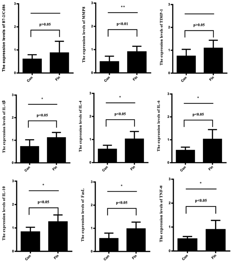
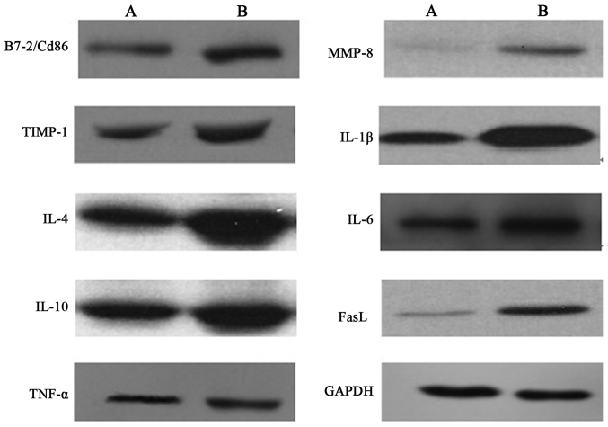
Highly expressed and inflammation-related cytokines in the lacrimal gland, including B7-2/Cd86, IL-1β, IL-4, IL-6, IL-10, MMP-8, FasL, TNF-α and TIMP-1, were selected as candidates for further Western blot analysis. Their expression levels are presented in Figs. 3 and 4. The IL-1β, IL-4, IL-6, IL-10, MMP-8, FasL, and TNF-α expression levels were significantly increased in the Fin group compared with the Con group (P<0.05 or P<0.01). B7-2/Cd86 and TIMP-1 were also increased, but the difference was not statistically significant.
Figure 3.
Western blot analysis of cytokines in the lacrimal gland of the three groups of rats. *P<0.05, **P<0.01. MMP, matrix metalloproteinase; TIMP, tissue-specific inhibitor of metalloproteinase; IL, interleukin; FasL, Fas ligand; TNF, tumor necrosis factor.
Figure 4.
Western blot analysis of secreted inflammatory factors from the lacrimal gland. (A) Con group; (B) Fin group. TIMP, tissue-specific inhibitor of metalloproteinase; IL, interleukin; TNF, tumor necrosis factor; MMP, matrix metalloproteinase; FasL, Fas ligand; GAPDH, glyceraldehyde-3-phosphate dehydrogenase.
RNA expression analysis
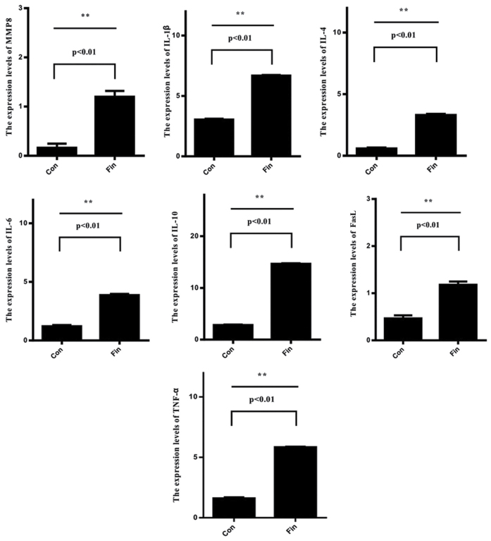
Quantitation of mRNA by real-time PCR is presented in Fig. 5. The RT-PCR results were consistent with those of western blots. Among the cytokines analyzed, IL-1β, IL-4, IL-6, IL-10, MMP-8, FasL, and TNF-α expression levels were significantly increased in the Fin group compared with the Con group (P<0.01).
Figure 5.
Real-time PCR analysis of cytokines in lacrimal glands from the three groups of rats. *P<0.05, **P<0.01. MMP, matrix metalloproteinase; IL, interleukin; FasL, Fas ligand; TNF, tumor necrosis factor.
Lacrimal gland histopathology
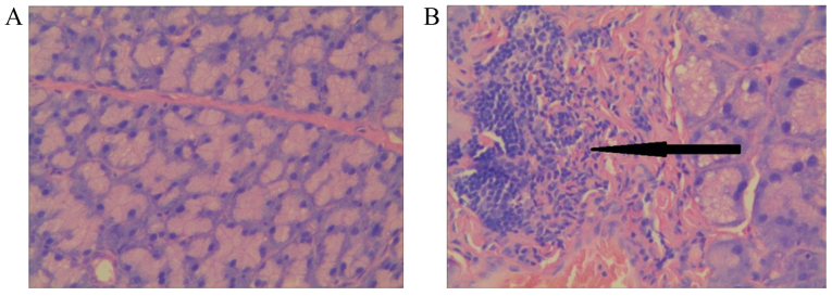
The pathologic changes of lacrimal glands in each group on day 28 are described in Fig. 6 (H&E staining). In the Con group, the normal acinar structure was well preserved, and no significant lymphocyte infiltration was noted (Fig. 6A). In the Fin group, a severe inflammatory response was noted in the lacrimal gland. A large number of lymphocytes had infiltrated the interlobular space and surrounded the acinar and ductal cells (Fig. 6B).
Figure 6.
Lacrimal gland histopathology (H&E staining, ×400). (A) Lacrimal gland from the Con group; (B) Lacrimal gland from the Fin group. A large number of lymphocytes had infiltrated the interlobular space and surrounded the acinar and ductal cells as indicated by the black arrow. H&E, hematoxylin and eosin.
Discussion
Dry eye is a common disease worldwide and an important public health issue. A few studies have reported the effect of dry eye on vision-related quality of life (10,11). The investigation of dry eye is necessary to determine its pathogenesis and relevant therapies. Recent findings from humans and animal models indicate that an inflammatory response occurs in the lacrimal gland and may significantly contribute to the pathophysiology of dry eye (12–15). Clinical evidence suggests that topical anti-inflammatory treatment of dry eye is effective (16–18).
Androgen-deficient rats develop dry eye. The characteristics of this type of dry eye include reduced tear secretion, ocular surface damage and inflammation of the lacrimal gland, and this animal model is used for the study of dry eye (19). Finasteride is a 5α-reductase inhibitor that specifically acts on type II and III isoenzymes. By inhibiting 5α-reductase, finasteride prevents conversion of testosterone to DHT via type II and III isoenzymes. In the present study, we noted that finasteride administration resulted in a significant reduction in tear flow and severe inflammation of the lacrimal gland. Tear secretion was significantly reduced in the Fin group compared with the Con group on days 14, 21, and 28. BUT values in the Fin group were significantly lower than those in the Con group on days 7, 14, 21, and 28. The scores of corneal fluorescein staining were significantly increased in the Fin group compared with the Con group on days 21 and 28. Therefore, oral administration of finasteride in normal rats produced a sex steroid-deficient dry eye model. In the current study, female rats were selected based on the high incidence rate of women who exhibit this clinical condition. A reduction in androgen levels is the main cause of dry eye. This situation is more common among women and increases with age because androgen levels decrease with age, especially after menopause. The results obtained by Singh et al also demonstrated that oral administration of finasteride in both sexes of normal rats produced an appropriate sex steroid-deficient dry eye model, but finasteride significantly downregulated the number of androgen receptors by 8-foldin the lacrimal glands of female rats compared with those of male rats (19). Correspondingly, finasteride-treated female rats exhibited a 49% reduction in tear flow, whereas male rats exhibited a 40% reduction in tear flow.
Identifying key cytokines and evaluating their expression may provide useful biomarkers of inflammationin diseases. In this respect, one or a group of cytokines present in the lacrimal gland throughout disease may be particularly useful for tracking disease. Similarly, knowledge of the tissue expression profile of such proteins can provide valuable information for distinguishing diagnostic markers and treatment targets of dry eye (20). In the present study, lacrimal glands from three groups of rats were screened for 35 different cytokines and cytokine-related proteins. Our results demonstrated that the lack of testosterone could influence cytokine expression in the lacrimal gland. Based on the results of an antibody array analysis, we observed that 22 cytokines in the lacrimal gland were significantly altered in the Fin group compared with the Con group. The expression levels of 15 cytokines were significantly increased, and most of these cytokines were inflammatory factors and chemokines. Inflammation-related cytokines highly expressed in the lacrimal gland were selected as candidates for further western blot and RT-PCR analyses. The RT-PCR results were consistent with the western blot analysis results. IL-1β, IL-4, IL-6, IL-10, MMP-8, FasL and TNF-α levels were significantly increased in the Fin group compared with the Con group. The levels of proinflammatory cytokines, including IL-1β, IL-6, GM-CSF, and TNF-α, were progressively upregulated in the Fin group compared with the Con group. In addition, the levels of anti-inflammatory cytokines, including IL-4 and IL-10, were upregulated in the Fin group compared with the Con group. These data suggested that inflammation in antiandrogenic models resulted from the balance between inflammatory and anti-inflammatory responses.
In conclusion, direct finasteride administration in normal rats produces a suitable androgen deficiency dry eye model that can be used to study the effect of topically applied pharmacological agents for the treatment of dry eye due to androgen deficiency. Our results demonstrated that the change in the ocular surface was related to changes in the expression of various proinflammatory and anti-inflammatory cytokines in the lacrimal gland, suggesting that a correlation between sex steroid deficiency and the inflammatory response may exist. Based on these findings, a global analysis of cytokine protein expression may contribute to the understanding the pathogenesis and diagnosis of androgen deficiency dry eye. Further investigations aimed at cytokine modulation as a therapeutic method for the treatment of androgen deficiency dry eye are warranted.
Acknowledgements
The study was supported by grants from Jiangsu Provincial Natural Science Funding of China (grant no. BK20141504), Six Talent Peaks Project in Jiangsu Province (grant no. WSW-048) and National Natural Science Foundation of China (grant no. 81173306).
References
- 1.The epidemiology of dry eye disease, corp-author. Report of the epidemiology subcommittee of the international dry eye workshop (2007) Ocul Surf. 2007;5:93–107. doi: 10.1016/S1542-0124(12)70082-4. [DOI] [PubMed] [Google Scholar]
- 2.Lemp MA. Report of the National Eye Institute/Industry Workshop on Clinical Trials in dry eyes. CLAO J. 1995;21:221–232. [PubMed] [Google Scholar]
- 3.Smith RE. The tear film complex: Pathogenesis and emerging therapies for dry eyes. Cornea. 2005;24:1–7. doi: 10.1097/01.ico.0000141486.56931.9b. [DOI] [PubMed] [Google Scholar]
- 4.Miljanović B, Dana R, Sullivan DA, Schaumberg DA. Impact of dry eye syndrome on vision-related quality of life. Am J Ophthalmol. 2007;143:409–415. doi: 10.1016/j.ajo.2006.11.060. [DOI] [PMC free article] [PubMed] [Google Scholar]
- 5.Johnson ME, Murphy PJ. Changes in the tear film and ocular surface from dry eye syndrome. Prog Retin Eye Res. 2004;23:449–474. doi: 10.1016/j.preteyeres.2004.04.003. [DOI] [PubMed] [Google Scholar]
- 6.Schaumberg DA, Sullivan DA, Buring JE, Dana MR. Prevalence of dry eye syndrome among US women. Am J Ophthalmol. 2003;136:318–326. doi: 10.1016/S0002-9394(03)00218-6. [DOI] [PubMed] [Google Scholar]
- 7.Serrander AM, Peek KE. Changes in contact lens comfort related to the menstrual cycle and menopause. A review of articles. J Am Optom Assoc. 1993;64:162–166. [PubMed] [Google Scholar]
- 8.Riemer C, Neidhold S, Burwinkel M, Schwarz A, Schultz J, Krätzschmar J, Mönning U, Baier M. Gene expression profiling of scrapie-infected brain tissue. Biochem Biophys Res Commun. 2004;323:556–564. doi: 10.1016/j.bbrc.2004.08.124. [DOI] [PubMed] [Google Scholar]
- 9.Zoukhri D, Macari E, Choi SH, Kublin CL. C-Jun NH2-terminal kinase mediates interleukin-1beta-induced inhibition of lacrimal gland secretion. J Neurochem. 2006;96:126–135. doi: 10.1111/j.1471-4159.2005.03529.x. [DOI] [PMC free article] [PubMed] [Google Scholar]
- 10.Goto E, Yagi Y, Matsumoto Y, Tsubota K. Impaired functional visual acuity of dry eye patients. Am J Ophthalmol. 2002;133:181–186. doi: 10.1016/S0002-9394(01)01365-4. [DOI] [PubMed] [Google Scholar]
- 11.Tong L, Waduthantri S, Wong TY, Saw SM, Wang JJ, Rosman M, Lamoureux E. Impact of symptomatic dry eye on vision-related daily activities: The Singapore Malay Eye Study. Eye (Lond) 2010;24:1486–1491. doi: 10.1038/eye.2010.67. [DOI] [PubMed] [Google Scholar]
- 12.Barabino S, Rolando M, Camicione P, Ravera G, Zanardi S, Giuffrida S, Calabria G. Systemic linoleic and gamma-linolenic acid therapy in dry eye syndrome with an inflammatory component. Cornea. 2003;22:97–101. doi: 10.1097/00003226-200303000-00002. [DOI] [PubMed] [Google Scholar]
- 13.Coursey TG, de Paiva CS. Managing Sjögren's syndrome and non-Sjögren syndrome dry eye with anti-inflammatory therapy. Clin Ophthalmol. 2014;8:1447–1458. doi: 10.2147/OPTH.S35685. [DOI] [PMC free article] [PubMed] [Google Scholar]
- 14.Stern ME, Schaumburg CS, Pflugfelder SC. Dry eye as a mucosal autoimmune disease. Int Rev Immunol. 2013;32:19–41. doi: 10.3109/08830185.2012.748052. [DOI] [PMC free article] [PubMed] [Google Scholar]
- 15.Stevenson W, Chauhan SK, Dana R. Dry eye disease: An immune-mediated ocular surface disorder. Arch Ophthalmol. 2012;130:90–100. doi: 10.1001/archophthalmol.2011.364. [DOI] [PMC free article] [PubMed] [Google Scholar]
- 16.Lee HK, Ryu IH, Seo KY, Hong S, Kim HC, Kim EK. Topical 0.1% prednisolone lowers nerve growth factor expression in keratoconjunctivitis sicca patients. Ophthalmology. 2006;113:198–205. doi: 10.1016/j.ophtha.2005.09.033. [DOI] [PubMed] [Google Scholar]
- 17.Marsh P, Pflugfelder SC. Topical nonpreserved methylprednisolone therapy for keratoconjunctivitis sicca in Sjögren syndrome. Ophthalmology. 1999;106:811–816. doi: 10.1016/S0161-6420(99)90171-9. [DOI] [PubMed] [Google Scholar]
- 18.Sall K, Stevenson OD, Mundorf TK, Reis BL. Two multicenter, randomized studies of the efficacy and safety of cyclosporine ophthalmic emulsion in moderate to severe dry eye disease. CsA Phase 3 Study Group. Ophthalmology. 2000;107:631–639. doi: 10.1016/S0161-6420(99)00176-1. [DOI] [PubMed] [Google Scholar]
- 19.Singh S, Moksha L, Sharma N, Titiyal JS, Biswas NR, Velpandian T. Development and evaluation of animal models for sex steroid deficient dry eye. J Pharmacol Toxicol Methods. 2014;70:29–34. doi: 10.1016/j.vascn.2014.03.004. [DOI] [PubMed] [Google Scholar]
- 20.Gu Q. High-throughput identification of molecular targets of brain disorders using antibody-based microarray analyses. Expert Rev Neurother. 2008;8:1281–1283. doi: 10.1586/14737175.8.9.1281. [DOI] [PubMed] [Google Scholar]


