Abstract
Sporothrix schenckii is a pathogenic dimorphic fungus with a global distribution. It grows in a multicellular hyphal form at 25°C and a unicellular yeast form at 37°C. The morphological switch from mold to yeast form is obligatory for establishing pathogenicity in S. schenckii. Two-component signaling systems are utilized by eukaryotes to sense and respond to external environmental changes. DRK1is a hybrid histidine kinase, which functions as a global regulator of dimorphism and virulence in Blastomyces dermatitidis and Histoplasma capsulatum. An intracellular soluble hybrid histidine kinase, homologous to DRK1 in B. dermatitidis, has previously been identified in S. schenckii and designated as SsDRK1. In the present study, the function of SsDRK1 was investigated using double stranded RNA interference mediated by Agrobacterium tumefaciens. SsDRK1 was demonstrated to be required for normal asexual development, yeast-phase cell formation, cell wall composition and integrity, melanin synthesis, transcription of the morphogenesis-associated gene Ste20 that is involved in the high osmolarity glycerol/mitogen-activated protein kinase pathway, and pathogenicity of S. schenckii in a murine model of cutaneous infection. Further investigations into the signals SsDRK1 responds to, and the interactions of upstream transmembrane hybrid histidine kinases with SsDRK1, are required to uncover novel targets for anti-fungal therapies.
Keywords: Sporothrix schenckii, hybrid histidine kinase DRK1, RNA interference, pathogenesis
Introduction
Sporothrix schenckii, which is the etiological agent of sporotrichosis, is a dimorphic fungus that produces lymphocutaneous lesions. It assumes a filamentous form in its saprophytic stage or when cultured at 25°C, composed of hyaline septate hyphae with conidiogenous cells arising from undifferentiated hyphae, which form conidia. This fungus is evident in human and animal tissues as budding yeasts. Yeast-like cells are round to oval in shape and are usually formed of elongated, cigar-shaped buds on a narrow base. The morphological switch from mold to yeast form can be induced in S. schenckii by culturing mycelia or conidia on brain heart infusion (BHI) medium between 35 and 37°C (1). This transition process also occurs when filamentous S. schenckii adapts to a novel niche and infects a mammalian host, and this stage is obligatory for establishing its pathogenicity (2).
Several signaling cascades have previously been demonstrated to be involved in the host-environmental response and the morphological switch in dimorphic fungi, including the cyclic adenosine monophosphate-dependent protein kinase signaling pathway, the Ca2+/calcineurin signaling pathway, the mitogen-activated protein kinase (MAPK) pathway and two-component regulatory systems (3–6). The two-component signaling system, which consists of a sensor histidine kinase (HK) and a response regulator (RR) receiver protein, is involved in sensing and responding to external environmental signals. In fungi, the HK and RR are fused into a single polypeptide, leading to the term hybrid histidine kinase. In 2006, a Blastomyces dermatitidis gene encoding a transmembrane hybrid histidine kinase, DRK1, was identified during an insertional mutagenesis screen for regulators of the yeast phase-specific gene Blastomyces adhesin 1 (BAD1) (7). DRK1 mutants of B. dermatitidis and Histoplasma capsulatum fail to convert to the yeast stage at 37°C and demonstrate reduced pathogenicity in a murine lethal pulmonary infection model (8). The typical DRK1 histidine kinase is a transmembrane receptor with an amino-terminal extracellular sensing domain that detects environmental signals and a carboxy-terminal cytosolic signaling domain (9); however, the homologue of B. dermatitidis DRK1 in S. schenckii has been revealed to be a soluble histidine kinase that lacks transmembrane segments, and carries GAF and PAS domains in the amino-terminal sensory domain (10). This raises questions regarding the function of DRK1 in S. schenckii and whether it differs from that of transmembrane DRK1 in other dimorphic fungi. Therefore, the present study aimed to investigate the function of the DRK1 homologue in S. schenckii.
Materials and methods
Fungal strains and media
The American Type Culture Collection (ATCC) 10268 strain of S. schenckii (ATCC, Manassas, VA, USA), maintained in the Research Center for Pathogenic Fungi of Dalian Medical University (Dalian, China), was used as the wild-type strain, as well as for the construction of the SsDRK1 RNA interference mutant. Mycelial colonies were cultured in sabouraud dextrose agar (SDA) medium (The First Affiliated Hospital of Dalian Medical University, Dalian, China) at 25°C and yeast colonies were grown in BHI medium (Hyclone; GE Healthcare Life Sciences, Logan, UT, USA) at 37°C. To achieve the switch from the mycelial to the yeast phase in S. schenckii, mycelial colonies were transferred to liquid BHI medium at 37°C and agitated at 100 rpm for 96 h. The EHA105 strain of Agrobacterium tumefaciens (Dalian University of Technology, Dalian, China) was cultured in lysogeny broth (LB) medium (Dalian University of Technology) containing 50 µg/ml kanamycin and 20 µg/ml rifampicin at 28°C to maintain the plasmids. To create an inductive medium for RNA interference, 200 µmol/lacetosyringone was added to the medium [agar 15 g/l, K-buffer 8 ml/l, Mn-buffer 20 ml/l, 1% CaCl2.2H2O 1 ml/l, 0.01% FeSO4 10 ml/l, 20% NH4NO3 2.5 ml/l, 50% glycerine 10 ml/l, 1M 2-(N-morpholino) ethanesulfonic acid 40 ml/l, 20% glucose 10 ml/l] as an inducer. Hygromycin (100 mg/l) and cefotaxime (200 µM) were also added to the inductive medium to create a DRK1-interference isolate screen. For the cell wall test, strains were grown for 5 days at 25°C on SDA medium with 20 µg/ml Congo red. To test resistance to zymolyase, strains were grown on SDA medium with 100 µl zymolyase20T (10 mg/ml−1 in 10 mM Tris/HCl, pH 7.5; Sigma-Aldrich; Merck KGaA, Darmstadt, Germany) at 25°C for 30 h.
Plasmid construction
All basic molecular biology procedures were performed as previously described by Sambrook et al (11). The SsDRK1 primer sequences used were as follows: DRK1, forward 5′-CANGANAAYGTVAAYACYATGGC-3′ and reverse 5′-CGRTCMACCATRTBRTTGATNGT-3′. They were designed to amplify the S. schenckii genomic cDNA sequence that encodes the conserved kinase domain of the SsDRK1 gene. Each primer was designed to introduce some endonuclease sites, resulting in silencing of the gene, and the digested fragment was ligated into pCB309 plasmids synthesized from pSilent-1 (the Fungal Genetics Stock Center, Manhattan, KS, USA), which were used for transformation. Following polymerase chain reaction (PCR), amplified DNA was cloned into pCB309 plasmids to construct the final SsDRK1 double stranded RNA interference (dsRNAi) plasmid, pCB309-pfgrt (11). It contained a 1.5 kb fragment of SsDRK1 sequence hairpin repeats and the hygromycin resistance gene marker. The SsDRK1 hairpin repeat sequence was placed under the control of the common fungal promoter, PtrpC. pCB309 plasmids lacking the SsDRK1 hairpin repeat sequence were used as a control (SsDRK-1-control group).
Construction of SsDRK1 interference (SsDRK1-i) mutants
The transformation procedure was performed as previously described by Zhang et al (12). A. tumefaciens containing pCB309-pfgrt with the transfer (T-)DNA fragment was incubated in LB medium with kanamycin and rifampicin for 48 h at 28°C. S. schenckii conidia were grown on the BHI medium. Following washing with sterile water, the conidia (5×10⁶/ml) and A. tumefaciens containing PCB309-pfgrt were mixed in the inductive medium. S. schenckii was subsequently transformed by the pCB309-pfgrt. The transformants were selected on medium containing hygromycin (100 mg/l) and cefotaxime (200 µM). The mycelial dry weight of the wild strain, SsDRK1-i strain and DsDRK1+ strain were obtained following culture, centrifugation at 4°C for 30 min at a speed of 20,000 × g, drying and weighing.
Microscopic appearance
Samples were prepared for transmission electron microscopy as previously described by Garrison et al (13). Electron micrographs were generated using a JEOL JEM-2000EX electron microscope (JEOL Ltd., Tokyo, Japan). Samples were prepared for scanning electron microscopy using a method previously described by Wang et al (14). Dried colonies were coated with platinum-vanadium and observed under a QUANTA 450 scanning electron microscope (FEI; Thermo Fisher Scientific, Inc., Hillsboro, OR, USA) at an accelerating voltage of 20.0 kV.
Analysis of cell wall composition
Congo red sensitivities were measured as previously described by Davis and De Serres (15). Spores of wild-type and SsDRK1-interference (SsDRK1-i) ATCC 10268 S. schenckii strains were concentrated to equivalent densities. Subsequently, 10-fold dilution series were made, with 5 ml of each dilution spotted onto SDA media plates containing 20 µg/ml Congo red. Plates were incubated for 5 days at 25°C. Zymolyase sensitivities were analyzed as previously described by De Nobel et al (16). Wild-type and SsDRK1-i strains were grown to equivalent optical densities (OD) and an OD530 unit was taken as measurement, followed by a cell count for confirmation. Cells were washed twice and resuspended in 900 µl 10 mM Tris/HCl (pH 7.5). The OD530 measurement was repeated 30 h following the addition of 100 µl zymolyase 20T.
Transcription analysis
Total RNA was isolated from wild-type, SsDRK1-i and SsDRK1-control hyphal cells grown at 25°C for 7 days in SDA liquid medium and yeast cells grown at 37°C for 7 days in BHI liquid medium. RNA was extracted using TRIzol reagent (Invitrogen; Thermo Fisher Scientific, Inc., Waltham, MA, USA) according to the manufacturer's protocol. RNA was treated with DNAase (Promega Corporation, Madison, WI, USA) prior to reverse transcription-quantitative PCR (RT-qPCR) analysis. An increasing number of cycles were used to ensure the amplification in the exponential phase, and the 18S ribosomal gene was used as a control. Transcription of DRK1 and Ste20 was determined by RT-qPCR, using the Thermal Cycler Dice Real Time system (Takara Bio, Inc., Otsu, Japan) with a SYBR Prime Script RT-PCR kit (Takara Biotechnology, Co., Ltd., Dalian, China). The thermocycling conditions were as follows: 40 cycles of initialization at 95°C for 30 sec, denaturation at 95°C for 5 sec, and annealing at 60°C for 30 sec. The following primer sequences were used: DRK1, forward 5′-TTGCTATTGAAGTAAACC-3′, reverse 5′-AGCTTGTCCTCCAAGCA-3′; Ste20, forward 5′- TCC TCA ACG GAA GCC AGT C-3′, reverse 5′- GCT GAT GTT GTT GTT GTG CTT G-3′ and18S, forward 5′-TGGTGCATGGCCGTTCTTA-3′ and reverse 5′-GGTCTCGTTCGTTATCGCAATT-3′. PCR results were quantified and analyzed using the 2−∆∆Cq method (17). The results were normalized using the Cq values obtained for the (18). S ribosomal gene RNA amplifications run in the same plate. Each reaction was analyzed ≥3 times.
Evaluation of in vivo infection in a murine model
Male Kunming mice (weight, 18–22 g; n=16) were obtained from the Experimental Animal Center of Dalian Medical University (Dalian, China). Mice were housed at a temperature of 20°C in 5% humidity in a 12-h light/dark cycle with free access to food and water. Mice were subjected to intraperitoneal injections of hydrocortisone (10 mg/kg) every other day for 2 weeks prior to inoculation, and once more following inoculation until the end of the present study. The mice were randomly divided into 2 groups, with one group receiving an intracutaneous inoculation of wild-type ATCC 10268 S. schenckii conidia and the other an intracutaneous inoculation of SsDRK1-i ATCC 10268 S. schenckii conidia. Conidial suspensions were prepared as previously described (16) at a density of 1×10⁸ conidia/ml. The mice were injected intracutaneously with 0.1 ml of conidial suspension at two different sites on the shaved abdomen. Following 1 week of observation, all mice were sacrificed by cervical dislocation, immersed in phenol for 5 min and washed 3 times with sterile distilled water. Following this, biopsies were sectioned (5-6-µm thick) and mounted onto slides using a Leica slicer. Slides were viewed under an electron microscope at magnification, ×40 and ×100.
Statistical analysis
Comparisons of inflammatory cell infiltration in the biopsied lesions between different groups were performed using unpaired Student's t-tests. SPSS 11.5 software (SPSS, Inc., Chicago, IL, USA) was used for statistical analysis. Data are presented as the mean ± standard deviation. P<0.05 was considered to indicate a statistically significant difference.
Results
SsDRK1 transcription was downregulated in the SsDRK1-imutant
In order to determine the function of SsDRK1, a SsDRK1 knock-down strain was constructed using dsRNAi mediated by A. tumefaciens. To assay silencing efficiency, RT-qPCR was used to evaluate target gene specificity. RT-qPCR analysis demonstrated that the degree of dsRNAi silencing was stable and efficient, with ~10% of the normal transcription level observed in the SsDRK1-i mutant (Fig. 1). SsDRK1 mRNA expression levels in the wild-type strain were similar to those in the SsDRK1-control strain at 37°C (Fig. 1).
Figure 1.
Reverse transcription-quantitative polymerase chain reaction was used to detect the mRNA expression levels of (A) DRK1 and (B) Ste20 in the wild-type, SsDRK1-i and SsDRK1-control groups during dimorphic switching from the mycelium (25°C) to the yeast (37°C) phase. Data are presented as the mean ± standard deviation. *P<0.05 vs. **P<0.01 vs. DRK1+ at the respective temperature. Ss, Sporothrix schenckii; i, interference.
Phenotypic characterization of SsDRK1-i mutants
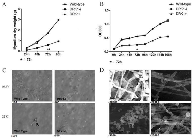
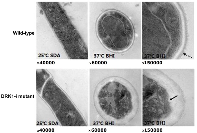
The macroscopic morphology of S. schenckii was monitored following dsRNAi, with the colony morphology of the wild-type, SsDRK1-i and SsDRK1-control strains displayed in Fig. 2A. SsDRK1-i mutants demonstrated altered phenotypic characteristics compared with the wild-type and the SsDRK1-control strains, exhibiting an obvious delay in growth, with reduced diameters (Fig. 2B) and reduced colony pigmentation (Fig. 2C) when cultured in SDA solid medium at 25°C and BHI solid medium at 37°C. The mycelial dry weight of the SsDRK1-i strain was markedly decreased compared with the wild-type and SsDRK1-control strains when cultured in SDA medium at 25°C for 96 h (P<0.05; Fig 3A). Compared with the wild-type and SsDRK1-control strains, the OD value of SsDRK1-i yeast cells was significantly decreased following 1 week of incubation on BHI liquid medium at 37°C (P<0.05; Fig 3B), indicating that interference with SsDRK1 resulted in reduced yeast cell germination. Regarding spore formation, SsDRK1-i conidia were scarce and wizened, whereas the wild-type strain displayed dense and round conidia and visible conidia formation at the tips and along the hyphae (Fig. 3C and D) To observe detailed morphological changes in the SsDRK1-i strain, the wild-type and SsDRK1-i strains were cultured for 5 days at 25°C or 37°C and examined by transmission electron microscopy. The SsDRK1-i yeast cells demonstrated plasma membrane invaginations, increased cell wall thickness and increased variability of cell wall thickness compared with the wild-type strain (Fig. 4). Furthermore, the plasma membrane invaginations observed in SsDRK1-i yeast cells were short and abundant compared with the wild-type, where they were scarce and longer; however, the plasma membrane of SsDRK1-i and wild-type hyphae were smooth and lacked invaginations. In addition, a conspicuous electron-dense outer layer was present along the outer limits of the wild-type yeast, whereas this was not observed on the surface of the SsDRK1-iyeast or the wild-type mycelial cell (Fig. 4).
Figure 2.
SsDRK1-i mutant displays defects in (A) colony morphology, (B) colony growth and (C) colony pigmentation, with the wild-type strain on the right and the SsDRK1-i strain on the left. Data are presented as the mean ± standard deviation of three experiments. *P<0.05 vs. wild-type. Ss, Sporothrix schenckii; i, interference; WT, wild-type.
Figure 3.
SsDRK1 interference affects hyphal growth and spore germination. Dry weight of (A) mycelium and (B) conidia of wild-type, SsDRK1-control and SsDRK1-i strains. (C) Wild-type and SsDRK1-i strains visualized under a microscope, with conidia formation at the tips and along the hyphae indicated by black arrowheads. (D) Wild-type and SsDRK1-i strains visualized under an electron microscope. Sparse hypha and scarce conidia are displayed in the SsDRK1-i mutant grown at 25°C and 37°C, respectively, compared with the wild-type. *P<0.05, **P<0.01 SsDRK1-I vs. wild-type and SsDRK1-control groups at 72 h. Ss, Sporothrix schenckii; i, interference; OD, optical density.
Figure 4.
Hyphal and conidial sections of wild-type and SsDRK1-i strains were observed by transmission electron microscopy. The SsDRK1-i strain displays cell walls with increased and variable thickness. The dashed line arrowhead indicates the electron-dense outer layer of wild-type yeast cells. The black arrowhead indicates the plasma membrane invaginations in the SsDRK1-i conidia. Ss, Sporothrix schenckii; i, interference; SDA, sabouraud dextrose agar; BHI, brain heart infusion.
SsDRK1 functions in cell wall synthesis
In the present study, cell wall characteristics were assessed in the SsDRK1-i strain using the cell wall binding chemical Congo red and the glucan-digesting enzyme zymolyase. The wild-type and SsDRK1-i strains were spotted onto SDA solid medium containing 20 µg/ml Congo red. SsDRK1-i strains displayed an increased sensitivity to Congo red on SDA medium (Fig. 5A). This result indicated that a reduction in SsDRK1 activity is associated with decreased polysaccharide levels in the cell wall. A similar conclusion is supported by the results of the zymolyase test. SsDRK1-i strains were more sensitive to zymolyase on SDA medium, and cell wall degradation was faster than wild-type strains (P<0.05; Fig. 5B). This may be associated with the alterations observed in the cell wall composition and structural arrangement.
Figure 5.
(A) Serial (10-fold) dilutions of wild-type and SsDRK1-i cells spotted onto sabouraud dextrose agar medium containing 20 µg/ml Congo red. (B) Effects of treatment with zymolase 20T on wild-type and SsDRK1-i strains. *P<0.05 vs. wild-type. Ss, Sporothrix schenckii; i, interference; OD, optical density.
SsDRK1 affects the transcription of the morphogenesis-associated gene, Ste20
In pathogenic fungi, the high osmolarity glycerol (HOG) pathway is involved in growth, morphology, differentiation and virulence factor regulation, including capsule and melanin synthesis. The HOG pathway in fungi consists of two major signaling modules: The MAPK module and the two-component system-like phospho-relay system. To assess the effects of SsDRK1 in the two-component system on the expression levels of key genes in the MAPK pathway, relative Ste20 mRNA expression levels in the wild-type and SsDRK1-i strains were measured using RT-qPCR. In the wild-type strain, the mRNA expression levels of Ste20 (Fig. 1B) and DRK1 (Fig. 1) in yeast cells were markedly increased compared with those in mycelia. Ste20 mRNA expression levels were also increased inSsDRK1-i cells during dimorphic switching from mycelia to yeast phase (P<0.05; Fig. 1B). However, Ste20 mRNA expression levels were markedly downregulated in SsDRK1-i yeast cells compared with wild-type yeast cells (Fig. 1B), thus suggesting that the DRK1 gene regulates Ste20 gene transcription.
SsDRK1 affects the pathogenicity of S
schenckii in a murine model
The level of inflammation in lesions of mice infected with wild-type S. schenckii strains was measured by granuloma formation. Reduced inflammatory cell infiltration was observed in the lesions of mice infected with SsDRK1-i mutants compared with the wild-type strain (Fig. 6A). Neutrophil infiltration in the lesions of the wild-type group (97.5±10/ten high magnification views) was significantly increased compared with the SsDRK1-i group (29.4±5/ten high magnification views; P<0.01; Fig. 6B). Mononuclear cell infiltration in the lesions of the wild-type group (90.2±8/ten high magnification views) was also significantly increased compared with the SsDRK1-i group (30.0±4/ten high magnification views; P<0.01; Fig. 6B).
Figure 6.
Double stranded RNA interference of DRK1 transcription affects the in vivo pathogenicity of Ss in a murine model of cutaneous infection. (A) Conidial suspensions of SsDRK1-i and wild-type strains were used to infect the mice. Local lesions were biopsied 1 week following infection. (B) Number of infiltrated neutrophils and mononuclear cells were counted in 10 fields per group/mouse. **P<0.01 vs. wild-type. Ss, Sporothrix schenckii; i, interference.
Discussion
Interference with DRK1 mRNA expression results in retarded growth of mycelia and yeast cells. Reduced colony diameter, dry weight of mycelia and optical density of spores were observed in the SsDRK1-i strain compared with the wild-type strain, demonstrating a delay in asexual development. The DRK1 homologues in B. dermatitidis (6), Botrytis cinerea (19), Aspergillus nidulans (20) and Penicilliummarneffei (21) have also been demonstrated to regulate the extent of asexual development, with DRK1 mutants producing no, or markedly reduced numbers of, conidia. In addition, the SsDRK1-i mutant displayed abnormalities in the ultrastructure of yeast cells, with variations observed in plasma membrane invaginations. Unlike wild-type yeast cells, invaginations in the SsDRK1-i yeast cells were short and abundant, being more similar to the conidia of the mycelia phase (22). This abnormality may account for the irregular morphology of spores in the SsDRK1-i strain, as detected by scanning electron microscopy.
The SsDRK1-i strain demonstrated abnormal sensitivity to Congo red and zymolyase; in addition, transmission electron microscopy revealed that the cell walls of the SsDRK1-istrain varied in thickness and lacked the electron-dense zone external to the cell wall observed in the wild-type strain. These results indicated that DRK1 is required for the deposition of cell wall materials in S. schenckii. It has previously been demonstrated that DRK1 homologues regulate cell wall composition in numerous species of pathogenic fungi. B. dermatitidis DRK1 mutants demonstrate aberrant chitin distribution and sensitivity to Congo red, a reduction in α-1, 3-glucan levels and an increase in β-1-6-glucan levels (8). Interference with NIK1, the DRK1 homologue of Candida albicans, results in reduced total cell wall hexose, a lower manno protein mass and upregulated expression of a subset of genes involved in mannan biosynthesis compared with the wild-type (23). DRKA, the DRK1 homolog in P. marneffei, has also been demonstrated to be required for cell wall integrity, with the ΔDRKA mutant displaying abnormal chitin deposits along the hyphae, abnormal cell lysis, increased sensitivity to Congo red, a decrease in the expression of genes involved in α-1, 3-glucan synthesis and variable thickness of cell walls (21). Therefore, the function of DRK1 homologues in the maintenance of cell wall integrity appears to be conserved across fungal species.
Previous research on human pathogenic fungi has revealed that the HOG pathway is involved in responses to diverse external stimuli, including osmotic shock, UV irradiation, oxidative and heavy metal stress, and high temperature (3,4). The HOG pathway is comprised of the MAPK module and the two-component system and is involved in the adaptation of fungi to the host niche, making its normal function essential for persistence of a fungus in a mammalian host (2). This conclusion is supported by the results of the present study, in which DRK1 and Ste20 mRNA expression levels were higher in yeast cells compared with mycelia in wild-type and SsDRK1-i strains. Furthermore, the present study demonstrated that dsRNAi with DRK1 resulted in significantly decreased Ste20 mRNA expression levels during the morphologic switch from the mycelia phase to the yeast phase. DRK1 is located in an upstream position in the HOG pathway, whose RR senses and relays environmental signals and subsequently activates the MAPK pathway; this location may explain decrease in Ste20 resulting from interference of DRK1 (4).
The SsDRK1-i strain demonstrated mild virulence in a murine model of cutaneous infection, and this may be associated with the observed deficiencies in yeast cell formation, cell wall synthesis, colony pigmentation and morphogenesis-associated gene expression. Yeast cell formation is essential for virulence in dimorphic fungi, including S. schenckii. For dimorphic fungal pathogens that grow in a multicellular hyphal form, the capacity to switch from the hyphal growth form to a unicellular yeast form is a central attribute that facilitates growth inside host cells without rapidly killing them. Conversion to the yeast form may also offer protection against killing by neutrophils, monocytes and macrophages. Drutz and Frey (24) demonstrated that B. dermatitidis yeast cells are too large to be ingested by polymorphonuclear neutrophils (PMNs), unlike the smaller conidia. Peripheral blood monocytes are also more efficient at killing conidia than yeast, similar to PMNs (25). In the present study, fewer spores and aberrant yeast cells, with morphology mostly consistent with the conidia of the mycelia phase rather than yeast cells, were observed in the SsDRK1-i strain, which may account for the mild virulence observed in SsDRK1-i strains.
dsRNAi of DRK1 in S. schenckii also affects colony pigmentation (Fig. 2). Melanin is an insoluble compound associated with virulence in several fungal pathogenic species (26). Melanization has been demonstrated to be involved in the pathogenesis of cutaneous sporotrichosis, with a melanized strain displaying increased formation of multifocal granulomas and greater invasive abilities compared with an albino mutant strain in an experimental rat model of sporotrichosis (27). Furthermore, conidial melanizationhas been observed to enhance the resistance of S. schenckii to macrophage phagocytosis, enhancing the first stages of infection, as these structures are usually the fungal infective particles (28). Morris-Jones et al (29) suggested that the melanin pigment in S. schenckii may reduce the efficacy of treatment in some sporotrichosis cases, particularly in cases of extracutaneous disease or in patients co-infected with human immunodeficiency virus (29). In the present study, SsDRK1-i strains failed to produce as much melanin as the spores of wild-type strains, and this may affect the pathogenic potential of this strain.
Defects that affect cell wall integrity have significant implications for fungal pathogenicity. Dysregulation of cell wall biosynthesis has previously been demonstrated to affect the initial stages of infection. The C. albicans CHK1 mutant displays altered cell wall composition and reduced adherence to human esophageal tissue, resulting in attenuated virulence (30). Defects in cell wall integrity also increase recognition by the innate immune system. The P. marneffei ΔDRKA mutant displays severe cell wall defects and an increased rate of phagocytosis (21). The abnormal cell wall components may also result in decreased virulence in S. schenckii.
Furthermore, the DRK1 gene has been identified as a regulator of phase-specific virulence factor expression in pathogenic fungi. RNA silencing of the DRK1 gene results in the reduction of the yeast-phase gene BAD1 in B. dermatitidis and the yeast-phase genes CBP1, α-glucan synthase (AGS1) and yps-3 in H. capsulatum (31). Since expression of BAD1, CBP1 and AGS1 is required for pathogenicity, Nemecek et al (8) hypothesized that silencing DRK1 expression would impair the virulence of B. dermatitidis and H. capsulatum. The reduced Ste20 mRNA expression levels observed in the SsDRK1-i strain in the present study may lead to a reduction of yeast-phase gene expression through regulating HOG/MAPK pathway signal transduction, and further inhibit the morphogenesis of the pathogenic yeast phase.
In conclusion, SsDRK1, an intracellular soluble histidine kinase, has been demonstrated to be required for normal mycelial growth, yeast cell formation, cell wall integrity, morphogenesis-associated gene expression and pathogenicity in S. schenckii. However, several lines of enquiry worthy of further investigation remain, including what environmental signals DRK1 senses and whether other upstream transmembrane histidine kinases act as external signal receptors and interact with SsDRK1. These results implicate SsDRK1 as a potential novel target for the development of anti-sporotrichosis therapies.
Acknowledgements
The present study was supported by the Shenzhen Innovation and Technology Commission Foundation (grant no. JCYJ20140414092023239) and the National Natural Science Foundation of China (grant nos. 81000699 and 81472891).
References
- 1.Morris-Jones R. Sporotrichosis. Clin Exp Dermatol. 2002;27:427–431. doi: 10.1046/j.1365-2230.2002.01087.x. [DOI] [PubMed] [Google Scholar]
- 2.Cooney NM, Klein BS. Fungal adaptation to the mammalian host: It is a new world, after all. Curr Opin Microbiol. 2008;11:511–516. doi: 10.1016/j.mib.2008.09.018. [DOI] [PMC free article] [PubMed] [Google Scholar]
- 3.Bahn YS, Xue C, Idnurm A, Rutherford JC, Heitman J, Cardenas ME. Sensing the environment: Lessons from fungi. Nat Rev Microbiol. 2007;5:57–69. doi: 10.1038/nrmicro1578. [DOI] [PubMed] [Google Scholar]
- 4.Bahn YS. Master and commander in fungal pathogens: The two-component system and the HOG signaling pathway. Eukaryot Cell. 2008;7:2017–2036. doi: 10.1128/EC.00323-08. [DOI] [PMC free article] [PubMed] [Google Scholar]
- 5.Brewster JL, de Valoir T, Dwyer ND, Winter E, Gustin MC. An osmosensing signal transduction pathway in yeast. Science. 1993;259:1760–1763. doi: 10.1126/science.7681220. [DOI] [PubMed] [Google Scholar]
- 6.Brown SM, Campbell LT, Lodge JK. Cryptococcus neoformans, a fungus under stress. Curr Opin Microbiol. 2007;10:320–325. doi: 10.1016/j.mib.2007.05.014. [DOI] [PMC free article] [PubMed] [Google Scholar]
- 7.Bahn YS, Kojima K, Cox GM, Heitman J. A unique fungal two-component system regulates stress responses, drug sensitivity, sexual development, and virulence of Cryptococcus neoformans. Mol Biol Cell. 2006;17:3122–3135. doi: 10.1091/mbc.E06-02-0113. [DOI] [PMC free article] [PubMed] [Google Scholar]
- 8.Nemecek JC, Wüthrich M, Klein BS. Global control of dimorphism and virulence in fungi. Science. 2006;312:583–588. doi: 10.1126/science.1124105. [DOI] [PubMed] [Google Scholar]
- 9.Cheung J, Hendrickson WA. Sensor domains of two-component regulatory systems. Curr Opin Microbiol. 2010;13:116–123. doi: 10.1016/j.mib.2010.01.016. [DOI] [PMC free article] [PubMed] [Google Scholar]
- 10.Hou B, Zhang Z, Zheng F, Liu X. Molecular cloning, characterization and differential expression of DRK1 in Sporothrix schenckii. Int J Mol Med. 2013;31:99–104. doi: 10.3892/ijmm.2012.1193. [DOI] [PubMed] [Google Scholar]
- 11.Sambrook J, Fritsch EF, Maniatis T, editors. Molecular cloning: A laboratory manual. 3rd edition. Cold spring harbor laboratory; New York, NY, USA: 2001. [Google Scholar]
- 12.Zhang P, Xu B, Wang Y, Li Y, Qian Z, Tang S, Huan S, Ren S. Agrobacterium tumefaciens-mediated transformation as a tool for insertional mutagenesis in the fungus Penicillium marneffei. Mycol Res. 2008;112:943–949. doi: 10.1016/j.mycres.2008.01.017. [DOI] [PubMed] [Google Scholar]
- 13.Garrison RG, Boyd KS, Mariat F. Ultrastructural studies of the mycelium-to yeast transformation of sporothrix schenckii. J Bacteriol. 1975;124:959–968. doi: 10.1128/jb.124.2.959-968.1975. [DOI] [PMC free article] [PubMed] [Google Scholar]
- 14.Wang F, Tao J, Qian Z, You S, Dong H, et al. A histidine kinase PmHHK1 regulates polar growth, sporulation and cell wall composition in the dimorphic fungus Penicillium marneffei. Mycological Research. 2009;113:915–923. doi: 10.1016/j.mycres.2009.03.006. [DOI] [PubMed] [Google Scholar]
- 15.Davis RH, De Serres FJ. Genetic and microbiological research techniques for neurospora crassa. Methods Enzymol. 1970;17:79–143. doi: 10.1016/0076-6879(71)17168-6. [DOI] [Google Scholar]
- 16.De Nobel JG, Klis FM, Priem J, Munnik T, van den Ende H. The glucanase-soluble mannoproteins limit cell wall porosity in Saccharomyces cerevisiae. Yeast. 1990;6:491–499. doi: 10.1002/yea.320060606. [DOI] [PubMed] [Google Scholar]
- 17.Livak KJ, Schmittgen TD. Analysis of relative gene expression data using real-time quantitative PCR and the 2(-Delta Delta C(T)) method. Methods. 2001;25:402–408. doi: 10.1006/meth.2001.1262. [DOI] [PubMed] [Google Scholar]
- 18.Kong X, Xiao T, Lin J, Wang Y, Chen HD. Relationships among genotypes, virulence and clinical forms of sporothrix schenckii infection. Clin Microbiol Infect. 2006;12:1077–1081. doi: 10.1111/j.1469-0691.2006.01519.x. [DOI] [PubMed] [Google Scholar]
- 19.Viaud M, Fillinger S, Liu W, Polepalli JS, Le Pêcheur P, Kunduru AR, Leroux P, Legendre L. A class III histidine kinase acts as a novel virulence factor in botrytis cinerea. Mol Plant Microbe Interact. 2006;19:1042–1050. doi: 10.1094/MPMI-19-1042. [DOI] [PubMed] [Google Scholar]
- 20.Vargas-Pérez I, Sánchez O, Kawasaki L, Georgellis D, Aguirre J. Response regulators SrrA and SskA are central components of a phosphorelay system involved in stress signal transduction and asexual sporulation in Aspergillus nidulans. Eukaryot Cell. 2007;6:1570–1583. doi: 10.1128/EC.00085-07. [DOI] [PMC free article] [PubMed] [Google Scholar]
- 21.Boyce KJ, Schreider L, Kirszenblat L, Andrianopoulos A. The two-component histidine kinases DrkA and SlnA are required for in vivo growth in the human pathogen Penicillium marneffei. Mol Microbiol. 2011;82:1164–1184. doi: 10.1111/j.1365-2958.2011.07878.x. [DOI] [PubMed] [Google Scholar]
- 22.Svoboda A, Trujillo-Gonzalez A. Sporothrix schenckii-a freeze-fracture study. J Basic Microbiol. 1990;30:371–378. doi: 10.1002/jobm.3620300514. [DOI] [PubMed] [Google Scholar]
- 23.Kruppa M, Jabra-Rizk MA, Meiller TF, Calderone R. The histidine kinases of Candida albicans: Regulation of cell wall mannan biosynthesis. FEMS Yeast Res. 2004;4:409–416. doi: 10.1016/S1567-1356(03)00201-0. [DOI] [PubMed] [Google Scholar]
- 24.Drutz DJ, Frey CL. Intracellular and extracellular defenses of human phagocytes against Blastomyces dermatitidis conidia and yeasts. J Lab Clin Med. 1985;105:737–750. [PubMed] [Google Scholar]
- 25.Klein BS, Tebbets B. Dimorphism and virulence in fungi. Curr Opin Microbiol. 2007;10:314–319. doi: 10.1016/j.mib.2007.04.002. [DOI] [PMC free article] [PubMed] [Google Scholar]
- 26.Revankar SG, Sutton DA. Melanized fungi in human disease. Clin Microbiol Rev. 2010;23:884–928. doi: 10.1128/CMR.00019-10. [DOI] [PMC free article] [PubMed] [Google Scholar]
- 27.Madrid H, Cano J, Gené J, Bonifaz A, Toriello C, Guarro J. Sporothrix globosa, a pathogenic fungus with widespread geographical distribution. Rev Iberoam Micol. 2009;26:218–222. doi: 10.1016/j.riam.2009.02.005. [DOI] [PubMed] [Google Scholar]
- 28.Romero-Martinez R, Wheeler M, Guerrero-Plata A, Rico G, Torres-Guerrero H. Biosynthesis and functions of melanin in Sporothrix schenckii. Infect Immun. 2000;68:3696–3703. doi: 10.1128/IAI.68.6.3696-3703.2000. [DOI] [PMC free article] [PubMed] [Google Scholar]
- 29.Morris-Jones R, Youngchim S, Gomez BL, Aisen P, Hay RJ, Nosanchuk JD, Casadevall A, Hamilton AJ. Synthesis of melanin-like pigments by Sporothrix schenckii in vitro and during mammalian infection. Infect Immun. 2003;71:4026–4033. doi: 10.1128/IAI.71.7.4026-4033.2003. [DOI] [PMC free article] [PubMed] [Google Scholar]
- 30.Bernhardt J, Herman D, Sheridan M, Calderone R. Adherence and invasion studies of Candidia albicans strains, utilizing in vitro models of esophageal candidiasis. J Infect Dis. 2001;184:1170–1175. doi: 10.1086/323807. [DOI] [PubMed] [Google Scholar]
- 31.Rappleye CA, Engle JT, Goldman WE. RNA interference in Histoplasma capsulatum demonstrates a role for alpha-(1,3)-glucan in virulence. Mol Microbiol. 2004;53:153–165. doi: 10.1111/j.1365-2958.2004.04131.x. [DOI] [PubMed] [Google Scholar]


