Abstract
Sensors are devices or systems able to detect, measure and convert magnitudes from any domain to an electrical one. Using light as a probe for optical sensing is one of the most efficient approaches for this purpose. The history of optical sensing using some methods based on absorbance, emissive and florescence properties date back to the 16th century. The field of optical sensors evolved during the following centuries, but it did not achieve maturity until the demonstration of the first laser in 1960. The unique properties of laser light become particularly important in the case of laser-based sensors, whose operation is entirely based upon the direct detection of laser light itself, without relying on any additional mediating device. However, compared with freely propagating light beams, artificially engineered optical fields are in increasing demand for probing samples with very small sizes and/or weak light–matter interaction. Optical fiber sensors constitute a subarea of optical sensors in which fiber technologies are employed. Different types of specialty and photonic crystal fibers provide improved performance and novel sensing concepts. Actually, structurization with wavelength or subwavelength feature size appears as the most efficient way to enhance sensor sensitivity and its detection limit. This leads to the area of micro- and nano-engineered optical sensors. It is expected that the combination of better fabrication techniques and new physical effects may open new and fascinating opportunities in this area. This roadmap on optical sensors addresses different technologies and application areas of the field. Fourteen contributions authored by experts from both industry and academia provide insights into the current state-of-the-art and the challenges faced by researchers currently. Two sections of this paper provide an overview of laser-based and frequency comb-based sensors. Three sections address the area of optical fiber sensors, encompassing both conventional, specialty and photonic crystal fibers. Several other sections are dedicated to micro- and nano-engineered sensors, including whispering-gallery mode and plasmonic sensors. The uses of optical sensors in chemical, biological and biomedical areas are described in other sections. Different approaches required to satisfy applications at visible, infrared and THz spectral regions are also discussed. Advances in science and technology required to meet challenges faced in each of these areas are addressed, together with suggestions on how the field could evolve in the near future.
Keywords: optical sensors, optical sensing, fiber sensors, micro and nano-engineered optical sensors, chemical sensing, biological sensing, biomedical sensing
1. Terahertz sensors and terahertz sensing applications
Enrique Castro-Camus
Centro de Investigaciones en Optica A.C
Status
The band of the electromagnetic spectrum that falls between the microwaves and the infrared has traditionally been referred to as the terahertz band. It corresponds to wavelengths between ~10 mm and 30 μm, frequencies between 30 GHz and 10 THz or photon energies between 0.1 meV and 40 meV. This band was particularly hard to access until the late 1980s when the invention of ultrafast mode-locked lasers together with some progress in the area of semiconductor devices allowed the creation of photoconductive emitters and detectors [1], which in turn gave rise to the technique known as terahertz time-domain spectroscopy [2] that remains the most widely used spectroscopic tool for this band of the spectrum.
In addition to terahertz time-domain spectroscopy, which relies on the generation and detection of broadband electromagnetic transients, there has been enormous progress on the development of other types of sensors for the terahertz band. For instance bolometric devices that now incorporate antennae structures are being integrated in planar arrays [3]. Additionally, photomixers that have similar structures to the photoconductive detectors, but that are CW-pumped, have been introduced [4]. Quantum-well structures with specifically engineered inter-sub-band transitions have been tested as potential photodetectors [5]. Field effect transistors that act as terahertz detectors that incorporate graphene (see figure 1) and other nanomaterials have been introduced too [6, 7]. Of course there has been an enormous amount of other progress that we cannot list exhaustively here.
Figure 1.
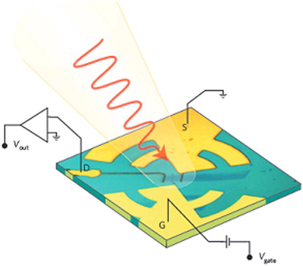
Graphene-based field effect transistor used as a sensor of terahertz radiation. Reprinted by permission from Macmillan Publishers Ltd: Nature Materials [6], Copyright (2012).
In addition to the rapid development that terahertz detector devices has seen in the last three decades, the availability of such devices has allowed the use of terahertz radiation as a powerful non-destructive and non-contact sensing tool in a variety of areas. For instance, terahertz technology is able to probe the conductivity of semiconductors with sub-picosecond time resolution [8]. It has also been used to determine the water content of biological tissues [9], the evolution of chemical processes [10], the complex dynamics of proteins [11], and as an inspection tool in industry [12] and cultural heritage [13], among many other applications.
Current and future challenges
While there has been enormous progress in the creation of terahertz devices and in the proof-of-concept of various applications, currently the main reason why terahertz technology is uncommon in ‘real world’ applications is its cost. Almost two decades ago the first commercial terahertz system was introduced. Currently the price of THz spectroscopy starts in the range of 50000 US dollars but can go up to several hundred thousand depending on their capabilities. Furthermore, although transportable, terahertz systems are still far from being hand-held devices. In addition, terahertz systems are still relatively unreliable, usually requiring regular minor ‘tweaking’ by qualified staff, and maintenance by specialized engineers every few months.
An application that will probably develop strongly in the coming years is the use of terahertz frequencies for high speed telecommunications. In this particular case, the development of appropriate high speed modulators and demodulators is probably the most clear challenge within the community.
Advances in science and technology to meet challenges
The main reason for the high cost of terahertz systems is the inclusion of expensive lasers and the need for relatively uncommon semiconductors for the fabrication of emitters and detectors. Although the terahertz community has made some progress in this sense, this aspect will probably only be resolved when the market requires terahertz technologies and they start being mass produced.
The robustness of terahertz time-domain systems has improved recently, especially with the introduction of fiber lasers, which are immune to misalignment problems and far less sensitive to environmental conditions. Another reason for the lack of robustness is the need for moving parts; these are being replaced by using clever solutions that involve using the jitter between pulses of two actively tuned laser cavities. In addition the traditional semiconductor used for the fabrication of terahertz detectors, low-temperature MBE-grown GaAs, is being replaced by ion implanted III–V materials and there are some geometric device designs that are being explored that could make it possible to simply use standard semi-insulating GaAs.
Potential physical phenomena that can lead to modulators for telecommunications are being explored. Devices based on metamaterials, metasurfaces, liquid crystals, ultrafast photoconductors and many other materials have been proposed, and some of them have shown to be promising for this application [14].
Concluding remarks
Terahertz sensors have evolved dramatically over the last three decades. Many of these technologies are still only available in research laboratories, and those that are commercially available remain relatively expensive. Terahertz has shown enormous potential as a sensing tool in a number of applications which range from characterization of semiconductor properties to industrial inspection. The market for high speed telecommunication technologies can clearly be foreseen as the driver of the development of economical and mass produced terahertz devices in the future.
2. Mid-infrared sensing
David J Ottaway
The University of Adelaide
Status
The mid-infrared is commonly defined as the part of the electromagnetic spectrum that has wavelengths between 3–8 μm. It offers significant opportunities for trace gas sensing since many strong absorption lines occur in this region (see figure 2). The absorption lines in this part of the spectrum can be two orders of magnitude stronger than the overtone lines that occur in the shorter end of the infrared. A significant number of sensing techniques have been developed in this shorter wavelength region, which are suitable to deployment in the mid-infrared.
Figure 2.
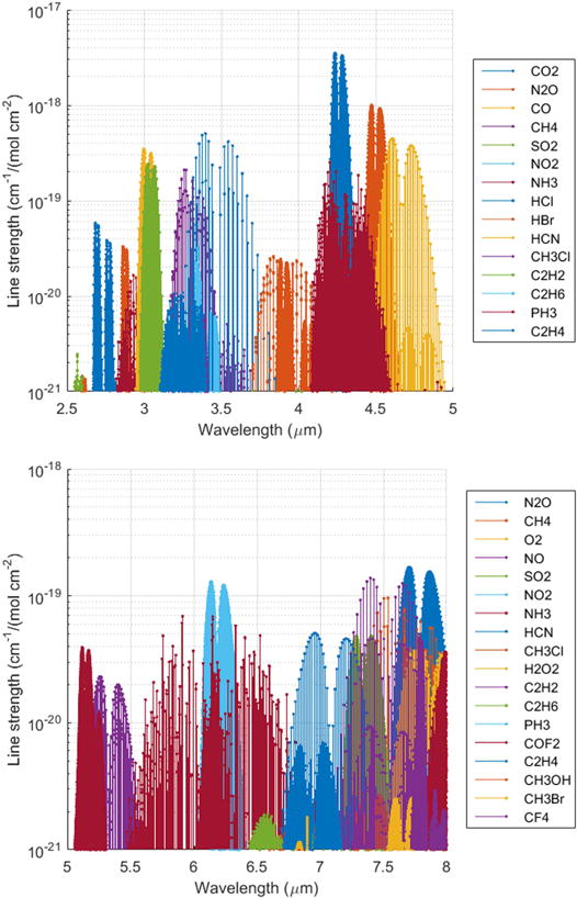
The mid-infrared contains many of the strongest absorption lines for important trace gases, data from www.spectralcalc.com/.
The sensitivity of a sensing platform is governed by the combination of its light source and its associated detection system. The availability of convenient sources of coherent radiation in the mid-infrared has been limited but there has been significant source development in recent years. Development in mid-infrared detectors has also occurred such that the performance of these detectors is approaching the limit set by thermal background radiation over much of the band.
Exploitation of the mid-infrared will accelerate once a number of new sensing techniques that were initially demonstrated in the visible and near infrared are adapted to longer wavelengths. These include suspended and exposed core fibers and techniques that make use of frequency combs such as dual-comb spectroscopy and virtual imaged planar array (VIPA) spectroscopy. These technologies should be readily adaptable into the mid-infrared once a few technological hurdles have been overcome.
Current and future challenges
To achieve faster and more accurate standoff detection of gases the product of the source spectral brightness and receiver area must be maximized. Significant development of compact and convenient mid-infrared sources with high spectral purity and beam quality is still required to fully exploit the mid-infrared.
The direct generation of laser light in the mid-infrared has been achieved using a number of different approaches including lasers based on semiconductor (quantum and interband cascade techniques), fiber and Cr/Fe ZnSe/S solid state platforms.
Nonlinear techniques have also been extensively used to generate mid-infrared radiation by shifting high brightness near-infrared sources into the mid-infrared. Such techniques include optical parametric oscillators/amplifiers, frequency difference generation and Raman shifting. Further, high beam quality fiber-based super continuum based sources have generated white light sources that cover the entire mid-infrared band [15]. Frequency combs that routinely achieve octave spanning performance in the near infrared are being pushed deeper into the mid-infrared [16].
Quantum cascade lasers were invented in 1994 and have made impressive advances across most of the mid-infrared band particularly at the longer end [17]. These lasers and interband cascade lasers provide a convenient platform for integration into compact trace gas sensing devices. These lasers have produced continuous power levels exceeding 1 W and can cover most of the mid-infrared band. Cascade lasers suffer from short upper state lifetimes, typically of picoseconds for quantum cascade lasers and sub-nanosecond for interband cascade lasers. This makes achieving peak power challenging since the maximum energy storage is low [18]. Initially thought to be a barrier for mode-locking, these lasers have generated mode-locked output using Kerr nonlinearity within the gain medium itself [19] or as a pump source for crystalline resonators but more work remains to increase power and wavelength coverage.
Fiber lasers have produced very high power and excellent beam quality in the near infrared (see figure 2 in [20]). Recently, the output of these lasers has been pushed into the shorter end of the mid-infrared with the potential to go further using either dual-wavelength pumped rare-earth doped lasers [20] or Raman based lasers [21]. Fiber lasers are compact, rugged and portable devices that are a promising candidate for generating significant peak power in the mid-infrared. This is possible because the sources benefit from long upper state lifetimes, which allow significant energy storage. Fiber lasers have delivered significant average power in both mode-locked and continuous-wave output in the near infrared. Recently, the direct generation of mode-locked power has been demonstrated near the short edge of the mid-infrared, which is a precursor to broad frequency combs [22, 23]. In Q-switched operation peak power approaching 1 kW has been demonstrated around 3 μm [24] in erbium and recent modeling results suggest that peak powers approaching this should be feasible from the 3.5 μm transition [20]. These high peak powers could be invaluable for long distance standoff detection.
There is considerable scope to push the output of fiber lasers further into the mid-infrared. The glass host will play a critical role here since the absorption losses and non-radiative quenching of upper lasing states will become really significant once wavelengths extend beyond 4 μm [25]. Two types of glasses, indium fluoride and chalcogenide, may have a significant role here as they have significantly lower phonon energy than ZBLAN. Indium fluoride has been shown to be more mechanically robust than ZBLAN with transmission to 6.5 μm [25] and shows real promise for enabling laser emission in the 4–5 μm range. For laser emission significantly beyond this chalcogenide certainly has the transmission and low phonon energy needed to push laser emission to the long edge of the mid-infrared band [25]. However, laser emission from a chalcogenide host has proven challenging and only a few reports of lasing in the near infrared have been made.
Frequency combs have allowed significant advances in lab based spectroscopy and remote sensing (see section 7). Remote trace gas sensing using dual-comb spectroscopy has been demonstrated over kilometer scales [26] and VIPA spectroscopy has enabled rapid high SNR spectral measurements [27]. The sensitivity of both these techniques could be increased if mid-infrared wavelengths were used. The detection side of these techniques pretty much exists for the mid-infrared but convenient combs still need to be improved. Recent work on silicon microresonators has demonstrated that convenient combs in the mid-infrared are feasible [16] but more work needs to be done to provide the convenient inexpensive sources that exist in the near infrared.
One of the holy grails of trace gas sensing is breath analysis for medical diagnostics from an inexpensive platform that could be deployed in medical clinics. Variants of mid-infrared absorption spectroscopy offer hope for this endeavor. To achieve the extremely high sensitivity needed, a long path length is required. Conventionally this has been achieved using resonant cavities or Herriot delay lines. One compact and potentially convenient alternative is microstructure fibers such as suspended core and hollow core fibers, which allow light to interact with a trace gas over extended distances [25].
Suspended core fiber sensing relies on the evanescent field to interact with the material under testing. The numerical aperture of an air–glass interface is very high which means that the cores need to be significantly less than 1 μm such that a significant fraction of the light propagates outside the core when operated in near IR/visible. The longer wavelength of the mid-infrared light means considerably more light is transmitted outside the core for a comparable core size. To date there has been very few microstructure fibers demonstrated in glass with significant mid-IR transmission. Hollow core type fibers offer significant hope here since the light travels predominantly in air and hence much less material interaction loss (see section 5). Exposed core fibers (see figure 3) [28] are a recent advance that allows for more convenient interaction between light and chemicals than fibers that need to be filled from one end. A future challenge is to develop microstructured fiber that has low loss deep into the mid-infrared.
Figure 3.
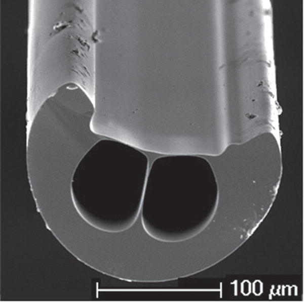
An exposed core fiber in fused-silica. Reproduced with permission from [28]. © 2012 OSA.
Advances in science and technology to meet challenges
The mid-infrared remains a tricky area to work in and currently there is not the proliferation of commercially available components that exist in the near infrared. Fortunately, this situation is changing. There still exists significant challenges regarding the fabrication of components that need to be solved to enable the development of sensing technologies deep in the mid-infrared.
Concluding remarks
The mid-infrared offers a fertile area for the development of new highly sensitive and selective trace gas sensing technologies. There have been significant advances in a wide variety of source developments in recent years but much remains to be done if the mid-infrared is to completely fulfill its promise.
Acknowledgments
We thank Steve Maddern, Andre Luiten, Heike Ebendorff-Heidepriem and Ori Henderson-Sapir for useful discussions.
3. Optical fiber sensors
José Miguel López-Higuera
University of Cantabria, CIBER-bbn and IDIVAL, Spain
Status
Sensors are devices or systems used to capture, quantify and faithfully translate magnitudes from any domain to the electrical one. When light sciences and technologies are used, the sensing using light (SuL) area of photonics appears. Optical fiber sensors (OFS) can be understood as a subarea of SuL in which fiber technologies are employed. In an OFS the measurand modulates characteristics of light in some part of an optical fiber system (the transducer), faithfully reproducing it in the electric domain [29]. Any OFS can be considered to be integrated by three main parts: transducer, channel and optoelectronic or interrogation units (figure 4).
Figure 4.
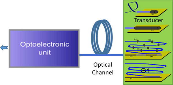
Schematic view of the three main parts of an optical fiber sensor system (optoelectronic or interrogation unit), the optical channel and the transducer part. Four main types of transducers according to the spatial distribution of the measurand: point (the upper one); integral; quasi-distributed and (below one) distributed.
It can be said that the development of OFS started in the latter half of the 1970s. As reported by Giallorenzy [30], relevant contributions were achieved on temperature, magnetic, acoustic and pressure point based sensors by 1982. Many techniques were explored such as those implementing interferometry (Michelson, Mach-Zehnder, Sagnac,..) for the development of current sensors (Faraday effect), hydrophones for the navy and also fiber optic gyroscopes (Sagnac effect) [31]. The possibility of multiplexing sensors in a given fiber (enabling OFS networks and reducing the cost-per-measurement-point) was demonstrated by 1986. Based on scattering and optical time-domain reflectometry (OTDR) techniques, distributed sensors appeared by the end of the 1980s. Linear (Rayleigh) and nonlinear back-scatterings in fibers (Raman and later Brillouin) were used. Techniques such as OTDR and DTS (distributed temperature sensing) based on Rayleigh and Raman effects, respectively, and also BOTDR and BOTDA (Brillouin optical time-domain reflectometry/analysis) with their respective variants enabled and nurtured the very important subarea of fiber distributed sensors. Both types of Brillouin sensing techniques became commercially available towards the end of the 1990s [32].
The fiber Bragg grating (FBG) structure, a key technology for sensing, was introduced in September of 1989 [33]. The appearance of fiber optical amplifiers in the 1990s enabled the increasing of the signal/noise (S/N) ratio in the optoelectronic units, the appearance of the fiber optic active networks [34], fiber lasers sensors and also enhanced distributed sensors with a more dynamic range (longer transducers) for a given resolution.
In 1996 the photonic crystal fiber was demonstrated [35] which, with many other specialty fibers and new and advanced optical devices (in many cases developed for the telecom industry), became a new key technological platform for sensing during the first two decades of the 21st century.
Because of the relevance of this sensing area the international Optical Fiber Sensors (OFS) conference was born (London, 1983). Later, in 1989 the second more relevant meeting on this sensing technology was created: EWOFS (European Workshop on Optical Fiber Sensors). Over 6000 conference papers were defended after the OFS 25th edition (May 2017, Jeju Island, Korea), in addition to the large amount of journal articles, books and patents already published. This constitutes a great source to study the history, evolution and state-of the-art of SuL technology.
Given that the final aim of a technology is to be exploited in real applications and to have commercial success, the market and customer needs should be known. Some comments on the OFS market need to be addressed to realistically foresee the near future.
OFS market forecast
Point sensors (including integrated) often work as single point indicators and are commonly used in gyro, civil infrastructure, industrial, military, medical and oil and gas applications, by this order of market relevance. A comparison of the evolution of revenues of both point and distributed sensor markets in the 21st century reveals that, while at the beginning of the century the point sensor market was larger, the remarkably higher compound annual growth rates (CAGRs) of the distributed market drove it such that by 2008 both were practically matched (about $330 million) and already by 2014 the distributed market turned out to approximately double the former. This evolution continues and at the end of the second decade, the distributed market is expected to be over two times bigger than the point sensor one. In this regard, the following will focus on the distributed market.
The oil and gas market sector experienced a contraction lasting around 2015 as illustrated in figure 5. The forecast for distributed OFS sensors over the next five years is strong expecting a conservative CAGR of around 11% with revenues approaching $1.4 billion by 2020. Oil and gas in-wells, followed by homeland security, infrastructure and military, are expected to be the four main markets. The oil and gas sector represents 46% of the total market.
Figure 5.
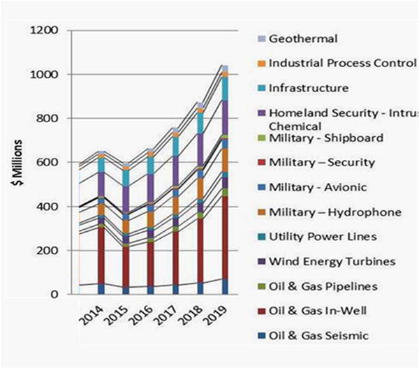
Distributed optical sensor market forecast by application sector and (inset) by technology type. Reproduced with permission from [36]. © Information Gatekeepers Inc. Data also courtesy of Alexis Mendez.
Concerning the technology used, the largest market share (33%) is expected for Raman scattering (DTS) sensors. They will be followed by quasi-distributed sensors based on FBGs (26%). DAS (Rayleigh scattering), which is rapidly gaining market share, is projected to be around 10% by 2019. However, Brillouin scattering sensors have less than 2% market share [36]. The total OFS market size in China is difficult to estimate, but could be about 25%–30% of the present size of the Western OFS market.
Current and future challenges
The main general challenge is still to guarantee that data from sensors represent the real behavior of the object to be sensed and is not corrupted due to any sensor malfunction. For that reason, it could be necessary to monitor the sensors themselves. This fact drives to the very challenging tasks of developing new techniques for: (a) sensor validation by itself or by means of reports on each other’s condition; (b) ‘fail-safe’ sensor networks: reliability.
If a sensor fails, the damage identification algorithms must be able to adapt to the network. This adaptive capability implies that a certain amount of redundancy must be built into the sensor system or network.
Challenging tasks on OFS are closely related to the previous ones and also: (i) to reduce the sensor cross-sensitivities; (ii) to improve the resolution and/or the dynamic range; (iii) to improve the stability, reliability in real situations and (iv) to achieve small size, light weight, low power consumption and cost-effective sensors.
On the other hand, to be used in real applications a key challenge is to ensure that the OFS sensor system (or sensor network) itself is not damaged when deployed in the field or during the working period of its expected lifetime.
Advances in science and technology to meet challenges
New concepts, techniques, technologies and fabrication processes are required to reach tiny fully integrated optical sensor systems to enable an extensive use of SuL approaches. The availability of fully integrated optical devices, circuits and subsystems will have a great impact on both the transducer and the optoelectronic units of point OFS systems with the exception of gyroscopes and distributed ones, where the impact will be on the optoelectronic unit. These facts will improve the technical characteristics and drive drastic reductions in the sensor costs.
With the exception of specific applications such as gyroscopes and PCFs-based sensors, point optical transducers will progressively emigrate towards integrated optics approaches, remaining in fiber technology the optical channel.
With no comparable performance with other available ‘classic’ sensors, nowadays the distributed fiber sensing technology is expected to be the stronger OFS one. So new methods, techniques and technologies to improve both the spatial and measurand resolutions, the dynamic ranges, and also materials to enable new transducer fiber cables able to reliably work in harsh environments, are all welcomed.
New concepts and advances that can be potentially used with advantages in sensing must be explored. Quantum optics [37] and metamaterials with exotic optical properties not found in nature, such as those where light waves can propagate with infinite phase velocity, corresponding to a refractive index of zero [38], and also using light to create sound in fibers able to develop innovative new chemical sensing methods, are three candidates [39].
New fiber technology platforms able to support the on-line trapping, propelling, stopping and enabling of both sensing and treatments of tiny particles or molecules using light, are very welcome. Developments based on new hollow core PCFs are clear candidates [40].
Concluding remarks
Integrated optical devices, circuits and subsystems are expected to improve their technical characteristics and drive drastic reductions in the sensor costs. In addition, in the coming years, new quantum based concepts and metamaterials with exotic optical properties, among others, will be explored and potentially developed for sensing purposes. The above-mentioned research lines will contribute to significantly increasing the real use of OFS technologies in real applications and hence to expand their market. As an example, progress will enable one to reach the ultimate challenge of gyros with inertial navigation performance of one nautical mile per month corresponding to a long-term bias stability of 10−5 °/h.
Acknowledgments
This work has been supported by the projects TEC2013-47264-C2-1-R, TEC2016-76021-C2-2-R of the Spanish CICYT and also cofunded with European FEDER funds.
4. Specialty fibers for sensing applications
Xian Feng
Beijing University of Technology
Status
The emergence of optical fiber sensor technology was largely driven by the telecommunication industry, in combination with low-cost optoelectronic components in 1970s and 1980s [41]. As technology has advanced, the need for optical fiber sensors has increased dramatically in areas such as construction, manufacturing, medicine, defense and aerospace. So far, optical fibers can be used as sensors to measure physical and chemical information such as temperature, strain, pressure, vibration, rotation, acceleration, electric fields, magnetic fields, chemical compositions of the surrounding liquids or gases and other quantities, which provide measurable modulation on the intensity, phase, polarization, wavelength or transit time of light in the fiber.
Typically fiber sensors are divided into two categories, intrinsic fiber sensors and extrinsic fiber sensors. The former is also referred to as all-fiber optic sensor, in which the sensing takes place within the fiber itself. The latter is often referred to as a hybrid fiber sensor, which is capable of sensing the characteristics taking place in the region outside the fiber. In comparison with the conventional telecommunication fiber, a specialty fiber is made either through modifying the fiber materials or through introducing micro-structures or nanostructures into the fiber core and/or cladding for achieving desired novel sensing functionalities, high sensitivity and low detection limit. Figure 6 illustrates the schematic of a traditional specialty optical fiber sensor. When the fiber is located inside the physical or chemical environment, the interaction between the optical field propagating along the fiber core and the external fields modulates the optical field. Due to the certain correlation between the differential optical signal through the fiber core and the surrounding field(s), the sensing function can be achieved. Specialty fiber sensors are advantageous due to their attributes such as low weight, small size, low power, environmental ruggedness, immunity to electromagnetic interference, good performance specifications and low cost. These are what an ideal sensor should possess. Hence, while the technology of other families of sensors grows rapidly, specialty fiber sensors with multiple novel non-optical functionalities plays an important role nowadays.
Figure 6.
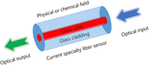
Schematic of traditional specialty optical fiber sensor.
Current and future challenges
The rapid development of modern sensors presents specialty fiber sensors with great challenges. For example, other families of sensors can offer similar sensing functions but with easily accessible readout systems; and other waveguide sensors, e.g., integrated photonic waveguide sensors on a silicon chip, could also provide compact, economic devices benefitting from the large-scale silicon manufacturing industry. A comprehensive coverage of the current and future challenges for specialty fiber sensors is not be possible due to the limited understanding and expertise of the author. Nevertheless, this section tries to address this issue by highlighting some recent hot subjects on sensors.
For example, in the case of biochemical sensors in order to realize single molecule detection or single cell manipulation, the main challenge is how to breach the diffraction limit and how to confine the light on the nanometer size, which is far below the optical wavelength scale. The sensitivity can be enhanced by increasing the light–matter interaction, which effectively increases the light fraction per unit area of the detection surface. This can be done by attaching functional nanomaterials or nanostructures onto the proximity of the guiding core. The localized dipolar surface plasmon resonance (LSPR) and surface-enhanced Raman scattering (SERS) can enhance the detected signal by several orders of magnitude [42, 43]. The underlying physical properties of nanophotonics can be seamlessly integrated with a wide range of chemical surface modification approaches, opto-microfluidics and biological interfacing biosensors with single molecule sensitivity and detection limits.
Generally speaking, recent research activities suggest a few promising directions for various novel sensors:
In order to largely enhance the sensitivities and detection limit, the sensor can be embedded with submicron-scale or nanoscale features, i.e., nanomaterials or nanostructures [42, 43];
Chemical gas/liquid (aqueous) sensors with fast response time and ultrahigh sensitivity (i.e., sub-ppm level or single molecule level) become an important subject due to the dramatic increasing demands from the areas of defense, homeland security, food safety, biomedicine and so on [44].
Integrating all components into a compact chip is a big challenge but a necessary step to allow research grade sensors move into the commercial market. For example, for a biosensor, such integration includes spectrometers, micro/nanofluidic channels, nanomechanical devices, microcontrollers and a readout system. Lab-on-a-fiber, originating from the concept of lab-on-a-chip, is expected to provide a solution for integrating multiple functionalities with multiplexing capabilities [45].
In comparison with a wide range of various sensors, the functionalities of optical fiber sensors are somewhat limited, because glass is a type of dielectric material and is intrinsically poor at, for example, sensing electric and magnetic fields, etc. Introducing multiple functional materials, such as conducting or semiconducting materials, into the fiber can effectively combine photonic, electric, mechanical, magnetic and other field sensing functions into a single fiber device [46, 47]. And it will be possible to realize the interchange between the optical signal and the electric signal within the specialty fiber. An electric-compatible platform is always more convenient than an optic platform, because ultimately the readout system in any sensor is an electric component.
The sensor can be compatible with mobile devices [48] and can be wearable [49].
Finally, it would be ideal if the sensor could be self-powered or with ultra-low power consumption, i.e., near zero power [50].
These are not only practical challenges but also great opportunities for the future development of the specialty fiber for sensing applications.
Advances in science and technology to meet challenges
Figure 7 describes a conceptual future integrated specialty fiber sensor from the personal viewpoint of the author. The input signal can be physical or chemical stimuli and may excite through the fiber side rather than through the fiber ends. The output may be optical or electric signals. The integrated fiberized multi-material sensor is expected to possess multiple functionalities covering physical and chemical applications. In traditional fiber optics, the specialty photonic fiber is seen as a type of fiberized waveguide. However, viewed from the material engineering side, photonic fiber is first a type of optical material in the fiber-type geometry. The combination of fiber materials, fiber design and fiber fabrication is the key to meet the future demands of multi-material specialty photonic fiber sensors with multiple functionalities. It is suggested that the next-generation of specialty fiber sensors possesses all-in-one integrated structures and multiple functionalities even beyond the photonic area.
Figure 7.
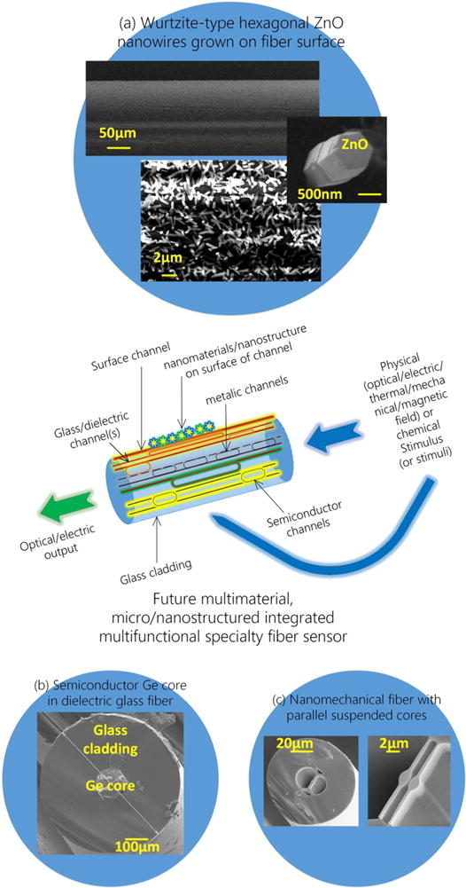
Conceptual future integrated multi-material multifunctional specialty fiber sensor. (a), (b) and (c) are our recent specialty fibers (all unpublished) with nanomaterials (ZnO nanowires), semiconductor (Ge) core and suspended nanomechanical cores, respectively.
Concluding remarks
The rapid development of modern sensors presents specialty fiber sensors with great challenges and opportunities. The solution for future specialty fiber sensors is to combine multiple materials, micro- or nano-scaled structures for an all-integrated compact fiberized multifunctional sensor.
Acknowledgments
This work is supported by the National Natural Science Foundation of China (NSFC, Nos. 61527822, 61235010 and 61307054). Xian Feng acknowledges the support from Beijing Overseas Talents Center.
5. Photonic crystal fibers for sensing applications
Wei Jin
The Hong Kong Polytechnic University
Status
Photonic crystal fibers (PCFs) refer to a class of optical fibers that have periodic transverse microstructures in the cladding [51]. Most PCFs reported so far are based on a single material, typically silica, with an array of air-holes running along the fiber length. These fibers have achieved transmission loss from below one to a few tens of dB/km, sufficient for many practical applications.
The flexibly patterned air-holes in PCFs enable novel optical, thermal and mechanical properties, providing new opportunities for photonic sensing. For example, the high air-filling fraction in the cladding allows tight confinement of the optical field to a small solid core via modified total internal reflection (figure 8(a)). Such fibers have very high nonlinear coefficients and novel dispersion properties, which enables super continuum generation over a very broad wavelength range (e.g., from 500–1700 nm) with relatively low-energy picoseconds or nanoseconds pump pulses and allows high resolution optical coherence tomography and spectroscopy systems with smaller-size and lower cost [51]. With proper air-hole diameter to spacing ratios, the fiber shown in figure 8(b) guides a single mode over a very large wavelength range, allowing for extremely broadband wavelength division multiplexing and networking of photonic sensors. By having asymmetric air-holes in the cladding, highly birefringent (Hi-Bi) polarization maintaining PCFs are made (figure 8(c)) and the single material property provides such PCFs with better thermal stability over conventional solid Hi-Bi fibers with stress rods, improving the performance of fiber sensors. PCFs with a thin suspended core (figure 8(d)) have a large evanescent field extended into the air-holes, enabling all-fiber evanescent wave spectroscopic and refractive index sensors and distributed sensing over the entire length of the fiber.
Figure 8.
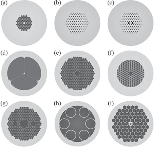
Different types of PCFs. (a) Highly nonlinear PCF, (b) endlessly single-mode PCF, (c) Hi-Bi PCF, (d) suspended core PCF, (e) photonic bandgap HC-PCF, (f) Kagomé HC-PCF, (g) polarization maintaining single-mode HC-PCF, (h) broadband single-mode HC-PCF and (i) PCF with a subwavelength air-hole in the core.
The microstructured claddings enable guiding of a light mode in a hollow core (HC) via photonic bandgap (figures 8(e) and (g)) or anti-resonant (figures 8(f) and (h)) effects, which minimizes the influence of material on light propagation and reduces the effect of thermal transient, optical (Kerr) nonlinearity and magneto-optic (Faraday) effects on the performance of sensors (gyroscopes) [52]. HC-PCFs with large air-fractions have demonstrated significantly enhanced phase sensitivity to acoustic waves, which could enable simplified acoustic sensor (hydrophone) arrays [53]. HC-PCFs also have better resistance to radiation damage as compared with conventional fibers and hence are better suited for applications in space or nuclear reactor environments.
The holey structures provide extra flexibility to modify the waveguide geometries (and hence optical properties) by controlled collapsing and inflating of the air-holes or filling selected air-holes with fluidic materials. A range of novel in-fiber devices, including long period gratings, polarizers, modal interferometers and polarimeters, as well as all-fiber photonic microcells, have been created, which can be used as compact sensing elements to measure strain, pressure, temperature, acceleration, twist and refractive index [53].
The air-holes in PCFs provide miniature compartments to contain a small volume of gas or liquid sample near the waveguide core or simultaneously confine the fluid sample and optical mode within the same HC over a long length. This enables strong light–fluid interaction over a long distance and hence sensitive spectroscopic and refractive index sensors [54, 55].
Current and future challenges
For wider spread applications of PCFs, a range of challenging issues need to be addressed. Based on the limited expertise of the author, three broad research topics are identified as follows:
Single-mode HC fibers. The guidance of light in the HC provides a range of sensing opportunities, including sensing physical parameters applied externally to the PCF (figure 9(a)) by detecting the intensity, phase, wavelength and polarization of the transmitted and/or reflected light, as well as sensors that exploit light–matter (e.g., gas) interaction inside the HC (figure 9(b)). However, current HC fibers are typically multi-mode, and coupling and interference between different modes result in noises that limit the sensor performance, especially when shorter (e.g., centimeters to meters) HC-PCFs are used [53]. A single-mode (and preferably polarization maintaining) operation would overcome such a problem but single-mode HC fibers are currently not available commercially.
PCF-based optofluidic systems. The holey nature of PCFs provides an ideal platform for strong light–fluid interaction directly within the fiber core or via an evanescent field, enabling the implementation of the so-called lab-in-fibers. Reliable and fast filling of fluid samples into hollow channels that partially or completely overlap with the optical mode, is important for efficient and cost-effective operation of the systems. Coupling light into/out from PCFs without being significantly affected by the fluid flow and connecting the PCF-based optofludic ‘chips’ to standard optical fiber pigtails [56] for more convenient and effective optical excitation and signal detection are important technical issues.
Novel sensing principles. Properly designed PCFs (e.g., HC-PCFs and suspended core PCFs) allow for very tight confinement of a propagating mode within or close to a hollow channel, enabling an extremely strong interaction of the light field with matter (atoms, molecules, particles, gas, liquid and so on) within the hollow channel. The local light intensity in the hollow channel can be made extremely high and the interaction length very long, which provides an opportunity for us to exploit new photonic sensing principles based on linear and nonlinear interaction between the light mode, matter filling the hollow channels and the external measurand fields, allowing for novel point and distributed sensors with unprecedented performance.
Figure 9.
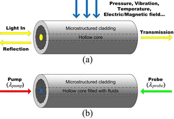
(a) PCF response to external stimuli (the HC can be filled with air or other materials). (b) Light matter interaction inside a PCF. HC-PCFs are shown here as examples but other types of PCFs can also be used.
Advances in science and technology to meet challenges
To achieve proper mode control, a good understanding of the physics responsible for mode selection and propagation is needed. The complex microstructure of HC-PCFs often means that analytically solving the wave equations is not practical and instead extensive numerical modeling and computation are often required, demanding advanced computational power as well as clever algorithms. Fabrication of micro-structures with the required high transverse precision and longitudinal uniformity also calls for improved fiber fabrication technologies. Recently, polarization maintaining single-mode (figure 8(g)) and broadband single-mode (figure 8(h)) operations of HC-PCFs were demonstrated by resonant filtering of unwanted modes [57, 58]. We look forward to further significant commercial progress in this area.
To implement practical PCF-based lab-in-fiber systems, advanced femtosecond laser micro-machining and other precision micro-fabrication technologies could play a significant role. These technologies have been used to create fluidic micro-channels and optical waveguides in glasses, which could be extended for use in PCF-based optofluidic systems. Fluid and thermal dynamics in fluid-filled micro-channels, interaction (and reaction) of fluids with the channel surface, and their effect on the light propagation are topics that need to be studied for efficient, reliable and reusable devices.
By taking the advantages of higher light intensity and longer interaction distance, ppb level gas detection has been demonstrated with photothermal interferometry spectroscopy in gas-filled HC-PCF (figure 9(b)) [55]. The same principle can be used for liquid detection and implemented with a suspended core or other types of PCFs. A range of processes, such as photoacoustic, stimulated Raman/Brillouin scattering and fluorescence, may be exploited for sensing a wider range of substances. Recently, a novel PCF with a subwave-length hole at the center of the core demonstrated significantly enhanced light intensity in the hole [59], enabling even stronger light–matter interaction in the hole and hence better detection sensitivity.
A new class of sensors based on flying particles within a HC-PCF was recently demonstrated [60]. This represents a new paradigm in fiber sensors and has the potential for sensing multiple physical quantities such as mechanical vibration (acceleration), electric and magnetic fields, temperature and ionizing radiation.
Concluding remarks
The development of PCFs enable fiber sensors with improved performance as well as novel sensing concepts that are not possible with conventional optical fibers. Some important progress has been made and further new advances are expected in the areas of lab-in-PCFs, nonlinear optofluidic sensors and novel sensing approaches such as flying-particle sensors. The practical realization of high performance PCF sensors requires advances in the fabrication of PCFs with better mode control, micro-fabrication and integration techniques to create reliable and efficient PCF-based sensing platforms and a better understanding of multidisciplinary sciences.
Acknowledgments
The author thanks C-Z Zhang and H-P Luo for drawing the diagrams. The support from the NSF of China through grants 61535004 and 61290313, and HK PolyU through grants 4-BCBE and 4-BCD1 are acknowledged.
6. Laser-based sensors: optical and quantum-enhanced sensing technologies
Yoonchan Jeong
Seoul National University
Status
Immediately after the demonstration of the first laser by Maiman in 1960, optical sensors started to exploit the unprecedented precision and accuracy offered by lasers [61]. Amongst them, laser-based sensors are distinguished from other optical sensors from the perspective that the measurement is entirely based upon the direct detection of laser light itself without relying on any external signal-transducing elements to the target object besides its ambient medium (e.g. see sections 3–5). Thus, the inherent properties of laser light are pivotal for laser-based sensing. While they can be represented from a variety of perspectives, the electric field of laser light is often represented in two distinctly different ways: (1) from the standpoint of the wave nature of light, it is given by a real vector E (see the upper part of figure 10). (2) From the standpoint of the particle nature of light, it is given by a field operator Ê together with a state vector|ψ> (see the lower part of figure 10). In this regard, the types of laser-based sensors can be classified into several groups as illustrated in figure 10, depending on which parameter of laser light and which standpoint of the nature of light is most exploited for sensing. No matter what sensing mechanisms are utilized, they all inherently allow for non-invasive measurements with high precision and accuracy as well as a fast response. Consequently, demands and challenges for laser-based sensors have never stopped growing in science and technology.
Figure 10.
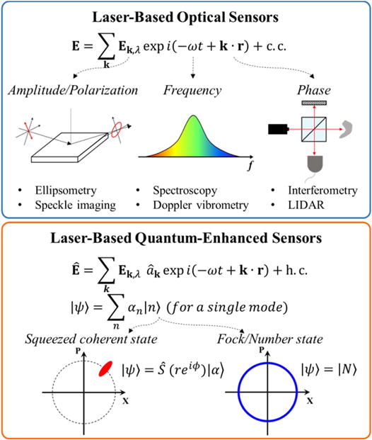
Classification of laser-based sensors.
Current and future challenges
Laser-based ‘optical’ sensing basically exploits the so-called ‘classical-like’ properties of lasers, while the amplification of light has nothing to do with classical phenomena at all. In general, laser light is approximated by a coherent state. Therefore, the precision of a measurement based upon the coherent state of light is ultimately limited by shot noise [37], which is proportional to or where I and N denote the intensity and average number of photons of the light, respectively. If I or N is sufficiently high, which is the case in most laser-based optical sensing, the actual precision of the measurement is not limited by shot noise but by some other uncertainties that may occur during the sensing process. In contrast, if I or N are supposed to be as minimal as possible for some reasons, such as probe-induced damage or disturbance issues, the consequence of the shot-noise limit becomes more crucial. In fact, such issues are the main technical concerns of laser-based ‘quantum-enhanced’ sensing [37]. The more detailed issues and challenges of the individual laser-based sensing technologies are overviewed in the following.
Laser ellipsometry
Laser ellipsometry is based upon detection of the changes in the strength and polarization state of laser light. Typically its sensitivity can go down to 0.01 nm or a sub-monolayer thickness [62]. However, such sensitivity can only be achieved after careful alignment and calibration of the system, which may also need the additional aid of electron microscopy, electron spectroscopy or scanning prove microscopy [62]. In addition, non-specular reflection or scattering can be a potential issue, depending on the surface roughness condition.
Laser speckle imaging
Laser speckle imaging is based upon detection of the contrast of speckle images produced on the target object by laser light. It can be a simple and powerful method for 2D mapping of flow or vibration. However, the spatial and temporal resolutions are relatively poor in comparison with other laser-based sensing techniques, and absolute velocity estimation or deep assessment beyond a few hundred μm remains an issue [63]. Further improvements in both theoretical models and measurement techniques are strongly in demand.
Laser spectroscopy
Laser spectroscopy is based upon detection of the changes in the spectral properties of laser light, utilizing a variety of different spectroscopic mechanisms, such as Raman scattering, Brillouin scattering, laser-induced breakdown, laser-induced fluorescence, etc. The availability of lasers that can operate at the desired wavelength matched for the material to be traced is a critical issue. In addition, the improvement of sensitivity remains a challenge in that the technology should be able to deal with a low concentration atomic or molecular sample with high fidelity [64].
Laser Doppler vibrometry
Laser Doppler vibrometry is based upon detection of the Doppler-shift frequency in the reflected laser light that is incurred by the vibration or movement of the target object. A state-of-the-art vibrometer offers a sub-Hz resolution [65]. The wavelength drift of laser light and laser speckle effects remain particular concerns. Technical advances for multidimensional and multi-channel measurements [65] in combination with increased spatial and temporal resolution are strongly in demand. The multi-channel capability for transient measurements will play a crucial role in various micro-scale applications, including micro-electromechanical systems (MEMS) and biomedical applications.
Laser interferometry
Laser interferometry is based upon detection of the change in the phase of laser light, utilizing an optical interferometer that includes the Mach-Zehnder interferometer, Michelson interferometer, etc. Since the phase change is caused by various physical origins, it has a wide range of applications, from gravitational wave detection to biomedicine. While the ultimate sensitivity of the phase detection is limited by shot noise [37], it is also severely affected by mechanical and thermal fluctuations of the components of the interferometer. In particular, the axial resolution of laser-based optical coherence tomography (OCT) is determined by the bandwidth or tuning range of the laser as well as the group velocity dispersion of the material, which is currently limited to a few μm [66].
Laser light detection and ranging (LIDAR)
Laser light detection and ranging (LIDAR) is based upon measurement of ‘time of flight’ of laser light, in general [61], which determines the distance of the target object, while the technology can also be combined with the other aforementioned sensing technologies. Typically, its resolution can be as small as tens of cm [67], depending on the distance range which can reach a hundred km. Since the technology is often implemented into airborne and spaceborne systems as well as ground-based systems, improvements in miniaturization and efficiency are invariably desired for both transmitters (i.e., lasers) and receivers (i.e., photodetectors or photon counters). Other issues and challenges include power scaling of lasers, reliable wavelength conversion, eye safety, improvement in the data compression and extraction, etc. In particular, diode-or fiber-laser-based sources will continue to receive great attention from the standpoint of miniaturization and efficiency. In addition, flash LIDAR technology will play an important role, considering the rapid growth and advances in autonomous driving technology expecting in upcoming years.
Laser-based quantum-enhanced sensing
Laser-based quantum-enhanced sensing can be regarded as quantum optical forms of all the aforementioned laser-based optical sensing technologies, exploiting non-classical states of light and their quantum optical nature. This can beat the classical diffraction or shot-noise limit, thereby eventually leading to super-resolution or super-sensitivity [37]. However, the main issues and challenges of such techniques include photon loss, dephasing noise, detector inefficiency, etc [37, 68] in that they substantially degrade the ideal precision that the quantum-enhanced sensing offers. There also remain inherent challenges, such as difficulty, complexity and inefficiency (low probability), in producing non-classical states of light.
Advances in science and technology to meet challenges
The use of as small a number of test photons as possible is invariably meaningful in that the consequence of the measurement onto the target object may be minimized. This is especially true if the measurement is done on delicate material systems, including atomic, biomolecular or nanoscale systems, the importance of which has been growing most rapidly in recent years. However, too small a number of test photons inevitably leads to inefficiency and low probability in sensing in case a fraction of the photons are lost during the sensing process [37, 68]. Consequently, advances in producing non-classical states of light, such as quantum-squeezed light or entangled photon pairs of a sufficiently high number will open up the opportunity for fully exploiting the quantum-enhanced effects. Figure 11 shows a schematic of quantum interferometric sensing when using a N00N state of light or an entangled coherent state (ECS) of light, either of which can beat the shot-noise limit [37, 68], but the latter is known to outperform the former when photon loss becomes relatively significant [68].
Figure 11.
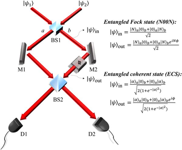
Quantum-enhanced interferometry based upon non-classical states of light, e.g., N00N state and ECS.
In fact, a laser as the source of light in a coherent state plays a pivotal role in whichever way it is used, directly for sensing or indirectly for creating non-classical states of light. Therefore, an in-depth understanding of light–matter interaction, particularly, at the mesoscopic level is also crucial for further success. In contrast, the selective elimination of coherence from laser light is another area to watch [69] because of its rich dynamics in light–matter interaction and great potential for novel sensing applications.
Concluding remarks
The definition of laser-based sensors has been clarified, which has remained unclear to date, and their types are classified into several groups, from the standpoint of what nature of laser light is most exploited for sensing. Their current and future challenges, and required advances in science and technology, have been overviewed and mapped out briefly. Since the laser property itself plays the most pivotal role in laser-based sensing, more effort must be paid to the innovation of laser science and technology. In particular, the development of more efficient and practical schemes for generating quantum-correlated photon pairs is an inevitable key to the success of future laser-based sensors.
Acknowledgments
The author acknowledges financial support under grant 10065150 (MOTIE, Korea), and also the useful discussion with K Park and H An.
7. Frequency comb-based sensors
Nathalie Picqué
Max-Planck Institute of Quantum Optics
Status
A laser frequency comb is an optical spectrum (figure 12(a)), which consists of phase-coherent equidistant narrow lines. The regular pulse train of a mode-locked femtosecond laser can produce such a comb spectrum of millions of laser modes with an equidistant spacing precisely controlled. Introduced in the late 1990s, laser frequency combs have revolutionized precise measurements of frequency and time by directly linking microwave and optical frequencies. In frequency metrology, frequency combs act like rulers in frequency space that can, for instance, be used to measure a large separation between two different optical frequencies. In recent years, laser frequency combs have found applications beyond their original purpose. In particular, they are becoming enabling tools for novel and innovative techniques of sensing, including broad-spectral-bandwidth optical spectroscopy, but also distance or strain measurements, vibrometry, optical coherence tomography etc. As the main application to sensing has been molecular spectroscopy, here we mostly emphasize some of the latest achievements and the current challenges and prospects of such research.
Figure 12.
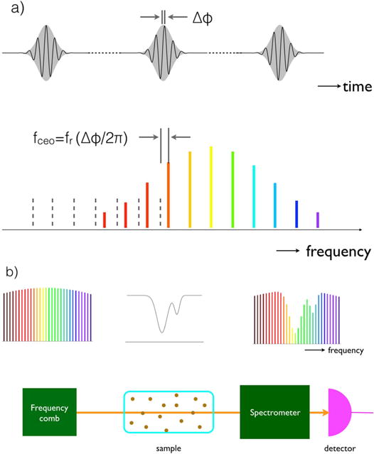
Most of the time frequency combs are generated by ultrashort pulse mode-locked lasers. The time-domain regular train of periodic optical waveforms (upper line) gives rise to a broad frequency-domain spectrum made of discrete comb lines (lower line) precisely separated by the pulse repetition frequency. (b) Frequency comb generators can serve as broadband light sources to interrogate the absorption of an atomic or molecular sample.
Current and future challenges
It was quickly realized that the broad spectrum of a laser frequency comb, made of phase-coherent narrow laser lines of precisely known positions, could be useful for directly interrogating the transitions of an atomic or molecular sample (figure 12(b)). A spectrometer is then required to analyze the spectrum. Several types of spectrometers have been successfully harnessed or devised: dispersive spectrometers (gratings or crossed dispersers) [70], interferential spectrometers (mostly Michelson-based Fourier transform spectrometers [70, 71]), spectrometers based on the analysis of the speckle pattern generated by the comb in a multi-mode optical fiber and a comb-enabled approach: dual-comb Fourier transform spectroscopy. Each approach has its own strengths and weaknesses, so that its suitability depends on the scientific and experimental requirements. There are however some common trends and challenges that are summarized below.
The use of laser beams does not make it possible to cover spectral spans as broad as with incoherent light sources, but it facilitates propagation over large distances in free space or in optical fibers, tight focusing (e.g. for microscopy applications) etc. The spectral resolution is limited by the comb line spacing (figure 13(b)) but interleaving approaches can overcome this limitation. Furthermore, the position of a spectral sample can be defined significantly more precisely than the resolution and the frequency scale can be self-referenced within the accuracy of an atomic clock. Finally, as femtosecond lasers involve intense laser pulses, nonlinear interactions can be harnessed extending the capabilities of the techniques much beyond linear absorption.
Figure 13.
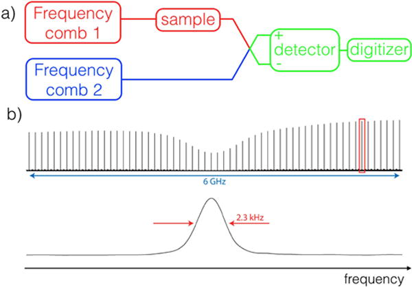
(a) Simple sketch of a dual-comb spectroscopy set-up probing the absorption and the dispersion of a sample. (b) Small portion of a near-infrared experimental dual-comb spectrum with resolved comb lines. The upper line shows that the Doppler-broadened Pe(27) line of the ν1 + ν3 band of acetylene is correctly sampled by combs of 100 MHz line spacing. The entire spectrum, which comprises 120 000 comb lines, is measured within 6 s and the comb line-width (lower line) is determined by the measurement time.
Although the prospects of spectroscopic sensing with frequency combs sound auspicious, numerous challenges must be addressed before the techniques realize their full potential.
First, impressive and convincing demonstrations of spectroscopic sensing have been performed in the telecommunication near-infrared region. The comb sources that emit in spectral domains of high relevance to molecular spectroscopy—terahertz (see also section 1), mid-infrared (see also section 2), ultraviolet, extreme ultraviolet—are under active development. Novel crystals for nonlinear frequency conversion, such as orientation-patterned gallium arsenide or phosphide, extend the domain of operation of the combs deeper in the mid-infrared [72], though the laser systems are still challenging to operate in the laboratory. Emerging quantum cascade lasers [73] hold much promise for versatile chip-scale frequency comb generators in the terahertz and mid-infrared regions. Microresonators, which span close to an octave in the mid-infrared region [74], are other intriguing comb generators of large line spacing. The technology of sub-ps mode-locked fiber lasers that directly emit in the mid-infrared region is significantly progressing. Electronic spectroscopy in the ultraviolet region has practically remained unexplored and the challenges of generating combs of sufficient power in the extreme ultraviolet range through high harmonic generation are daunting, though the scientific prospects in this spectral region, which is hard to access to any type of high resolution spectroscopy, are extremely exciting.
Broad-spectral-bandwidth frequency combs of small (<10 MHz) or large (>10 GHz) line spacing will benefit sub-Doppler and condensed-matter phase [74–76] spectroscopy, respectively. For the latter, a recent proof-of-principle demonstration [74] of a mid-infrared micro-resonator-based dual-comb source with a line spacing of about 120 GHz shows intriguing prospects for real-time vibrational spectroscopy: although this appears extremely challenging because of the poor sensitivity of mid-infrared detectors and the limited dynamic range of fast electronics, mid-infrared vibrational spectra spanning one octave could in theory be recorded on the time scale of a few nanoseconds with refresh rates of several hundreds of MHz. In the long term, one can envision spectroscopy laboratories on chips (see also section 8) able to detect and analyze in real time, with label-free and high-throughput detection, and very low quantities of molecules.
So far, the samples have mostly been molecules in the gas phase, in which transitions were Doppler- or collision-broadened. The sampling techniques and the nature of the samples will diversify to answer new questions in molecular physics and physical chemistry and to provide field sensors suited, for example, to analytical chemistry or the atmospheric sciences. Some groups are actively pursuing such goals, extending the scope of direct frequency comb spectroscopy to, for example, combustion diagnostics, the detection of greenhouse gases in the atmosphere [77], evanescent sensing, chemical kinetics in microfluidic devices or spectro-microscopy. Novel schemes for nonlinear spectroscopy, and perhaps even for multidimensional nonlinear spectroscopy, might shed light on some new aspects of the structure of matter.
Dual-comb interferometers without moving parts seem to be the instruments that are currently attracting the highest number of contributions. We therefore end this short contribution by an overview of their particular features. Dual-comb systems have mostly been developed and used for molecular spectroscopy, but they can be used for any applications where a two-beam interferometer is required. One of the most widespread configurations (figure 13(a)) is to interrogate the linear absorption and dispersion of the sample. A laser frequency comb is transmitted through the sample. It is combined on a beam mixer with a second comb of slightly different repetition frequency, which serves as a local oscillator. The time-domain interference between the two lasers, the interferogram, is recorded on a fast photodetector. The complex Fourier transform (amplitude and phase) of the interferogram reveals the absorption and dispersion spectra of the sample.
Such a simple concept holds much promise for new schemes of broadband spectroscopy in the gas, liquid and solid phases: although its physical principle is the same as that of a Michelson interferometer, the absence of moving parts makes the recordings potentially much shorter and higher resolution may be reached. As any multiplex technique, a single photodetector is needed. This improves the overall consistency of the spectra and facilitates the operation of the spectrometers in spectral regions where detector arrays are not conveniently available. Moreover, nonlinear dual-comb spectroscopy has been demonstrated with stimulated and coherent Raman effects [75], two-photon Doppler-free spectroscopy and double resonance Doppler-free spectroscopy, but many additional promising schemes can be explored. A basic instrumental challenge is to maintain the coherence between the two combs on long time scales, outside the frequency metrology lab. This stimulates much of the current research, with simplified schemes involving mode-locked lasers [77], innovations for generating two asynchronous trains of pulses from a single laser oscillator or novel dual-comb systems based, for example, on electro-optic modulators [78], or on microresonators [74, 79], pumped by a single continuous-wave laser. With the increasing size of the community involved in direct frequency comb spectroscopy and especially in dual-comb spectroscopy, other clever solutions can be expected and dual-comb spectroscopy might evolve one day into a turn-key instrument like the Michelson-based Fourier transform spectrometer. Portable and even on-chip spectrometers would benefit the development of sensing applications.
Concluding remarks
Although the field is its infancy, intriguing and unforeseen approaches to sensing with laser frequency combs have already been demonstrated in the past few years. With continued progress to frequency comb technology and with the exploration of novel and innovative insights, molecular sensing is likely to be deeply impacted and one can envision that applications beyond spectroscopy will also mature and gain in importance.
Acknowledgments
Financial support by the European Research Council (Grant Agreement 267854) is acknowledged.
8. Micro and nano-engineered sensors
Limin Tong
Zhejiang University
Status
Using a beam of light as a probe for optical sensing is one of the most efficient approaches for detecting unknowns, especially for those well beyond our reach ranging from astronomical scale to atomic size. Historically, light from a distant star was used to sense the gravitational bending of light predicted by the general relativity, in which the ‘sample’ to be measured was so large that no engineering on the probing light is required or possible. Compared with freely propagating light beams, artificially engineered optical fields are in increasing demand for probing samples with much smaller sizes and/or weaker light–matter interaction. For example, for a biomolecule with a scattering cross-section of tens of nanometers that is well below the wavelength of the probing light or the optical diffraction limit, the signal or the signal-to-noise ratio will be very low when using a free-space probing light. As the wavelength of the probing light is usually within visible to near-infrared spectral range (i.e., hundreds of nanometers to about one micrometer), the most efficient way of tailoring light for optical sensing is structurization with wavelength or subwavelength feature size, which leads to micro- and nano-engineered optical sensors.
Concerning the way for light manipulation, micro- and nano-engineered sensors can be classified into two categories: using propagation light (usually guided by non-resonant structures) or localized optical fields (usually confined by resonant structures), as shown in figure 14. Mature platforms for the non-resonant approach are fiber- and chip-based structures. In fiber-based systems, the fiber Bragg gratings (FBG) represents the most successful sensing element with subwavelength or nano-engineered periodical structures in a single optical fiber [80], and has been widely used for measuring strain, temperature and concentration in both laboratories and industry. To obtain better light manipulation and greater versatility in fiber-based micro and nano-engineered optical sensors, reducing the feature size of the fiber itself is an effective approach. The photonic crystal fiber (PCF), or more generally the microstructure optical fiber (MOF) is an example for measuring liquids or gases with high robustness, compact structure and less requirement on samples [81]. More recently, optical sensing with optical micro- or nanofibers (MNFs) or nanowires, showed that, by reducing the diameter of a fiber-like waveguide to the subwavelength scale, it is possible to generate tightly confined high-fractional evanescent fields for optical sensing with miniaturized footprint and high sensitivity [82]. In addition, grating structures can also be fabricated in an MNF or a nanowaveguide alike for further enhancing of the sensitivity [83]. Chip-based optofluidic systems are another excellent platform for micro and nano-engineered optical sensors. Using microfluidic channels to confine and deliver the liquid sample and/or the probing light, it is possible to operate the sensor with much less amount of samples, and perfect isolation from environmental disturbance. Also, fiber-based structures, such as a MNF can be integrated into the lab-on-chip system for better light manipulation. For example, by embedding a 800 nm diameter nanofiber into a microfluidic chip with a 5 μm-wide detection channel, Zhang et al successfully demonstrated a nanofiber optical sensor for sensitive and fast detection of femtoliter samples [84].
Figure 14.
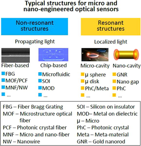
Typical structures for micro and nano-engineered optical sensors.
For the resonant type, in terms of feature size and material used for light localization, there are typically two kinds of platforms: photonic microcavities and plasmonic nanocavities. The microcavities, usually made of micro- or nano-engineered dielectric structures, can offer high quality (high-Q) resonance and consequently high sensitivity for both particle and bulk samples [85]. To date, a variety of microcavities such as microspheres, microdisks, microtoroids, optofluidic resonators and photonic crystal cavities have been demonstrated for optical sensing. Relying on localized surface plasmonic resonance (LSPR) in metal nanostructures (e.g., metallic nanoparticles and nanogaps), the nanocavity can provide a mode size much smaller than the vacuum wavelength of the light and comparable with the cross-section of biomolecules, and is therefore highly sensitive in detecting nanoscale particle samples. Moreover, as a result of tight confinement, the significant field enhancement in the LSPR structure is also highly desired for applications such as surface-enhanced Raman scattering.
Overall, micro or nanoscale structurization is a highly efficient approach to better light manipulation for better optical sensors with higher sensitivity, smaller footprints, smaller amount of sample, higher resolution and greater versatility, which have been one of the main driving forces in the advances of optical sensors in recent years. However, the physics and techniques of reducing the structure and mode sizes below the wavelength of light have their own limitations, which present challenges for pushing the limits of optical sensors.
Current and future challenges
The first challenge comes from the fabrication side—as the feature sizes go down to subwavelength scale, issues related to the accuracy and cost of the micro or nanofabrication are realized. In both propagation and resonant structures, low surface roughness is critical to handling light with low optical loss, which is in many cases critical to achieve high sensor performances such as sensitivity and resolution. So far, with the exception of the pristine surface frozen form melting glasses (e.g., glass fibers or microspheres), most of the other structures, especially those fabricated by top-down lithography, exhibit surface roughness much higher than 1 nm, if there is no sophisticated and costly smoothing processes afterward. Secondly, for nanocavities, although the LSPR is able to confine optical frequency electromagnetic fields down to the nanometer scale, the loss of this kind of resonance is very high (e.g., indicated by the 10 fs level relaxation time), resulting in a resonance band much broader than those of dielectric microcavities, which is undesirable for achieving high sensitivity or resolution by measuring the LSPR spectral shift. Thirdly, although the amount of sample can be significantly reduced by reducing the feature size in a nano-engineered sensor, the signal intensity or signal-to-noise ratio will also be reduced at the same time, which may degrade the overall performance of the sensor, or require additional sensing time. Therefore, the balance between the performance and the size (or sometimes the complexity) of a micro- and nano-engineered sensor should be optimized from case to case. Finally, for practical applications, compared with other types of sensors, besides their higher price, optical sensors are more likely to suffer from the coupled measurands. For example, the output of a fiber optic strain sensor may depend on both the strain and the temperature, which means additional compensation should be included. The above-mentioned issue also exists in micro- or nano-engineered optical sensors, in which decoupling techniques should be considered for real applications.
Advances in science and technology to meet challenges
For nanofabrication, low-cost high-throughput fabrication techniques, such as nanoimprint, may be one of the possible solutions, if the surface roughness and material diversity can be improved. For higher-accuracy fabrication, the continuous progress in lithography techniques, for example, the recently emerging focused ion beam milling using helium rather than gallium ions has shown great potential for better nanofabrication [86]. Also, compared with bulk structures, materials with lower dimension usually show higher mechanical strength and flexibility. For example, the strain allowed in a silica nanofiber is much larger than that in a standard silica fiber, providing the possibility of realizing strain sensors with larger dynamic range and lower detection limit.
From the physics side, new mechanisms or effects could be introduced into optical sensors with micro- or nano-engineered structures. For example, the bandwidth of the LSPR could be significantly reduced when strong coupling occurs between the LSPR mode and a high-Q photonic cavity mode [87, 88], offering the possibility of enhancing the sensitivity of the LSPR to the same level of the propagation surface plasmon polaritons. In addition, it is worth noting that, with decreasing diameter, the fraction of evanescent fields around a waveguiding plasmonic nanowire does not change a lot [89], offering an opportunity to combine the ultra-tight confinement and propagation features of optical fields in the same nanostructure for optical nanosensors.
Concluding remarks
Introducing micro- and nano-engineered structures has greatly enhanced the performance of optical sensors. Although there are challenges in pushing the limits of current sensors in both fabrication and optics aspects, the combination of a better fabrication technique and new physical effects may open new opportunities for future micro- and nano-engineered optical sensors.
Acknowledgments
This research was supported by the National Key Basic Research Program of China (2013CB328703), National Natural Science Foundation of China (61475136) and Fundamental Research Funds for the Central Universities.
9. Plasmonic nanoparticles for sensing, imaging and manipulating cellular processes
Björn M Reinhard
Boston University
Status
Coherent oscillations of the conduction band electrons in noble metal nanoparticles (NPs), so-called plasmons, give rise to large resonant optical scattering and absorption cross-sections. As a consequence, individual gold and silver NPs can be detected in a variety of optical imaging schemes. Noble metal NPs with diameters ⩾40 nm can be imaged in differential interference contrast microscopy or in scattering microscopy under darkfield or total internal reflection illumination [90]. NPs smaller than 40 nm can be visualized through photothermal imaging. Small metal NPs retain relatively large absorption cross-sections that allow for an efficient heating of the immediate ambient environment [91]. The resulting local changes in the refractive index can be detected optically even for NP diameters as small as 5 nm and below. Unlike fluorescence emitters, noble metal NPs do not blink or bleach. Given their extreme photophysical stability, noble metal NPs are desirable labels for imaging and tracking applications that require long observation times. Interestingly, as the exact resonance conditions depends on the refractive index of the environment, the spectrum of the metal NPs can also provide information about the local environment of the NPs.
Another advantage of noble metal NPs as labels is that the large E-field enhancement associated with the resonant charge density oscillations in individual NPs and their clusters can strongly enhance inelastic scattering from molecules located in their electromagnetic ‘hot spots’. This has led to a large number of biomedically relevant cellular sensing concepts based on surface-enhanced Raman spectroscopy (SERS). Prominent examples include the detection of bacterial pathogens with rationally designed SERS substrates [92], as well as the molecular characterization of disease states of mammalian cells [93]. Furthermore, as vibrational transitions have much narrower bandwidths than electronic transitions, SERS imaging of dye-functionalized noble metal NPs has unique advantages for multiplexed imaging.
One other unique property of plasmonic NPs is that the plasmons of the individual NPs couple when the particles approach each other to below one NP diameter. The distance-dependent plasmon coupling in this separation range induces changes of the resonance wavelength, spectral shape, intensity and polarization [90]. These observables encode information about interparticle separations as well as the number of interacting NPs and their spatial arrangement in plasmon coupling microscopy (figure 15). As the spectral properties of the coupled NPs are quantifiable through far-field optical measurements, plasmon coupling between NP labels bound to specific surface features provides unique opportunities for mapping their spatial clustering on sub-diffraction length scales across an entire cell as well as for monitoring their trafficking and determining their intracellular fate [94, 95].
Figure 15.
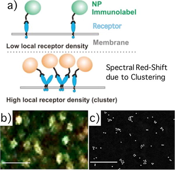
(a) Concept of plasmon coupling microscopy. Clustering of NPs bound to a target receptor results in spectral shifts. (b) Color image of 40 nm diameter gold NP targeted to CD44 surface proteins on a MCF7 cell. (c) SEM image of NP immunolabels targeted to CD24. Adapted with permission from [96]. Copyright (2013) American Chemical Society.
A further appeal of plasmonic NPs is that they provide interesting photothermal, photocatalytic and other material properties that—in combination with the superb imaging capabilities—make it possible to perform some therapeutic procedures in a rational and controlled fashion. Well-known examples are the application of gold NPs in photothermal and photodynamic therapy.
Current and future challenges
The use of NP labels in a cellular environment brings about a number of challenges. First, the size of the NPs needs to be chosen to be compatible with the problem of interest. Fortunately, different imaging techniques for different sizes of plasmonic NPs are already available. SERS sensing/imaging and plasmon coupling microscopy require NPs with diameters of approximately 40 nm and above. Particles of this size are useful, for instance, for mapping the self-organization of receptors in the plasma membrane on length scales of tens to hundreds of nanometers [94]. In addition, the size of the NPs is not per se a disadvantage. For some applications, such as probing the molecular foundation underlying virus–cell interactions, the size of the NPs, together with the rational tunability of the surface properties, are in fact advantages for generating appropriate model systems [97]. In addition, the investigation of NP–cell interactions by itself is important. For those applications that cannot tolerate NP sizes of ⩾40 nm, photothermal imaging of much smaller noble metal NPs is indicated.
Another challenge, especially, for intracellular applications of NPs in living cells is the need to access the cytoplasm and to selectively bind specific intracellular targets. Although different strategies to target intracellular structures with NPs are available, they require careful optimization on a case-by-case basis. The development of alternative universal strategies based on intrinsic NP properties (e.g. photothermal melting of the membrane) remains important. Once NPs have entered the cytoplasm, they can bind to specific intracellular structures, provided they are equipped with targeting functionalities that retain their functionality in the cell. Unfortunately, specific binding interactions facilitated by nanoconjugated ligands can be compromised in biological media through the rapid nonspecific attachment of proteins (corona formation) or through degradation. The development of NP surface strategies or self-cleaning procedures that maintain the functionality of surface ligands remains a priority.
Cellular structures absorb and scatter light, so especially in the single particle limit there is a need for further enhancing the sensitivity of NP detection in complex biological media.
Advances in science and technology to meet challenges
Recent advances in the biomimetic design of NPs resulted in the implementation of membrane wrapped plasmonic NPs (artificial virus NPs, figure 16) that successfully recapitulate viral glycoprotein-independent virus uptake and trafficking [97]. The successful mimicry of evolutionarily optimized viral behavior with plasmonic NPs paves the path to overcoming key challenges associated with transferring NPs across the plasma membrane into the cytoplasm. Many lipids are zwitterionic. A zwitterionic wrap around plasmonic NPs also provides opportunities for minimizing protein corona formation [98].
Figure 16.
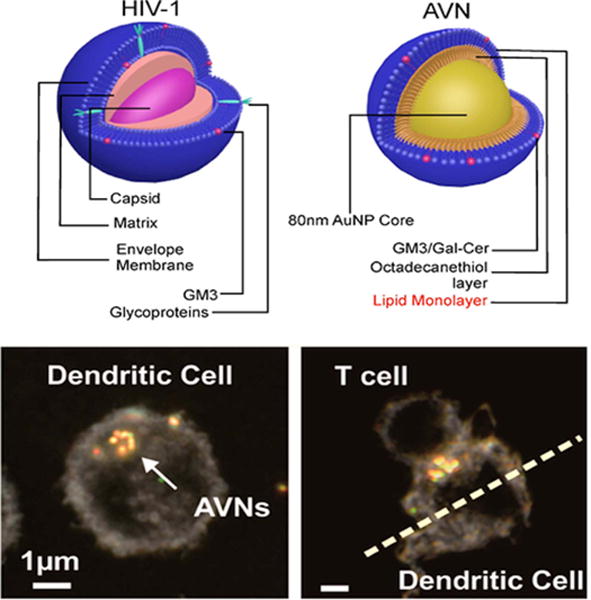
Comparison of enveloped virus (top-left) and artificial virus NP (AVN, top-right). Reprinted by permission from Macmillan Publishers Ltd: Nature Communications [97], Copyright (2014). AVNs are collected in unique compartments in dendritic cells and enrich at the T-cell dendritic cell interface. Adapted with permission from [99]. Copyright (2015) American Chemical Society.
Emerging new plasmonic materials, such as doped semiconductors, promise a more rational tunability of plasmon resonances by means of doping. As the resonances of these materials lie in the near- to mid-IR where interesting molecular vibrations lie, the NPs can lead to improved chemical imaging modalities. These semiconductor materials can have less electronic damping and lead to sharper spectral lineshapes when compared with metal NPs.
The potential for advancement is not limited to NPs and surface functionalization. There is also tremendous potential for improving imaging and sensing strategies. For instance, the development of effective light sources for frequency entangled photons has the potential to greatly aid the suppression of uncorrelated background signal in plasmonic NP imaging and sensing [100].
We also envision that in the future NPs will find applicability as a tool not only to image cellular processes but also to directly manipulate and control them. Recent studies have confirmed that ligand functionalized NPs provide a unique means for controlling cellular signaling processes [101].
Concluding remarks
Plasmonic NPs have superb photophysical properties that make them versatile optical labels. Their E-field enhancing and tunable photothermal and photocatalytic properties provide opportunities not only for cellular sensing but also for manipulating cellular processes in a controlled fashion while monitoring with high temporal and spatial resolution.
Acknowledgments
This work was supported by the National Institutes of Health (NIH) through grant R01CA138509.
10. Optical chemical sensors
Paul M Pellegrino
US Army Research Laboratory
Status
Definition and structure
In general terms, a sensor is a device that detects some physical property and records, indicates or otherwise responds to it. This definition can be coupled with a fitting definition of a chemical sensor derived from the so-called ‘Cambridge definition’: chemical sensors are miniaturized devices that can deliver real-time and on-line information on the presence of specific compounds or ions in even complex samples. This combination sets the stage for discussions of optically based chemical sensors [102]. Obviously, the final defining element of the use of light as a transduction is self-evident, but important line of demarcation as well. As in biosensors, chemical sensors share the similar core functioning elements such as sample or analyte introduction, transduction signal or signature, signal processing, and finally quantitation or indication of target chemical presence. Figure 17 pictorially represents this methodology. The other core feature or element of the sensor is the recognition of the target chemical, but depending on the technique being used as transduction this feature can appear in either or both, the transduction stage or the signal processing stage of the sensory pathway.
Figure 17.
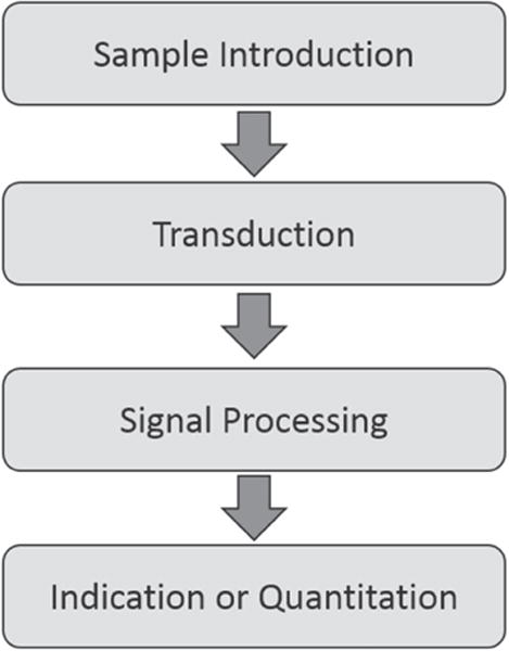
Methodology for optical chemical sensor.
Discussions on transduction methods inevitably bring about a bifurcation of these methods into direct and indirect or mediated methods. Stalwart spectroscopic methods such as fluorescence, absorbance (visible and infrared) and Raman are clearly classified as direct, while other methods such as color-change (colorimetric), emissive changes due to the environment of the fluorophore (lifetime and wavelength) or capture which can sometimes produce a luminescent product or be generally sensed using change in optical property such as index of refraction, are examples of indirect sensing [103].
Brief history and status of the field
The history of optical observations using certain direct methods based on infrared absorbance, emissive properties (flame spectroscopy) and florescence date back considerably to the 16th and 17th centuries, but it was not until the 19th century that the necessary connections to chemical composition were firmly established. Prior to the 19th century there were only occasional references to essential components like salts or mineral composition as the cause for wavelength shifted luminescence. In the 19th century studies of sunlight and its ‘dark bands’ or lack of light in certain regions by Fraunhofer and others set the stage for continued study. Contributions by two generations of the Hershel family (William and John) identified infrared emission and subsequently produced the first spectra [104]. Fluorescence took a similar development pathway with Hershel and Stokes paving the way for the capture of the first spectra. The foundation of linking chemicals to optical emission or absorbance was finally cemented as we turned into the 20th century as Colblenz and others categorized materials with their spectroscopic features [105]. Although the foundation was laid and instrumentation continued to be developed, the modern optical chemical sensor would not truly achieve maturity until the advent of the laser in the early 1960s. The activity in the development of optically based chemical sensors spiked during the period after new direct methods such as Raman and laser-induced breakdown had been enabled accompanied by increases in other older spectroscopic methods and mediated methods entering the mainstream [106]. Another dramatic increase was seen when fiber optics reached maturity and provided numerous avenues to manipulate and control light and light interactions. Fiber optic sensors have been the pursuit of numerous research and developmental efforts and have now entered into the application fields of sensing gases and vapors, medical and chemical analysis, environmental analysis, industrial production monitoring and the automotive industry [102, 103, 107].
All of these methods both direct and indirect have solidified optical sensing as a mainstream mechanism that now resides with other analytical techniques as a critical part of chemical identification in a modern laboratory and industrial monitoring and analysis. Penetration of these methods into fully mature and commercialized sensor platforms that truly act as responsive or indication sensors is less developed and remains one of the last technical hurdles for optically based chemical sensors. This brief section of the roadmap will confine its discussion to challenges of point optical chemical sensors and will not address the more ambitious effort for standoff chemical sensing or optical sensing of biomolecular compounds such as DNA or proteins.
Current and future challenges
In terms of current and future challenges in the field there are at least two difficulties that are set apart from the rest. One is based on the maturation of the optical sensor platforms that would make optical sensors for chemicals more commonplace and affordable, and the other resides more in the information provided through the optical sensing. Even after the arrival of the laser and its new semiconductor cousins, the fields of fiber optic sensors and planar waveguide sensors have failed to produce sensors with lasting and commercial impact. There are areas where these sensor platforms have excelled in the detection of volatile organic compound (VOC) detection for certain industries, but full ubiquitous use has not occurred. Clearly new sources, detectors and material developments such as quantum cascade lasers (QCLs) and plasmonics have become more routine, but the full translation of these more compact embodiments has yet to produce optical chemical sensors with deep market or widespread commercial use. One possible explanation of the popularity could be linked to the ease with which these platforms can control and manipulate light. Fiber optics has clear restrictions in its geometries with either distal or fiber exposure needed for appropriate interactions, while planar waveguides, although more attuned to semiconductor platform geometries, clearly have mechanisms to insert and manipulate light effectively. The other and possibly more fundamental challenge stems from the lack of information afforded by certain optically based sensor architectures. Despite all their shortcomings and difficulties, certain types of direct methods (e.g. Raman) have succeeded due solely to their ability to provide clear information for chemical identification that is fundamental and thereby adaptable to multitudes of targets. This type of information linked to fundamental properties is exactly what is lacking in certain optical chemical sensors, which rely on the mediation of the chemical being sensed. There are few platforms that are capable of sensing a wide variety of targets with any one mediated sensing modality. Nominally, reactive polymers and other linking or capture mechanisms are used, but they can tend to be specific or alternatively they are non-specific but used in an array format in an attempt to glean specificity through a combinatorial response. The final challenge is not linked to the technology itself, but it is more attributable to the scientific community that develops optical chemical sensors. This community is inherently segregated by the disciplines of chemistry, physics and engineering. In general, there are few cross-over disciplines that capture the necessary skills needed to detect and identify chemicals in a meaningful way while understanding the platform limitations needed to develop sensible sensor systems. The tendency is for physicists and engineers to develop sensitive and complex transduction mechanisms without regard for the chemical information needed or mediation technology used. In contrast, chemists known for their knowledge of chemical reactions and spectroscopy have a lack of depth when confronted with realistic engineering challenges presented in the technology maturation necessary. The one exception to this trend would be a small cross-section of analytical chemists who focus on development of instrumentation.
Advances in science and technology to meet challenges
One of the major advances that one sees in the horizon is the availability of photonics and specifically photonic integrated circuits (PICs) to the mainstream academic, industry and government communities. Public–private partnerships such as IMEC in Europe and American Institute for Manufacturing Integrated (AIM) Photonics have set in motion the ability for individual institutions to access mainstream silicon photonics design rules, processing capability and mating electronics and packaging capabilities. These capabilities are enabled by the introduction of education through educational outreach, availability of process design kits (PDKs) and most importantly through multi-project wafer (MPW) runs where large multi-inch wafers are sub allocated for individual projects if they conform to identified design rules. Pictured above in figure 18 is a notional PIC for biosensing with fiber optic input, microfluidic interface and electronic readout. Numerous other design architectures are available with varying degrees of on-chip integration including light sources and photodetectors. Clearly, integrated photonics can provide a platform to previous work on planar waveguide sensors and open up new avenues for continued examination of micro-ring resonators and other optical structures such as Bragg gratings and interferometric configurations. Moreover, advances in semiconductor sources at several wavelength regimes throughout the ultraviolet, near infrared and infrared can be envisioned in the future. Although progress is not guaranteed, it looks as if PICs are starting to mirror the path taken by electronic integrated circuits (ICs) so long ago. This seems to suggest that one of the challenges for the wide proliferation of optical chemical sensors can be overcome. What remains to be seen is whether groups of dedicated multidisciplinary scientists and engineers can produce methodologies with intelligence built into the sensor transduction, such as molecularly imprinted polymers, that can address the selectivity and adaptability demonstrated by direct methods.
Figure 18.
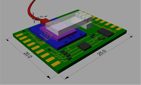
Graphical representation of a photonic integrated circuit.
Concluding remarks
In summary, optically based chemical sensors represent a class of sensors that have been established through direct measurement methods based on spectroscopic techniques with extensions to fiber optic technology. Advances in source, detector and planar waveguide technology have opened up new avenues for development, but to date have not engendered widespread acceptance of optically based chemical sensors as a compact and economical sensor type. Exciting new developments in the field of PICs provide an invaluable opportunity for the sensors to evolve in the near future, but strategic investment and multidisciplinary efforts will be required to ensure sensor architectures with the appropriate specificity, adaptability, sensitivity and most importantly commercial viability are pursued.
Acknowledgments
Appreciation and credit for the creation of figure 18 is extended to Justin Bickford, US Army Research Laboratory.
11. Biomedical optical sensors
Alexis Méndez
MCH Engineering LLC
Status
With a growing global population requiring healthcare and the need for ever more sophisticated diagnostic tools, clinicians worldwide are increasingly relying on advanced biomedical instrumentation and sensors as necessary and effective tools for patient diagnosis, monitoring, treatment and overall care. Many of the medical instruments in use today rely on optics (and optical components) to perform their functions, while optical and photonic-based sensors have been attracting attention in the healthcare industry in recent years leading to a wave of research activity and development of new products. Biomedical sensors present some unique design challenges. Sensors need to be safe, reliable, highly stable, biocompatible, amenable to sterilization and autoclaving, not prone to biologic rejection, and not require calibration or at least maintain it for prolonged times. In particular, sensor packaging is an especially critical aspect. It is highly desirable that sensors be as small as possible—particularly those for implanting or indwelling purposes.
In particular, fiber optic based sensors are ideally suited for a broad variety of—invasive and non-invasive—applications in the life sciences, clinical research, medical monitoring and diagnostics, ranging from optical coherence tomography (OCT) probes, to force-sensing catheters in robotic surgery, to intra-aortic pressure probes. Since the early days of medicine, optics has been a useful and powerful technology to assist doctors and all types of healthcare practitioners to carry out examination and diagnosis of their patients. This is so because one of the basic aspects of medicine is the use of visual and manual auscultations to diagnose a patient’s health. As such, optics are an ideal technology to assist doctors in gaining better visual examination capabilities by providing improved illumination, image magnification, or access to small or internal body cavities. But, in reality, it is light and its interaction with living tissues that is at the center of what makes optics (and photonics) in biomedicine possible. Light possesses energy and is capable of interacting with biological cells, tissues and organs. Such interaction can be used to probe the state of such living matter for sensing, diagnostic and analytical purposes, or, it could be used to induce changes on the same living systems and be exploited for therapeutic purposes.
The interrelation between optics and light in medicine is ever present and it could be said that more significant advances in biophotonics are now due to the availability of more powerful, concentrated and multi-spectral light sources which have been available only in the last 50 years. Historically, ambient light was the illumination source, which precluded performing exams late in the day or during certain hours in the winter time. With the development of semiconductor lasers and LEDs in the 1960s, modern medical optics began to take shape and, coupled with the availability of optical fibers, a new generation of optical biomedical instruments, sensors and techniques began to be developed. For instance, optical fibers have been used in the medical industry even before their adoption in data communications. The advantages of optical fibers were recognized by the medical community long ago [108]. Their initial and still most successful biomedical application has been in the field of endoscopic imaging, with the first fiber optic endoscope demonstrated in 1957 [109]. Then, during the 1980s and 1990s, extensive research was conducted to develop fiber-based biological, chemical and medical sensors [110]. To date, biomedical sensors based on external cavity Fabry–Perot interferometers (EFPI), fiber Bragg gratings (FBG) and spectroscopic types based on light absorption and fluorescence, are the most commonly researched and developed into commercial products [111]. For example, figure 19 shows a picture of a miniature FP fiber optic medical temperature sensor. Fiber optic biomedical sensors often rely on the use of special coatings or small cavities holding a specific reagent that can detect a given biochemical analyte of interest. This is a common practice in the so-called optrodes, as well as in the use of tilted FBGs.
Figure 19.
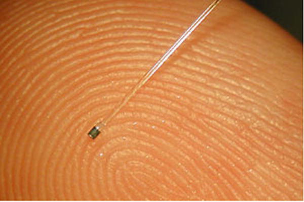
Aspect of a miniature fiber optic Fabry–Perot biomedical pressure sensor. Reproduced with permission from FISO Technologies.
Besides fiber-based devices, integrated optic planar devices are also an attractive and effective platform to develop biomedical sensors. The combination of microfluidics with different surface sensing optical effects such as plasmons and surface-enhanced Raman scattering (SERS), make it possible to detect multiple biochemical substances in rapid time, using small size devices. Other optical platforms exploited are ring resonators and photonic crystals.
Biomedical optical sensors can be categorized into four main types: physical, imaging, chemical and biological. Physical sensors are used to measure a broad variety of different physiological parameters such as body temperature, blood pressure, respiration, heart rate, blood flow, muscle displacement, cerebral activity, etc. Imaging sensors encompass both endoscope devices for internal observation and imaging, as well as more advanced techniques such as OCT, photoacoustic imaging and others, where internal scans and visualization can be made non-intrusively. Chemical sensors rely on fluorescence, spectroscopic and indicator techniques to measure and identify the presence of particular chemical compounds and metabolic variables (pH, blood oxygen, glucose, etc), detecting specific chemical species for diagnostic purposes, as well as monitoring the body’s chemical reactions and activity for diagnostic and therapeutic applications. For their part, biological sensors tend to be more complex and rely on biologic recognition reactions—such as enzyme–substrate, antigen–antibody or ligand–receptor—to identify and quantify specific biochemical molecules of interest.
Current and future challenges
We all need medical care—from the day we are born, until the day we day we die. However, this need has been dramatically accentuated in recent years by a convergence at a global level of diverse social, demographic, economic, environmental and political trends. Among these are the growing number of chronic diseases, such as obesity, arteriosclerosis, diabetes and cancer, which have become leading causes of death and disability. Another key trend is the increase in the world’s population, now estimated to be in excess of 7.3 billion and growing at annual rate of ~1.1%, and expected to reach 9.7 billion in 2050 [112]. Furthermore, medical personnel are increasingly relying on advanced biomedical instrumentation and sensors as tools for patient diagnosis, monitoring, treatment and care. In addition, advances in minimally invasive surgery (MIS) coupled with the advent of medical robotics and computer-assisted surgical systems (MRCAS) are demanding the development of smaller disposable sensing catheters and sensing probes, for which optical fibers are ideally suited. In addition, there is a desire for analytical instruments and sensors that can provide almost immediate results on blood and other sample analyzes, which can facilitate on-the-spot actionable diagnosis.
Given such challenges, optics and photonics are powerful, versatile and enabling technologies for the development of present and future generations of biomedical sensors, instruments and techniques for diagnostic, therapy and surgical applications.
Advances in science and technology to meet challenges
In the future, advances in the development of ever smaller and thinner medical probes and catheters should be expected, as well as broad utilization of OCT devices to become as common as ultrasound scanning devices are in today’s society. Other novel capabilities brought on by optics will be in the form of the so-called lab-on-a-fiber (LOF) [113], where functionalized thin layers of micro- and nanoparticle materials are deposited on the tip of an optical fiber (see figure 20). Specific biochemical analytes are detected by the light–matter interaction with the surrounding environment, which produces specific optical resonance, surface plasmon or SERS effects.
Figure 20.
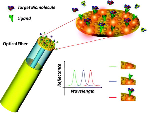
Illustration of the concept of a biomedical lab-on-fiber multi-analyte sensor.
Future innovations can already be witnessed today in the form of optical devices used in combination with smart phones. For example, there is the CellScope [114], which is a microscope that attaches to a camera-equipped cell phone and allows for both brightfield and fluorescence microscopy to be performed on the spot on a patient. Several other innovations can be expected in which the smartphone acts as the interrogating instrument to an external, passive, fiber optic or planar biomedical sensor.
Concluding remarks
Optics and photonics are attractive and versatile enabling technologies for the development of present and future generations of novel biomedical sensors and techniques for diagnostic, therapy and surgical applications. Biomedical optical sensor development is not trivial, and proper design, materials selection, biocompatibility, patient safety and other issues must be taken into account. Nevertheless, the biomedical area is an attractive and growing field to generate new R&D and commercial opportunities for optical sensors.
12. Spectral histopathology: a novel mid-infrared imaging based cancer diagnostic methodology
Max Diem
Northeastern University and Cireca, LLC
Status
The past 15 years have seen the introduction of a novel optical method for tissue diagnostics and classification, based on mid-infrared spectral imaging and subsequent analysis of hyperspectral data sets by methods of multivariate statistics. The resulting technology, referred to as ‘spectral histopathology’ (SHP), offers a new diagnostic and prognostic approach for tissue biopsies that is observer independent, reproducible and compatible with the workflow in standard pathology and highly accurate. In addition, it preserves the sample and—in contrast to methods such as proteomic or genetic analysis—requires minimal sample amounts.
The methodology utilizes commercial Fourier transform (FT) or tunable laser-based infrared micro-spectrophotometers that acquire infrared spectral signatures from tissue pixels ca. 5–6 μm on the edge and a thickness determined by the thickness of the biopsy section, generally 5 μm. A biopsy core from a tissue microarray (TMA) typically measures ca. 1.5 mm in diameter (see figure 21) and yields between 10000 and 50000 pixel spectra. The methodology works equally well for tissue sections that were flash frozen and cryomicrotomed, or from sections cut from paraffin-embedded tissue blocks and subsequently de-paraffinized.
Figure 21.
Information flow in SHP. Image of unstained (a) and H&E-stained (b) tissue core. (c) Schematic of spectral data acquisition from microscopic pixels. (d) HCA image.
Each of these spectra, collected from an area smaller than the size of an individual cell, contains a snapshot of the tissue voxel’s biochemical composition. This composition is a sensitive indicator of cell type, the cell’s metabolic state and state of disease. Typically, an infrared spectrum of a (soft tissue) cell is dominated by protein signals, but also contains information on nucleic acids (mostly RNA), lipids as well as carbohydrates. Since the spectral features of these cellular components overlap to some extent, methods of signal decomposition or spectral correlation are used for analysis of the data.
Figure 21 depicts the information flow for SHP. First, spectral data are acquired from the unstained tissue; typically, in the 800–1800 cm−1 spectral fingerprint region. Initial data analysis may be carried out by unsupervised spectral correlation methods such as hierarchical cluster analysis (HCA) that segments the dataset into clusters of spectra of high similarity. The HCA results may be depicted as pseudo-color images of the tissue where regions shown in the same color denote spectra similarity. Subsequently, the tissue section may be stained using standard histopathology stains such as hematoxylin/eosin (H&E). A comparison of the visual and the HCA-based spectral image immediately reveals the ability of infrared spectral imaging to detect compositional differences in the sample tissue that correlates to the features detected in visual pathology after H&E staining.
In order to utilize SHP as a stand-alone classifier with diagnostic and prognostic ability, the spectral features corresponding to tissue types or disease states need to be extracted from spectral images from multiple patients, and incorporated into a database that can be used for training and testing of diagnostic algorithms. To this end, special software was developed at Cireca, LLC that allows a pathologist to select regions of tissue that can be assigned unambiguously to a disease state or stage, or tissue type. Spectra from these regions are extracted from the data sets, and used to train support vector machine (SVM)-based algorithms for subsequent classification of unknown data.
At Cireca, SHP was applied to the classification of lung cancers. Using data from a pilot study (80 cases) [115], a large-scale, statistically significant follow-up study (ca. 480 cases) [116, 117], and a presently ongoing validation study at a national cancer center (440 cases), a lung cancer database was established that is now sufficiently large enough to allow for the following conclusions to be made.
SHP can distinguish normal and abnormal lung tissue with an accuracy of ca. 99%.
Benign and malignant abnormal tissue can be classified with high accuracy.
Small cell lung cancer (SCLC), squamous cell carcinomas (SqCC) and adenocarcinomas (ADC) can be classified with accuracies ranging from ca. 94%–90%.
Inflammatory response can be detected.
Distal and cancer adjacent normal tissue can be distinguished.
One of the most exciting aspects of SHP is its ability to distinguish with high accuracy truly normal tissue types from normal tissue in close proximity of a cancerous lesion. Detection of such a cancer ‘field effect’ may have enormous implication in delineating cancer margins and a tumor’s propensity to metastasize. Furthermore, even when a cancerous lesion is not apparent in a tissue section, the detection of cancer proximal (adjacent) normal tissue provides an indication of a serious threat (see figure 22).
Figure 22.
(a) H&E-stained tissue core sample of lung squamous cell carcinoma (b) SHP prediction of same spot by trained (supervised) SVM algorithm.
Current and future challenges
At Cireca, LLC., SHP has shown enormous promise for the fast and reliable classification of lung cancer and non-malignant lung lesions. Similar studies have been carried out for different organs by several other research groups [118–120], and classification accuracies similar to the those described above have been achieved in these studies as well. SHP has proven to be particularly useful in studies where patient samples are available in TMA format, since the infrared data acquisition can be carried out in a few minutes. Also, the size of the resulting data sets, a few hundred MBytes, presents no problems for modern workstations.
Typical clinical biopsies, however, can measure more than several cm2 in area, and would presently require many hours of data acquisition time and produce data sets of hundreds of GBytes. While the analysis of data sets of this size presents no major difficulty, the data acquisition times constitute a serious bottleneck. This is particularly true since one of the most promising applications of SHP is for the detection of margins of recession during surgery. This would require fast data acquisition of frozen biopsy sections that can measure several cm in length and width.
Thus, the present status of the work in SHP can be summarized as follows. The technology (infrared microspectral imaging) is mature, and several commercial instrument platforms exist that have been used successfully for SHP. The spectral results can be presorted with high reliability by methods of multivariate image analysis. Indeed, many of the algorithms used routinely for SHP were originally developed by the remote sensing community. For the final classification via supervised algorithms, SVMs, artificial neural networks (ANNs) and random forests (RFs) have been used very successfully. The bottleneck to be addressed, as pointed out above, is a technical matter of instrument optimization to reduce data acquisition times while improving the signal quality of the data.
Advances in science and technology to meet challenges
Recent efforts by the author have revealed that classical, highly optimized interferometric micro-spectrometers with suitable HgCgTe detector arrays can produce signals at about ten times the rate of present FT micro-spectrometers, at the same signal quality and spatial resolution. However, the future will most likely belong to infrared microscopes equipped with a quantum cascade laser (QCL) or other tunable laser sources. Presently available QCL-based instruments use room temperature infrared detector arrays— another huge advantage over the cryogenic detectors used in present FT instrumentation. In the view of the author, the development of affordable, rapid-scan, high power (>100 mW) infrared lasers covering the 5–12 μm range will be the breakthrough this field requires for broad applications.
Concluding remarks
This short section introduced a highly promising optical method for tissue biopsy analysis that rivals IHC in classification accuracy but fits better into the routine pathology workflow and requires no extra sample sections, nor expensive IHC stains.
Acknowledgments
This work was originally funded (2003–2013) by several grants from the NIH and the American Cancer Foundation.
13. Whispering-gallery mode sensors
Frank Vollmer
University of Exeter
Status
Whispering-gallery mode (WGM) sensors are emerging as a platform technology with applications as varied as sensing physical, chemical and biological entities [121]. The exploding interest in the WGM platform is driven by its versatility and its extreme sensitivity. These attributes duly enable precision measurements for various applications in sensing. This section presents a roadmap for the future of WGM sensors.
By far the most accessible WGM sensor is a ~100 μm glass microsphere that recirculates coherent light by total internal reflection (figures 23(a), (b)). The prolonged total internal reflection on many thousands of roundtrips results in strong interference if the resonance condition is met. At resonance, an exact integer number of wavelengths fit the optical path at the circumference of the bead and a sharp (i.e. high quality factor) spectral response emerges. The resonance wavelength or frequency of the WGM can be determined with great precision by laser interferometry.
Figure 23.
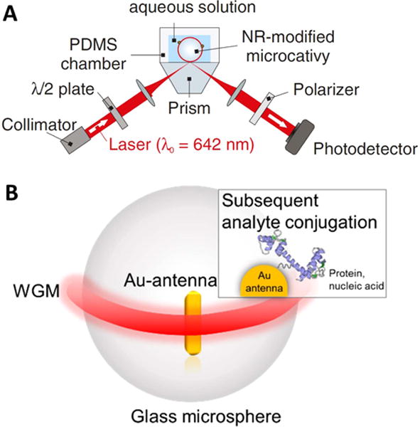
(a) WGM sensor. A glass microsphere is used as optical microcavity, to sense physical, chemical and biological entities. (b) Near total internal reflection of light results in an optical resonance (WGM, shown in red). The WGM couples to a nano-antenna where it excites plasmon resonance. Within the plasmonic hot spots, various nanoscale detection tasks are accomplished, such as protein and nucleic acid detection. [122] John Wiley & Sons. © 2016 Wiley-VCH Verlag GmbH & Co. KGaA, Weinheim.
Laser interferometry is at the heart of all precision measurements, gravitational wave detection being a prominent example. WGM sensing brings this type of metrology to the nanoscale. Sensing at the nanoscale is accomplished by attaching a plasmonic nanoparticle to the WGM microcavity. The resulting hybrid WGM sensor exploits the local field overlap of the plasmonic nanoparticle and a biomolecule to provide a heightened detection signal (figure 23(b)). Within the local field, WGM biosensing is accomplished with extreme sensitivity as to detect and analyze single virus particles, single biomolecules, surface reactions, and even single atomic ions [121, 128].
The WGM biosensor platform can be designed and fabricated from different materials and in different geometries, examples of which include silicon rings, glass toroids and glass capillaries. Efforts are underway to integrate these platforms onto portable devices as to, for example, create chip-scale biosensors [129]. Implantable sensors [130] are pursued, with the goal of enclosing WGM biosensors inside organisms and single cells.
Current and future challenges for WGM sensors
-
Exploring the physics of hybrid sensors.
A hybrid WGM sensor results, for instance, from the coupling of photonic WGMs to localized surface plasmon modes in nanoparticles [131, 132]. Another example of a hybrid sensor is an optical WGM coupled to a mechanical resonance [133]. Different physical effects contribute to the signal in a hybrid WGM sensor and, by exploring the intricate physics involved, we can reach yet unprecedented levels of sensitivity and conceive novel sensing modalities.
-
Adding single-molecule spectroscopy to sensing.
Single-molecule spectroscopy, such as Raman spectroscopy, suffers from a weak detection signal. This signal could be enhanced in hybrid, photonic–plasmonic WGM sensors. Adding spectroscopy to single-molecule detection schemes would provide a refined analytical tool for detecting and analyzing molecules in complex environments, hence leading to the real-time monitoring of structural and chemical changes within single molecules.
-
Monitoring the structural dynamics of single biomolecules.
Proteins are macromolecules with complex 3D structure. Proteins are not static entities. They gain biological function from their ability to change their structure. Their structural dynamics are central to catalysis, molecular recognition and signaling. WGM sensors can observe and analyze the dynamics of such intricate systems [134]. Developing such capability is a breakthrough in optical detection technology. Currently available techniques can only track the motion of parts of a protein when the protein is chemically modified, i.e. attachment of a chemical label. Without the need for a label, WGM biosensors will provide a universal tool for the unabated exploration of structural dynamics in individual proteins and biomolecular complexes. It will establish the cornerstone for an optical biosensor technology that is capable of harnessing the extreme speed, selectivity and specificity of the biological nanoworld.
-
Taking detection to the limit.
WGM sensors have not yet reached their physical detection limit. A future challenge is to implement techniques from laser interferometry, optical precision measurements and quantum optics to come closer to this limit. WGM sensors will benchmark nanoscale metrology and those with ultimate sensitivity will provide a tool for uncovering novel nanoscale physics and biophysics.
-
Real-time detection of signals from biomolecular networks in cells and organisms.
WGM sensors integrated with cells or organisms can track biomolecular signals in real time. This can enable new sensing modalities for organ-on-chip applications and for implantable biosensors. These sensing modalities will exploit single-molecule detection capability, i.e. in hormone sensing, enzyme-based assays and for detection tasks with membrane proteins. Integrated in the complex environment of an organism or a cell, WGM biosensors can monitor the specific signaling outputs from a network of biomolecules.
-
Engineering the abiotic-biotic optical interface.
Optogenetics provides a toolset for controlling cells with light. We can exploit this capability with optical WGM biosensors. The WGM sensor interface can be engineered to sense and manipulate the response of cells.
Advances in science and technology to meet challenges
Advances from optical precision measurements in atomic optics can be applied to WGM sensing.
Laser technology is improving, providing stable and ultra-narrow tunable sources for laser interferometry with WGM microcavities.
Single-photon detection capabilities from quantum optics can be combined with WGM sensors. Fairly recently, in the context of quantum logic structures, strong entanglement of photons (i.e. a 2-photon phase shift near π) was demonstrated in bottle resonators interacting with single atoms [135].
Advances in materials science can enable microcavities with novel sensing modalities, exploiting the linear and nonlinear optical effects of certain glasses and polymers. One could construct a sensor using MgF2 [136], TiO2 [137] or paraffin oil [138] to tune material loss for a desirable line-width, to alter the mode profile or to optimize nonlinearity coefficients. As in the case of a magneto-optical material such as yttrium iron garnet, fabricated WGM cavities have been shown to meet a triple-resonance condition for enhanced Brillouin scattering [139].
Bioengineering capabilities are advancing to print cells together with WGM sensors in complex organ-on-chips sensor structures.
Conclusions
There is an increasingly pervasive enthusiasm for advancements in WGM sensing. The versatility and sensitivity of the platform has not yet reached any bounds, thus it is up to the creativity of the experimenter to unlock or discover novel physical mechanisms for WGM sensing. Furthermore, the physics of nanoscale devices is a burgeoning field. With WGM sensors, nanoscale physics and biophysics can be studied with great precision. The detection of single atomic ions is just a first step towards exploring the ultimate limits of detection. By implementing advanced metrology and techniques from laser interferometry and atomic optics, further breakthroughs in nanoscale precision measurements are anticipated. Techniques from quantum optics combined with novel WGM materials may enable yet unexplored sensing strategies.
WGM biosensors can help us understand how our bodies work at the nanoscale level, where individual biomolecules such as enzymes take on the role of nano-machines, and where parts of a protein move similar to the pistons of an engine. Without the need for a label, WGM sensors will provide a universal tool for the unabated exploration of structural dynamics and shape-changes in individual proteins. Structural dynamics in proteins give rise to particular functions such as metabolism, signaling, energy harvesting, power strokes and many more. They constitute the basis of living matter in cells and organisms. Investigating these structural dynamics is of fundamental interest and will deliver novel approaches to nanomedicine.
14. Nanobiosensors for drug discovery
Qimin Quan
Rowland Institute at Harvard University
Status
Optical measurement features various advantages for biomedical applications. Unlike micro- or nanomechanical sensors, optical sensors are not compromised due to viscous damping in fluids. They are also immune to ionic screening— one of the biggest challenges for nanoelectronic sensors— and, therefore, are more compatible with physiological conditions. It is also possible to obtain molecular information of the test subject via Raman or absorption spectroscopy, in addition to the conventional biological recognition specificity offered by antibodies, aptamers, oligonucleotides, etc. Furthermore, optical measurements have a natural way to scale up by expanding a single biosensor into arrays and implementing imaging modalities.
Optical biosensors typically transduce biological signals based on changes in the electromagnetic field amplitude or phase, while interference and the evanescent field can increase their sensitivities. Nevertheless, as constrained by the light diffraction limit, traditional optical biosensors generally exhibit inferior sensitivity as compared to their electronic, mechanical or MEMS (micro-electro-mechanical systems) counterparts. The integration and packaging are also challenging due to more strict requirements for light coupling than electronic wiring.
The rapid advances in nanotechnologies have significantly boosted the performance of traditional optical biosensors, offering a new level of sensitivity (see section 13), enhanced specificity (see section 12), as well as improved integration capability [140]. This section will discuss the current challenges and future opportunities of nanobiosensing technologies in drug discovery. We refer the reader to other recent reviews for topics on point-of-care diagnostics [140], implantable devices [141] and therapeutic monitoring [142], etc.
Current and future challenges
New pharmaceutical drugs are discovered through a process including primary screening, secondary screening (i.e., hit validation and characterization), lead optimization, preclinical development (i.e., pharmacokinetics and toxicity), clinical phases and eventually regulatory approval. Target-based drug discovery (TDD) and phenotypic drug discovery (PDD) are two strategies being used before the potential drug candidates could enter the clinical phases. TDD starts from screening the effective compounds on a purified target via in vitro assays, while PDD looks at phenotypic effects at the system level, including but not limited to disease cell lines, patient-derived primary cells, tissues and animal models.
The screening pool size of TDD can be up to the order of millions, thus high-throughput screening technologies are essential. Fragment-based drug discovery has been gaining increasing interest in recent years. Compared to high-throughput screening, the pool size of fragment screening is less (a few thousands versus millions), however, much better sensitivity is demanded as the affinity of the fragments are significantly lower than the entire structure (μM-mM versus nM). The type of information gathered during the screening process includes binding kinetics, binding thermodynamics, stoichiometry, as well as information on the binding domains, ideally atomic structures. Fluorescent labeling techniques, such as fluorescence polarization assays and fluorescence thermal shift assays, are widely used high-throughput methods. However, the labeling process demands assay development and brings extra cost. Additionally, fluorescent labels may interfere with the compound-target interactions. It is also not uncommon in drug design for the compounds to be absorptive or fluorescent, which would complicate the signals from labels. Therefore, label-free methods are gaining more popularity [143]. Surface plasmon resonance (SPR) sensors have been established as a label-free, sensitive and high-throughput method that provides information on the binding kinetics and thermodynamics. Alternative techniques to SPR sensors include optical gratings, interferometers, photonic crystals, etc. Once the hit is identified, it is usually validated and studied using orthogonal technologies such as nuclear magnetic resonance (NMR) and isothermal titration calorimetry (ITC), which provide additional information on binding domains and stoichiometry. NMR and ITC are also direct binding assays, thus ideal for ruling out potential false positives due to the immobilization effect. However, NMR and ITC have low throughput and require a high sample amount. Mass spectrometry can be configured into either high-throughput mode or high-content mode to provide information on binding affinity and stoichiometry. Therefore, a wise combination of different technologies will not only save the cost, but also increase the speed to hit potential candidates. Modern drug discovery also takes advantage of the increasingly powerful computational techniques, such as Molecular Mechanics and Molecular Dynamics. Once the three-dimensional structure of the target is determined by x-ray crystallography, the structure-based lead optimization can provide rational insights to the drug candidates.
On the other hand, PDD screens drug candidates by their abilities to modify phenotypes at the system level. Historically, PDD has been the mainstream of drug discovery, which often starts from identifying the active ingredients of traditional remedies. TDD becomes popular as molecular biology offers the capability of rapidly synthesizing a large number of purified molecules to construct the screening pool. However, there are cases where the TDD hits fail to show system efficacy, despite demonstrating robust target binding ability. The recent slowdown in drug discovery also leads to the reflection that the reductionist approach such as hypothesis-driven TDD may limit the breadth of new findings. The phenotypic assays can evaluate various parameters, including cell morphology, motility, differentiation and certain hallmark features (gene and protein expression) that are specific to the disease. High-content screening technologies have evolved from small-scale biochemical assays to high-throughput imaging analysis using confocal or wide-field fluorescent microscopes. The lead needs to be optimized for its pharmacokinetics properties, known as absorption, distribution, metabolism and excretion (ADME). This can be achieved by using in vitro assays in combination with ADME models to extract the half-life properties and optimal dosages of drugs. Recent development of organ-on-chip models provides an alternative method that has shown better correlation in circulation and toxicity evaluation. They also recapitulate better the important intracellular and extracellular disease features. It is thus important to integrate the imaging capabilities and other optical sensing technologies in these systems.
Advances in science and technology to meet challenges
There are very few recent drug discovery success stories based purely on PDD or TDD. A holistic integration of both strategies is necessary and important. Besides throughput, the breadth and depth of information obtained from each measurement are becoming increasingly valuable. The marriage of nanotechnology with current biosensing and imaging approaches will reframe our current capability in drug discovery.
Single molecule methods
Current high-throughput screening methods are ensemble measurements. Interpreting binding kinetics (e.g. SPR data) is frequently carried out by assuming a one-step dynamic interaction. However, information on multiple binding domains and conformations is obscured in the ensemble model. The slow dissociation process from targets, which is the ideal drug characteristics, is also challenging to quantify due to limited stability in ensemble assays. The rapid development of nanosensors with significantly improved detection sensitivity may lead us to drug screening at the single molecule level (figures 24(a) and (b)) [123, 124].
Figure 24.
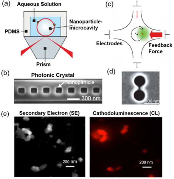
(a) Nanoparticle-microsphere [123] and (b) nanoparticle-photonic crystal [124] single molecule sensing platforms. [123] John Wiley & Sons. © 2016 Wiley-VCH Verlag GmbH & Co. KGaA, Weinheim. (c) Anti-Brownian electrokinetic traps. Reprinted with permission from [125]. Copyright (2012) American Chemical Society. (d) Double-nanohole plasmonic traps. Reprinted with permission from [126] Copyright (2012) American Chemical Society. (e) Nanodiamond [127] for dual mode electron and optical microscopy. Reprinted by permission from Macmillan Publishers Ltd: Scientific Reports [127], Copyright (2012). CC BY 3.0.
Native interactions
Current high-throughput methods require immobilization of the molecules on the sensor surface, which may affect their native interaction. The recent development of molecular trapping methods (such as electrophoretic (figure 24(c)) [125] or optical trapping (figure 24(d)) [126]) at the single to few molecule level will ultimately allow us to extract information of native interactions of molecules.
Microfluidics and MEMS Integration
Besides kinetic information, chemodynamic and thermodynamic information are also valuable. To extract this information in a high-throughput format, microfluidics and micro- to nano-electromechanical systems may be integrated to enable efficient liquid handling and precise thermodynamic control on each reaction.
Intracellular and sub-cellular nanosensors
Phenotypic information is predominantly analyzed by fluorescent microscopes. Label-free nanosensors will be extremely useful for patient-derived primary cell models or organ-on-chip models, where measurements on gene expressions, protein levels and metabolic events at targeted sub-cellular positions are desirable, without the need for transfection or genetic models [144].
New imaging technologies
Fluorescent microscopy provides specific functional information, but structural information is limited by the optical resolution. On the other hand, electron microscopy provides structural details without functional information. The development of new fluorescent and cathodoluminescent nanoprobes (e.g. nanodiamonds (figure 24(e)) [127], rare-earth nanocrystals [145]) compatible with both optical and electron microscopes might ultimately lead to an in-depth functional-structural understanding of the cellular machinery.
Concluding remarks
Besides technological development, it is also important to move beyond proof-of-concept demonstrations, and characterize the robustness, efficiency and weakness of the emerging technologies in relevant preclinical models to address practical biomedical problems. Strong collaboration with the drug discovery industry and early engagement in regulatory considerations may also help de-risk the translation from lab technologies to practical applications.
References
- 1.Auston DH, Smith PR. Generation and detection of millimeter waves by picosecond photoconductivity. Appl Phys Lett. 1983;43:631–3. [Google Scholar]
- 2.Cheung KP, Auston DHA. Novel technique for measuring far-infrared absorption and dispersion. Infrared Phys. 1986;26:23. [Google Scholar]
- 3.Nguyen D-T, Simoens F, Ouvrier-Buffet J-L, Meilhan J, Coutaz J-L. Broadband THz uncooled antenna-coupled microbolometer array—electromagnetic design, simulations and measurements. IEEE Trans Terahertz Sci Technol. 2012;2:299–305. [Google Scholar]
- 4.Preu S, Dohler GH, Malzer S, Wang LJ, Gossard AC. Tunable, continuous-wave terahertz photomixer sources and applications. J Appl Phys. 2011;109:061301. [Google Scholar]
- 5.Graf M, Scalari G, Hofstetter D, Faist J, Beere H, Linfield E, Ritchie D, Davies G. Terahertz range quantum well infrared photodetector. Appl Phys Lett. 2004;84:475–7. [Google Scholar]
- 6.Vicarelli L, Vitiello M, Coquillat D, Lombardo A, Ferrari A, Knap W, Polini M, Pellegrini V, Tredicucci A. Graphene field-effect transistors as room-temperature terahertz detectors. Nat Mater. 2012;11:865–71. doi: 10.1038/nmat3417. [DOI] [PubMed] [Google Scholar]
- 7.Vitiello MS, Coquillat D, Viti L, Ercolani D, Teppe F, Pitanti A, Beltram F, Sorba L, Knap W, Tredicucci A. Room-temperature terahertz detectors based on semiconductor nanowire field-effect transistors. Nano Letter. 2011;12:96–101. doi: 10.1021/nl2030486. [DOI] [PubMed] [Google Scholar]
- 8.Huber R, Tauser F, Brodschelm A, Bichler M, Abstreiter G, Leitenstorfer A. How many-particle interactions develop after ultrafast excitation of an electron-hole plasma. Nature. 2001;414:286–9. doi: 10.1038/35104522. [DOI] [PubMed] [Google Scholar]
- 9.Castro-Camus E, Palomar M, Covarrubias A. Leaf water dynamics of Arabidopsis thaliana monitored in vivo using terahertz time-domain spectroscopy. Sci Rep. 2013;3:2910. doi: 10.1038/srep02910. [DOI] [PMC free article] [PubMed] [Google Scholar]
- 10.Probst T, Sommer S, Soltani A, Kraus E, Baudrit B, Town G, Koch M. Monitoring the polymerization of two-component epoxy adhesives using a terahertz time domain reflection system. J Infrared Millim Terahertz Waves. 2015;36:569–77. [Google Scholar]
- 11.Falconer RJ, Markelz AG. Terahertz spectroscopic analysis of peptides and proteins. J Infrared Millim Terahertz Waves. 2012;33:973–88. [Google Scholar]
- 12.Stecher M, Jördens C, Krumbholz N, Jansen C, Scheller M, Wilk R, Peters O, Scherger B, Ewers B, Koch M. Towards Industrial Inspection with THz Systems Ultrashort Pulse Laser Technology: Laser Sources and Applications. New York: Springer; 2015. pp. 311–35. [Google Scholar]
- 13.Fukunaga K. THz Technology Applied to Cultural Heritage in Practice Cultural Heritage Science. Tokyo: Springer; 2016. https://doi.org/10.1007/978-4-431-55885-9. [Google Scholar]
- 14.Sensale-Rodriguez B, et al. Broadband graphene terahertz modulators enabled by intraband transitions. Nat Commun. 2012;3:780. doi: 10.1038/ncomms1787. [DOI] [PubMed] [Google Scholar]
- 15.Petersen CR, et al. Mid-infrared supercontinuum covering the 1.4-13.3 μ m molecular fingerprint region using ultra-high NA chalcogenide step-index fibre. Nat Photon. 2014;8:830–4. [Google Scholar]
- 16.Yu M, Okawachi Y, Griffith AG, Lipson M, Gaeta AL. Modelocked mid-infrared frequency combs in a silicon microresonator. Optica. 2016;3:854–60. doi: 10.1364/OE.24.013044. [DOI] [PubMed] [Google Scholar]
- 17.Yao Y, Hoffman AJ, Gmachl CF. Mid-infrared quantum cascade lasers. Nat Photon. 2012;6:432–9. [Google Scholar]
- 18.Vurgaftman I, Bewley WW, Canedy CL, Kim CS, Kim M, Merritt CD, Abell J, Meyer JR. Interband cascade lasers with low threshold powers and high output powers. IEEE J Sel Top Quantum Electron. 2013;19:1200210. [Google Scholar]
- 19.Hugi A, Villares G, Blaser S, Liu HC, Faist J. Mid-infrared frequency comb based on a quantum cascade laser. Nature. 2012;492:229–33. doi: 10.1038/nature11620. [DOI] [PubMed] [Google Scholar]
- 20.Henderson-Sapir O, Malouf A, Bawden N, Munch J, Jackson S, Ottaway DJ. Recent advances in 3.5 μm erbium doped mid-infrared fiber lasers. IEEE J Sel Top Quantum Electron. 2016;23:0900509. [Google Scholar]
- 21.Bernier M, Fortin V, El-Amraoui M, Messaddeq Y, Vallée R. 3.77 μm fiber laser based on cascaded Raman gain in a chalcogenide glass fiber. Opt Lett. 2014;39:2052–5. doi: 10.1364/OL.39.002052. [DOI] [PubMed] [Google Scholar]
- 22.Hu T, Jackson SD, Hudson DD. Ultrafast pulses from a mid-infrared fiber laser. Opt Lett. 2015;40:4226–8. doi: 10.1364/OL.40.004226. [DOI] [PubMed] [Google Scholar]
- 23.Duval S, Bernier M, Fortin V, Genest J, Piché M, Vallée R. Femtosecond fiber lasers reach the mid-infrared. Optica. 2015;2:623–6. [Google Scholar]
- 24.Tokita S, Murakami M, Shimizu S, Hashida M, Sakabe S. 12 WQ-switched Er: ZBLAN fiber laser at 2.8 μm. Opt Lett. 2011;36:2812–4. doi: 10.1364/OL.36.002812. [DOI] [PubMed] [Google Scholar]
- 25.Tao G, Ebendorff-Heidepriem H, Stolyarov AM, Danto S, Badding JV, Fink Y, Ballato J, Abouraddy AF. Infrared fibers. Adv Opt Photonics. 2015;7:379–458. [Google Scholar]
- 26.Rieker GB, et al. Frequency-comb-based remote sensing of greenhouse gases over kilometer air paths. Optica. 2014;1:290–8. [Google Scholar]
- 27.Hébert NB, Scholten SK, White RT, Genest J, Luiten AN, Anstie JD. A quantitative mode-resolved frequency comb spectrometer. Opt Express. 2015;23:13991–4001. doi: 10.1364/OE.23.013991. [DOI] [PubMed] [Google Scholar]
- 28.Kostecki R, Ebendorff-Heidepriem H, Davis C, McAdam G, Warren-Smith SC, Monro TM. Silica exposed-core microstructured optical fibers. Opt Mater Express. 2012;2:1538–47. [Google Scholar]
- 29.López-Higuera JM, Cobo LR, Quintela A, Cobo A. Fiber optic sensors in structural health monitoring. J Lightwave Technol. 2011;29:587–608. [Google Scholar]
- 30.Giallorenzi TG, Bucaro JA, Dandridge A, Sigel GH, Cole JH, Rashleigh SC. Optical fiber sensor technology. IEEE Microw Theory and Techniques Society. 1982;30:472–511. [Google Scholar]
- 31.Arditty HJ, Lefèvre HC. Sagnac effect in fiber gyroscopes. Opt Lett. 1981;6:401–3. doi: 10.1364/ol.6.000401. [DOI] [PubMed] [Google Scholar]; Lefèvre HC. The fiber-optic gyroscope: challenges to become the ultimate rotation-sensing technology. Opt Fiber Technol. 2013;19:828–32. [Google Scholar]
- 32.Rogers AJ. Distributed optical fiber sensors. J Phys D: Appl Phys. 1986;19:2237–55. [Google Scholar]; Rogers A. Distributed optical sensing Handbook of Optical Fibre Sensing Technology. New York: Wiley; 2002. ch 14. [Google Scholar]
- 33.Proceedings of OFS9, Paris, France; Collected Papers of the International Conferences on OFS. 1983–1997 http://spie.org/x648.html?product_id=316034.
- 34.Lopez-Amo M, Lopez-Higuera JM. Fiber Bragg Gratings Sensors: Recent Advancements, Industrial Applications and Market Exploitation. Potomac, MD: Bentham Science; 2011. Multiplexing techniques for FBG sensors; pp. 99–115. [Google Scholar]
- 35.Knight JC, Birks TA, Russell PSJ, Atkin DM. Optical Fiber Communications Conf. San Jose, California: 1996. Pure silica single-mode fiber with hexagonal photonic crystal cladding. [Google Scholar]
- 36.Gatekeepers Inc. Information. Photonic Sensor Consortium Market Survey Report Light Wave Venture LLC 2015 [Google Scholar]
- 37.Dowling J, Seshadreesan KP. Quantum technologies for sensing, metrology and imaging. J Lightwave Technol. 2014;33:2359–70. [Google Scholar]
- 38.Mazur E. Int School on Light Sciences and Technologies. Santander; Spain: 2017. Jun 19–23, Less is more: extreme optics with zero refractive index. [Google Scholar]
- 39.Antman Y, Clain A, London Y, Zadok A. Optomechanical sensing of liquids outside standard fiber using forward stimulated Brillouin scattering. Optica. 2016;3:510. [Google Scholar]
- 40.Russell P. Int School of Light Sciences and Technologies, UIMP. Santander; Spain: 2016. Jun 20, The multi-faceted world of photonic crystal fibers. [Google Scholar]
- 41.Udd E, Spillman WB., Jr . Fiber Optic Sensors: An Introduction for Engineers and Scientists. 2nd. New York: Wiley; 2011. [Google Scholar]
- 42.Willets KA, Van Duyne RP. Localized surface plasmon resonance spectroscopy and sensing. Annu Rev Phys Chem. 2007;58:267–97. doi: 10.1146/annurev.physchem.58.032806.104607. [DOI] [PubMed] [Google Scholar]
- 43.Oo MKK, Han Y, Kanka J, Sukhishvili S, Du H. Structure fits the purpose: photonic crystal fibers for evanescent-field surface-enhanced Raman spectroscopy. Opt Lett. 2010;35:466–8. doi: 10.1364/OL.35.000466. [DOI] [PubMed] [Google Scholar]
- 44.Leung A, Mohana Shankar P, Mutharasan R. A review of fiber-optic biosensors. Sensors Actuators B. 2007;125:688–703. [Google Scholar]
- 45.Blue R, Uttamchandani D. Recent advances in optical fiber devices for microfluidics integration. J Biophotonics. 2016;9:13–25. doi: 10.1002/jbio.201500170. [DOI] [PubMed] [Google Scholar]
- 46.Abouraddy AF, Bayindir M, Benoit G, Hart SD, Kuriki K, Orf N, Shapira O, Sorin F, Temelkuran B, Fink Y. Towards multimaterial multifunctional fibres that see, hear, sense and communicate. Nat Mater. 2007;6:336–47. doi: 10.1038/nmat1889. [DOI] [PubMed] [Google Scholar]
- 47.Lian Z, et al. Nanomechanical optical fiber. Opt Express. 2012;20:29386–94. doi: 10.1364/OE.20.029386. [DOI] [PubMed] [Google Scholar]
- 48.Hossain MA, Canning J, Cook K, Jamalipour A. Optical fiber smartphone spectrometer. Opt Lett. 2016;41:2237–40. doi: 10.1364/OL.41.002237. [DOI] [PubMed] [Google Scholar]
- 49.Rantala J, Hännikäinen J, Vanhala J. Fiber optic sensors for wearable applications. Pers Ubiquitous Comput. 2011;15:85–96. [Google Scholar]
- 50.Olsson T. Event driven persistent sensing: overcoming the energy and lifetime limitations in unattended wireless sensors. IEEE SENSORS (Orlando, FL, USA, 30 October–2 November) 2016 [Google Scholar]
- 51.Russell PSJ. Photonic-crystal fibers. J Lightwave Technol. 2006;24:4729–49. [Google Scholar]
- 52.Digonnet M, Blin S, Kim HK, Dangui V, Kino G. Sensitivity and stability of an air-core fibre-optic gyroscope. Meas Sci Technol. 2007;18:3089–97. [Google Scholar]
- 53.Jin W, Xuan HF, Ho HL. Sensing with hollow-core photonic bandgap fibers. Meas Sci Technol. 2010;21:094014. [Google Scholar]
- 54.Fini JM. Microstructure fibres for optical sensing in gases and liquids. Meas Sci Technol. 2004;15:1120–8. [Google Scholar]
- 55.Jin W, Cao Y, Yang F, Ho HL. Ultra-sensitive all-fiber photothermal spectroscopy with large dynamic range. Nat Commun. 2015;6:6767. doi: 10.1038/ncomms7767. [DOI] [PMC free article] [PubMed] [Google Scholar]
- 56.Xiao L, Demokan MS, Jin W, Wang Y, Zhao CL. Fusion splicing photonic crystal fibers and conventional single-mode fibers: microhole collapse effect. J Lightwave Technol. 2007;25:3563–74. [Google Scholar]
- 57.Fini JM, Nicholson JW, Mangan B, Meng L, Windeler RS, Monberg EM, DeSantolo A, DiMarcello FV, Mukasa K. Polarization maintaining single-mode low-loss hollow-core fibres. Nat Commun. 2014;5:5085. doi: 10.1038/ncomms6085. [DOI] [PubMed] [Google Scholar]
- 58.Uebel P, Gunendi MC, Frosz MH, Ahmed G, Edavalath MN, Menard J-M, Russell PSTJ. Broadband robustly single-mode hollow-core PCF by resonant filtering of higher-order modes. Opt Lett. 2016;41:1961–4. doi: 10.1364/OL.41.001961. [DOI] [PubMed] [Google Scholar]
- 59.Wiederhecker GS, Cordeiro CMB, Couny F, Benabid F, Maier SA, Knight JC, Cruz CHB, Fragnito HL. Field enhancement within an optical fibre with a subwavelength air core. Nat Photon. 2007;1:115–8. [Google Scholar]
- 60.Bykov DS, Schmidt OA, Euser TG, Russell PSJ. Flying particle sensors in hollow-core photonic crystal fibre. Nat Photon. 2015;9:461–6. [Google Scholar]
- 61.Smullin LD, Fiocco G. Optical echoes from the Moon. Nature. 1962;194:1267. [Google Scholar]
- 62.Irene EA. A brief history and state of the art of ellipsometry. In: Losurdo M, Hingerl K, editors. Ellipsometry at the Nanoscale. Berlin: Springer; 2013. pp. 1–30. [Google Scholar]
- 63.Vaz PG, Humeau-Heurtier A, Figueiras E, Correia C, Cardoso J. Laser speckle imaging to monitor microvascular blood flow: a review. IEEE Rev Biomed Eng. 2016;9:106–20. doi: 10.1109/RBME.2016.2532598. [DOI] [PubMed] [Google Scholar]
- 64.Butler HJ, et al. Using Raman spectroscopy to characterize biological materials. Nat Protocols. 2016;11:664–87. doi: 10.1038/nprot.2016.036. [DOI] [PubMed] [Google Scholar]
- 65.Rothberg SJ, et al. An international review of laser Doppler vibrometry: making light work of vibration measurement. Opt Lasers Eng. 2017 (to appear) [Google Scholar]
- 66.Jian Y, Lee S, Ju MJ, Heisler M, Ding W, Zawadzki RJ, Bonora S, Sarunic MV. Lens-based wavefront sensorless adaptive optics swept source OCT. Sci Rep. 2016;6:27620. doi: 10.1038/srep27620. [DOI] [PMC free article] [PubMed] [Google Scholar]
- 67.Hancock S, Anderson K, Disney M, Gaston KJ. Measurement of fine-spatial-resolution 3D vegetation structure with airborne waveform LIDAR: calibration and validation with voxelised terrestrial lidar. Remote Sens Environ. 2017;188:37–50. [Google Scholar]
- 68.Joo J, Munro WJ, Spiller TP. Quantum metrology with entangled coherent states. Phys Rev Lett. 2011;107:083601. doi: 10.1103/PhysRevLett.107.083601. [DOI] [PubMed] [Google Scholar]
- 69.Jeong Y, Vazquez-Zuniga LA, Lee S, Kwon Y. On the formation of noise-like pulses in fiber ring cavity configurations. Opt Fiber Technol. 2014;20:575–92. [Google Scholar]
- 70.Khodabakhsh A, Ramaiah-Badarla V, Rutkowski L, Johansson AC, Lee KF, Jiang J, Mohr C, Fermann ME, Foltynowicz A. Fourier transform and Vernier spectroscopy using an optical frequency comb at 3.54 μm. Opt Lett. 2016;41:2541–4. doi: 10.1364/OL.41.002541. [DOI] [PubMed] [Google Scholar]
- 71.Mandon J, Guelachvili G, Picqué N. Fourier transform spectroscopy with a laser frequency comb. Nat Photon. 2009;3:99–102. [Google Scholar]
- 72.Schliesser A, Picqué N, Hänsch TW. Mid-infrared frequency combs. Nat Photon. 2012;6:440–9. [Google Scholar]
- 73.Faist J, Villares G, Scalari G, Rösch M, Bonzon C, Hugi A, Beck M. Quantum cascade laser frequency combs. Nanophotonics. 2016;5:272–91. [Google Scholar]
- 74.Yu M, Okawachi Y, Griffith AG, Picqué N, Lipson M, Gaeta AL. Silicon-chip-based mid-infrared dual-comb spectroscopy. 2016 doi: 10.1038/s41467-018-04350-1. arXiv:1610.01121. [DOI] [PMC free article] [PubMed] [Google Scholar]
- 75.Ideguchi T, Holzner S, Bernhardt B, Guelachvili G, Picqué N, Hänsch TW. Coherent Raman spectro-imaging with laser frequency combs. Nature. 2013;502:355–8. doi: 10.1038/nature12607. [DOI] [PubMed] [Google Scholar]
- 76.Asahara A, Nishiyama A, Yoshida S, Kondo K, Nakajima Y, Minoshima K. Dual-comb spectroscopy for rapid characterization of complex optical properties of solids. Opt Lett. 2016;41:4971–4. doi: 10.1364/OL.41.004971. [DOI] [PubMed] [Google Scholar]
- 77.Truong GW, Waxman EM, Cossel KC, Baumann E, Klose A, Giorgetta FR, Swann WC, Newbury NR, Coddington I. Accurate frequency referencing for fieldable dual-comb spectroscopy. Opt Express. 2016;24:30495–504. doi: 10.1364/OE.24.030495. [DOI] [PubMed] [Google Scholar]
- 78.Millot G, Pitois S, Yan M, Hovannysyan T, Bendahmane A, Hänsch TW, Picqué N. Frequency-agile dual-comb spectroscopy. Nat Photon. 2016;10:27–30. [Google Scholar]
- 79.Suh MG, Yang QF, Yang KY, Yi X, Vahala KJ. Microresonator soliton dual-comb spectroscopy. Science. 2016;354:600–3. doi: 10.1126/science.aah6516. [DOI] [PubMed] [Google Scholar]
- 80.Rao YJ. In-fibre Bragg grating sensors. Meas Sci Technol. 1997;8:355–75. [Google Scholar]
- 81.Frazao O, Santos JL, Araujo FM, Ferreira LA. Optical sensing with photonic crystal fibers. Laser Photon Rev. 2008;2:449–59. [Google Scholar]
- 82.Guo X, Ying YB, Tong LM. Photonic nanowires: from subwavelength waveguides to optical sensors. Acc Chem Res. 2014;47:656–66. doi: 10.1021/ar400232h. [DOI] [PubMed] [Google Scholar]
- 83.Fang X, Liao CR, Wang DN. Femtosecond laser fabricated fiber Bragg grating in microfiber for refractive index sensing. Opt Lett. 2010;35:1007–9. doi: 10.1364/OL.35.001007. [DOI] [PubMed] [Google Scholar]
- 84.Zhang L, Li ZY, Yu SJ, Mu JX, Fang W, Tong LM. Femtoliter-scale optical nanofiber sensors. Opt Express. 2015;23:28408–15. doi: 10.1364/OE.23.028408. [DOI] [PubMed] [Google Scholar]
- 85.Vollmer F, Yang L. Label-free detection with high-Q microcavities: a review of biosensing mechanisms for integrated devices. Nanophotonics. 2012;1:267–91. doi: 10.1515/nanoph-2012-0021. [DOI] [PMC free article] [PubMed] [Google Scholar]
- 86.Kollmann H, et al. Toward plasmonics with nanometer precision: nonlinear optics of helium-ion milled gold nanoantennas. Nano Lett. 2014;14:4778–84. doi: 10.1021/nl5019589. [DOI] [PubMed] [Google Scholar]
- 87.Ameling R, Giessen H. Microcavity plasmonics: strong coupling of photonic cavities and plasmons. Laser Photon Rev. 2013;7:141–69. [Google Scholar]
- 88.Wang P, et al. Single-band 2 nm-linewidth plasmon resonance in a strongly coupled Au nanorod. Nano Lett. 2015;15:7581–6. doi: 10.1021/acs.nanolett.5b03330. [DOI] [PubMed] [Google Scholar]
- 89.Wang YP, Ma YG, Guo X, Tong LM. Single-mode plasmonic waveguiding properties of metal nanowires with dielectric substrates. Opt Express. 2012;20:19006–15. doi: 10.1364/OE.20.019006. [DOI] [PubMed] [Google Scholar]
- 90.Wu L, Reinhard BM. Probing subdiffraction limit separations with plasmon coupling microscopy: concepts and applications. Chem Soc Rev. 2014;43:3884. doi: 10.1039/c3cs60340g. [DOI] [PMC free article] [PubMed] [Google Scholar]
- 91.Boyer D, Tamarat P, Maali A, Lounis B, Orrit M. Photothermal imaging of nanometer-sized metal particles among scatterers. Science. 2002;297:1160. doi: 10.1126/science.1073765. [DOI] [PubMed] [Google Scholar]
- 92.Yang L, Yan B, Premasiri RW, Ziegler LD, Dal NegroL, Reinhard BM. Engineering nanoparticle cluster arrays for bacterial biosensing: the role of the building block in multiscale SERS substrates. Adv Funct Mater. 2010;20:2619. [Google Scholar]
- 93.Yan B, Reinhard BM. Identification of tumor cells through spectroscopic profiling of the cellular surface chemistry. J Phys Chem Lett. 2010;1:1595. [Google Scholar]
- 94.Wang J, Yu X, Boriskina SV, Reinhard BM. Quantification of differential ErbB1 and ErbB2 cell surface expression and spatial nanoclustering through plasmon coupling. Nano Lett. 2012;12:3231. doi: 10.1021/nl3012227. [DOI] [PMC free article] [PubMed] [Google Scholar]
- 95.Wang H, Wu L, Reinhard BM. Scavenger receptor mediated endocytosis of silver nanoparticles into J774A.1 macrophages is heterogeneous. ACS Nano. 2012;6:7122. doi: 10.1021/nn302186n. [DOI] [PMC free article] [PubMed] [Google Scholar]
- 96.Xinwei Y, Wang J, Feizpour A, Reinhard BM. Illuminating the lateral organization of cell-surface CD24 and CD44 through plasmon coupling between Au nanoparticle immunolabels. Anal Chem. 2013;85:1290. doi: 10.1021/ac303310j. [DOI] [PMC free article] [PubMed] [Google Scholar]
- 97.Yu X, Feizpour A, Ramirez N-GP, Wu L, Akiyama H, Yu X, Gummuluru S, Reinhard BM. Glycosphingolipid-functionalized nanoparticles recapitulate CD169-dependent HIV-1 uptake and trafficking in dendritic cells. Nat Commun. 2014;5:4136. doi: 10.1038/ncomms5136. [DOI] [PMC free article] [PubMed] [Google Scholar]
- 98.Xu F, Reiser M, Yu X, Gummuluru S, Wetzler L, Reinhard BM. Lipid mediated targeting with membrane-wrapped nanoparticles in the presence of corona formation. ACS Nano. 2015;10:1189. doi: 10.1021/acsnano.5b06501. [DOI] [PMC free article] [PubMed] [Google Scholar]
- 99.Xinwei Y, Fangda X, Nora-Guadalupe PR, Suzanne DGK, Akiyama H, Gummuluru S, Reinhard BM. Dressing up nanoparticles: a membrane wrap to induce formation of the virological synapse. ACS Nano. 2015;9:4182. doi: 10.1021/acsnano.5b00415. [DOI] [PMC free article] [PubMed] [Google Scholar]
- 100.Nasr MB, Goode DP, Nguyen N, Rong GX, Yang LL, Reinhard BM, Saleh BEA, Teich MC. Quantum optical coherence tomography of a biological sample. Opt Commun. 2009;282:1154. [Google Scholar]
- 101.Wu L, Xu F, Reinhard BM. Nanoconjugation prolongs endosomal signaling of the epidermal growth factor receptor and enhances apoptosis. Nanoscale. 2016;8:13755. doi: 10.1039/c6nr02974d. [DOI] [PMC free article] [PubMed] [Google Scholar]
- 102.McDonagh C, Burke CS, McCraith BD. Optical chemical sensors. Chem Rev. 2008;108:400–22. doi: 10.1021/cr068102g. [DOI] [PubMed] [Google Scholar]
- 103.Wang X-D, Wolfbeis OS. Fiber-optic chemical sensors and biosensors 2013–2015. Anal Chem. 2016;88:203–27. doi: 10.1021/acs.analchem.5b04298. [DOI] [PubMed] [Google Scholar]
- 104.Barbieri B Short History of Fluorescence. https://fluorescence-foundation.org/lectures/madrid2010/lecture1.pdf.
- 105.French R S The History of Infrared Spectroscopy, HET607. Swinburne Astronomy Online, http://rfrench.org/astro/papers/P44-HET607-RobertFrench.pdf.
- 106.Radziemski L, Cremers D. A brief history of laser-induced breakdown spectroscopy: from the concept of atoms to LIBS (2012) Spectrochim Acta B. 2013;87:3–10. [Google Scholar]
- 107.Qazi HH, bin Mohammad AB, Akram M. Recent progress in optical chemical sensors. Sensors. 2012;12:16522–56. doi: 10.3390/s121216522. [DOI] [PMC free article] [PubMed] [Google Scholar]
- 108.Katzir A. Selected Papers on Optical Fibers in Medicine (SPIE Milestone Series vol MS 11) Bellingham, WA: SPIE; 1990. [Google Scholar]
- 109.Hirschowitz B. A personal history of the fiberscope. Gastroenterology. 1979;76:864–9. [PubMed] [Google Scholar]
- 110.Mignani MAG, Baldini F. Biomedical sensors using optical fibres. Rep Prog Phys. 1996;59:1–28. [Google Scholar]
- 111.Mendez A. Optical fiber sees growth as medical sensors. Laser Focus World. 2011;47:91–4. [Google Scholar]
- 112.United Nations DESA. World Population Prospects: The 2015 Revision 2015 [Google Scholar]
- 113.Cusano A, Consales M, Crescitelli A, Ricciardi A. Lab-on-Fiber Technology. Berlin: Springer; 2015. [Google Scholar]
- 114.Breslauer DN, et al. Mobile phone based clinical microscopy for global health applications. PLOS One. 2009;4:e6320. doi: 10.1371/journal.pone.0006320. [DOI] [PMC free article] [PubMed] [Google Scholar]
- 115.Bird B, Miljković M, Remiszewski S, Akalin A, Kon M, Diem M. Infrared spectral histopathology (SHP): a novel diagnostic tool for the accurate classification of lung cancers. Lab Invest. 2012;92:1358–73. doi: 10.1038/labinvest.2012.101. [DOI] [PubMed] [Google Scholar]
- 116.Akalin A, Mu X, Kon M, Ergin A, Remiszewski S, Thompson C, Raz D, Bird B, Miljković M, Diem M. Classification of malignant and benign tumors of the lung by infrared spectral histopathology (SHP) Lab Invest. 2015;95:406–21. doi: 10.1038/labinvest.2015.1. [DOI] [PubMed] [Google Scholar]
- 117.Mu X, Kon M, Ergin A, Remiszewski S, Akalin A, Thompson C, Diem M. Statistical analysis of a lung cancer spectral histopathology (SHP) data set. Analyst. 2015;140:2449–64. doi: 10.1039/c4an01832j. [DOI] [PubMed] [Google Scholar]
- 118.Pounder FN, Bhargava R. Vibrational Spectroscopic Imaging for Biomedical Applications. New York: McGraw Hill; 2010. Toward automated breast histopathology using mid-IR spectroscopic imaging; pp. 1–26. [Google Scholar]
- 119.Bassan P, Sachdeva A, Shanks JH, Brown MD, Clarke NW, Gardner P. Automated high-throughput assessment of prostate biopsy tissue using infrared spectroscopic chemical imaging. Proc SPIE. 2014;9041(12):1–10. [Google Scholar]
- 120.Großerüschkamp F, Kallenbach-Thieltges A, Behrens T, Brüning T, Altmayer M, Stamatis G, Theegarten D, Gerwert K. Marker-free automated histopathological annotation of lung tumour subtypes by FTIR imaging. Analyst. 2015;140:2114–20. doi: 10.1039/c4an01978d. [DOI] [PubMed] [Google Scholar]
- 121.Foreman MR, Swaim JD, Vollmer F. Whispering gallery mode sensors. Adv Opt Photon. 2015;7:168–240. doi: 10.1364/AOP.7.000168. [DOI] [PMC free article] [PubMed] [Google Scholar]
- 122.Kim E, Baaske MD, Vollmer F. In situ observation of single-molecule surface reactions from low to high affinities. Adv Mater. 2016;28:9941–8. doi: 10.1002/adma.201603153. [DOI] [PubMed] [Google Scholar]
- 123.Kim E, Baaske MD, Vollmer F. In situ observation of single-molecule surface reactions from low to high affinities. Adv Mater. 2016;28:9941–8. doi: 10.1002/adma.201603153. [DOI] [PubMed] [Google Scholar]
- 124.Liang F, Guo Y, Hou S, Quan Q. Photonic–plasmonic hybrid single-molecule nanosensor measures the effect of fluorescent labels on DNA–protein dynamics. Sci Adv. 2017;3:e1602991. doi: 10.1126/sciadv.1602991. [DOI] [PMC free article] [PubMed] [Google Scholar]
- 125.Wang Q, Goldsmith RH, Jiang Y, Bockenhauer SD, Moerner WE. Probing single biomolecules in solution using the Anti-Brownian Electrokinetic (ABEL) Trap. Accounts Chem Res. 2012;45:1955–64. doi: 10.1021/ar200304t. [DOI] [PMC free article] [PubMed] [Google Scholar]
- 126.Pang YJ, Gordon R. Optical trapping of a single protein. Nano Lett. 2012;12:402–6. doi: 10.1021/nl203719v. [DOI] [PubMed] [Google Scholar]
- 127.Glenn DR, Zhang H, Kasthuri N, Schalek R, Lo PK, Trifonov AS, Park H, Lichtman JW, Walsworth RL. Correlative light and electron microscopy using cathodoluminescence from nanoparticles with distinguishable colours. Sci Rep. 2012;2:865. doi: 10.1038/srep00865. [DOI] [PMC free article] [PubMed] [Google Scholar]
- 128.Baaske MD, Vollmer F. Optical observation of single atomic ions interacting with plasmonic nanorods in aqueous solution. Nat Photonics. 2016;10:733–9. [Google Scholar]
- 129.Eugene Kim MB, Vollmer F. Towards next-generation label-free biosensors: recent advances in whispering gallery mode sensors. Lab Chip. 2017;17:1190–205. doi: 10.1039/c6lc01595f. [DOI] [PubMed] [Google Scholar]
- 130.Humar M, Yun SH. Intracellular microlasers. Nat Photonics. 2015;9:572–6. doi: 10.1038/nphoton.2015.129. [DOI] [PMC free article] [PubMed] [Google Scholar]
- 131.Baaske MD, Foreman MR, Vollmer F. Single-molecule nucleic acid interactions monitored on a label-free microcavity biosensor platform. Nat Nano. 2014;9:933–9. doi: 10.1038/nnano.2014.180. [DOI] [PubMed] [Google Scholar]
- 132.Swaim JD, Knittel J, Bowen WP. Detection limits in whispering gallery biosensors with plasmonic enhancement. Appl Phys Lett. 2011:99. [Google Scholar]
- 133.Yu W, Jiang WC, Lin Q, Lu T. Cavity optomechanical spring sensing of single molecules. Nat Commun. 2016;7:12311. doi: 10.1038/ncomms12311. [DOI] [PMC free article] [PubMed] [Google Scholar]
- 134.Kim E, Baaske MD, Schuldes I, Wilsch PS, Vollmer F. Label-free optical detection of single enzyme-reactant reactions and associated conformational changes. Sci Adv. 2017;3:e1603044. doi: 10.1126/sciadv.1603044. [DOI] [PMC free article] [PubMed] [Google Scholar]
- 135.Volz J, Scheucher M, Junge C, Rauschenbeutel A. Nonlinear [pi] phase shift for single fibre-guided photons interacting with a single resonator-enhanced atom. Nat Photonics. 2014;8:965–70. [Google Scholar]
- 136.Alnis J, Schliesser A, Wang CY, Hofer J, Kippenberg TJ, Hänsch TW. Thermal-noise-limited crystalline whispering-gallery-mode resonator for laser stabilization. Phys Rev A. 2011;84:011804. doi: 10.1364/OE.20.019185. [DOI] [PubMed] [Google Scholar]
- 137.Park J, Ozdemir SK, Monifi F, Chadha T, Huang SH, Biswas P, Yang L. Titanium dioxide whispering gallery microcavities. Adv Opt Mat. 2014;2:711–7. [Google Scholar]
- 138.Foreman MR, Avino S, Zullo R, Loock HP, Vollmer F, Gagliardi G. Enhanced nanoparticle detection with liquid droplet resonators. Eur Phys J Spec Top. 2014;223:1971–88. [Google Scholar]
- 139.Haigh JA, Nunnenkamp A, Ramsay AJ, Ferguson AJ. Triple-resonant Brillouin light scattering in magneto-optical cavities. Phys Rev Lett. 2016;117:133602. doi: 10.1103/PhysRevLett.117.133602. [DOI] [PubMed] [Google Scholar]
- 140.Lopez GA, Estevez M-C, Soler M, Lechuga LM. Recent advances in nanoplasmonic biosensors: applications and lab-on-a-chip integration. Nanophotonics. 2017;6:123–36. [Google Scholar]
- 141.Humar M, Kwok SJ, Choi M, Yetisen AK, Cho S, Yun S-H. Toward biomaterial-based implantable photonic devices. Nanophotonics. 2016;6:414. [Google Scholar]
- 142.Salvati E, Stellacci F, Krol S. Nanosensors for early cancer detection and for therapeutic drug monitoring. Nanomedicine. 2015;10:3495–512. doi: 10.2217/nnm.15.180. [DOI] [PubMed] [Google Scholar]
- 143.Cooper MA. Optical biosensors in drug discovery. Nat Rev Drug Discovery. 2002;1:515–28. doi: 10.1038/nrd838. [DOI] [PubMed] [Google Scholar]
- 144.Liang F, Zhang YY, Hong WY, Dong YL, Xie ZC, Quan QM. Direct tracking of amyloid and tau dynamics in neuroblastoma cells using nanoplasmonic fiber tip probes. Nano Lett. 2016;16:3989–94. doi: 10.1021/acs.nanolett.6b00320. [DOI] [PMC free article] [PubMed] [Google Scholar]
- 145.Fukushima S, et al. Correlative near-infrared light and cathodoluminescence microscopy using Y2O3:Ln, Yb (Ln = Tm, Er) nanophosphors for multiscale, multicolour bioimaging. Sci Rep. 2016;6:25950. doi: 10.1038/srep25950. [DOI] [PMC free article] [PubMed] [Google Scholar]


