Abstract
Hotspot relaxation time (τth) is one of the essential parameter which defines the maximum count rate of superconducting nanowire single-photon detectors (SNSPDs). We studied the τth for NbN-based SNSPDs on various substrates using the two-photon detection method based on the pump-probe spectroscopy technique. We observed that τth strongly increased with increasing bias current in the two-photon detection regime. In addition, the minimum hotspot relaxation time (τth)min was not significantly affected by the bath temperature; this is different from the previous observations reported for WSi SNSPDs. In addition, a strong dependency of (τth)min on the substrate was found. The minimum (τth)min was 11.6 ps for SNSPDs made of 5.5-nm-thick NbN on MgO (100), whereas the maximum (τth)min was 34.5 ps for SNSPDs made of 7.5-nm-thick NbN on Si (100). We presented a direct correlation between the values of τth and degrees of disorder of NbN films grown on different substrates.
Introduction
Superconducting nanowire single-photon detector (SNSPD) is one of the most promising detectors in the near-infrared region owing to its unique combination of high speed, high detection efficiency, small timing jitter, and low dark count rate1,2. These features make SNSPD attractive for a variety of applications, especially, quantum information processing3,4. In recent years, many experimental and theoretical studies have investigated the detection mechanism to further enhance the count rates5–7. The difficulty in achieving high-count-rate SNSPDs stems from their unique internal mechanism of photon detection and the external readout circuit.
A typical SNSPD comprises a meandered nanowire patterned from few-nanometer-thin superconducting films such as NbN or WSi8,9. Absorption of a photon in a superconducting nanowire resulted in a non-equilibrium distribution of quasiparticles, with highly energetic hot electrons at a higher temperature than the Cooper pairs. The energy of the excited quasiparticles is redistributed within the electron and phonon system, which lead to an accumulation of quasiparticle excitations just above the energy gap and bring on the formation of a growing hotspot. The phonons in the nanowire dissipate the excess energy into the substrate and relax to the bath temperature; the equilibrium is restored throughout the entire superconducting sample then the hotspot disappears. The process of energy redistributed after the initial absorption of a photon is very fast (~fs) in disordered superconductors. Therefore, the energy transfer and down-conversion process following the absorption of a photon related to the formation and evolution of hotspot (hotspot relaxation process) can be characterized by a single time constant τth (hotspot relaxation time). The minimum time of SNSPD required for recovery to superconducting state is determined by the hotspot relaxation process. NbN-based SNSPDs have demonstrated intrinsic response times down to a few tens of ps, allowing the count rates to approach the GHz regime10. However, being limited by their large kinetic inductance (Lk) and the input impedance of the readout circuit (Rs), the devices have reset times that are several orders of magnitudes larger (~ns and larger) than their intrinsic response time. Reducing Lk and/or increasing Rs lead to a high count rate; however, the improvement in count rate is further restricted by the negative electro-thermal feedback11. In addition, for superconducting materials with slow hotspot relaxation process, devices would latch into a finite voltage state instead of self-resetting to the superconducting state after detecting a photon12. Therefore, τth is an important parameter that defines the maximum achievable count rate.
To quantitatively study hotspot relaxation process, several experiments have been conducted. In 2012, Heeres et al. proposed a quantum pump-probe technique based on the use of correlated photons from spontaneous parametric down-conversion (SPDC); they derived that the τth of an approximately 4-nm-thick NbN film grown on a sapphire substrate is ~15 ps and independent of the SPDC pump power and bias current13. In 2013, Zhou et al. demonstrated a novel method to measure ultrasensitive N-photon interferometric autocorrelation and determined that the τth of an approximately 4.3-nm-thick NbN film prepared on GaAs substrates is ~20 ps14. In 2016, Marsili et al. proposed a new technique that combined the pump-probe detection technology and Mach-Zehnder interferometer. They found that the τth of ~5-nm-thick WSi film on SiO2/Si substrates varied from ~80 ps to ~800 ps with increasing bias current (Ib), bath temperature (TB), or photon energy15.
To identify the key factors affecting the relaxation time of NbN SNSPDs, we systematically studied the τth of NbN SNSPDs via two-photon detection experiments based on the pump-probe spectroscopy technique. Devices with various geometric parameters (e.g., film thickness and nanowire linewidth) were fabricated on different substrates: MgO (100), MgF2 (110), Al2O3 (0001), Si (100), and SiO2/Si (100). The τth values of different SNSPDs were measured and compared. Besides, the temperature dependence (2.15–5 K) of τth was also analyzed.
Results
Determination of τth
SNSPD devices with different nanowire linewidth/space ratio (w/s) were fabricated from NbN films deposited on different substrates and cooled down for the characterization (see Table 1). Detailed information concerning device preparation, characterization and measurement setup can be found in the methods’ section below. The single-photon regime in SNSPD can be achieved when Ib is biased close to the switching current (Isw). When lowering Ib, the energy of a single photon is insufficient to create a response pulse and the detector only responds to two or more photons10,14,15. Herein, we introduced the two-photon detection method, wherein the response pulse is triggered when the two incident photons separated by the time delay (tD) generate two hotspots that overlap spatially and temporally. According to recent research, the probability of having two absorbed photons of incident photons can be given by (ημ)2/2, where η is the two-photon detection efficiency and μ is the average number of photons per pulse15,16. When tD was longer than τth, the response probability is approximately equal to the sum of each pulse of (ημ)2. When tD was shorter than τth, the increased mean photon number doubled the response probability to 2 (ημ)2. Consequently, the evolution of hotspot relaxation process differed from that of SNSPD recovery.
Table 1.
Substrate material, film thickness (d), nanowire linewidth (w), space (s), switching current (Isw), switching current density (Jsw), and minimum hotspot relaxation time (τth)min for the seven SNSPDs.
| Sample | Substrate material | d (nm) | w (nm) | s (nm) | Isw (μA) | Jsw (MA/cm2) | (τth)min (ps) |
|---|---|---|---|---|---|---|---|
| #1 | MgO (100) | 5.5 | 90 | 110 | 33 | 6.7 | 11.6 ± 0.1 |
| #2 | MgO (100) | 5.5 | 105 | 110 | 40 | 6.9 | 12.2 ± 0.1 |
| #3 | MgO (100) | 5.5 | 120 | 110 | 51.5 | 7.8 | 12.6 ± 0.1 |
| #4 | MgF2 (110) | 4.5 | 90 | 110 | 22 | 5.4 | 19.7 ± 0.1 |
| #5 | Al2O3 (0001) | 7 | 130 | 70 | 21.5 | 2.4 | 22.5 ± 0.7 |
| #6 | SiO2/Si (100) | 7 | 90 | 110 | 21 | 3.3 | 22.7 ± 0.1 |
| #7 | Si (100) | 7.5 | 90 | 110 | 19 | 2.8 | 34.5 ± 0.3 |
First, Sample #6 was tested (7-nm-thick NbN/SiO2/Si (100) and 90-nm-wide nanowire with a 110-nm space) at TB of 2.15 K. The Isw of the device was approximately 21 μA. To distinguish the bias current range for two-photon detection, the count rates were measured as a function of the light power (average photon number per pulse) at several Ib values of 12.5 μA, 12 μA, 11 μA, 10 μA, and 7 μA. As shown in Fig. 1(a), the solid lines are fits to the measured data in the log-log scale. At Ib = 12.5 μA, and 7 μA, the fitted slope of 1.02 ± 0.01, and 3.05 ± 0.02 indicating that the detector operates in the single, and three-photon detection regime. At Ib of 12 μA, 11 μA, and 10 μA, the slopes of the fitting lines are 1.91 ± 0.02, 1.99 ± 0.01, and 2.04 ± 0.01, respectively. Therefore, the detector is dominated by two-photon detection for bias current 10 μA ≤ Ib ≤ 12 μA. We note that, there is no sharp boundary between either single or two-photon detection regime (~12 μA), and two-photon and three-photon regime (~10 μA). However, with the current lower than 10 μA, the count rate is too low to study with the fixed average photon number. With the current higher than 12.0 μA, the single-photon detection will be dominant so we cannot obtain the accurateτth based on Lorentz function fitting (see Fig. 1(b)). Therefore, the bias range of 10–12 μA is suitable for studying the τth. Figure 1(b) illustrates the photon-response count rate (PCR) as a function of tD at different Ib values from 10 μA to 12.5 μA. The measured data were fitted with a Lorentz function and normalized to 2 at tD = 0 ps, ignoring the optical interference in the range −2 ps ≤ tD ≤ 2 ps. At Ib = 12.5 μA, the PCR was irrelevant to tD as the detector was nearly working in the one-photon detection regime. When Ib was lowered to 10 μA, the normalized PCR declined from 2 to ~1, with tD increasing from 0 ps to ~150 ps15,17. In particular, the half width of this peak directly corresponds to τth. Figure 1(c) shows τth as a function of Ib extracted from the data in Fig. 1(b). The minimum value of the hotspot relaxation time (τth)min was 22.7 ps ± 0.1 ps at Ib = 10 μA. Additionally, τth increased to 79.5 ps ± 1.5 ps, whereas Ib increased to 12 μA. These trends have also been reported for WSi SNSPDs and NbN waveguide-integrated SNSPDs, for which self-recombination rather than diffusive expansion explained the strong dependence of τth on Ib18.
Figure 1.
(a) Count rates as a function of light power (average photon number per pulse) at five different Ib values. The measured points, denoted by the open circles, were fitted with straight lines in the log–log scale. The slopes of the fitting lines are 1.02 ± 0.01, 1.91 ± 0.02, 1.99 ± 0.01, 2.04 ± 0.01, and 3.05 ± 0.02, respectively. (b) Normalized PCR as a function of tD, which is measured when the detector operated in the two-photon detection regime at Ib values of 10, 10.5, 11.5, 11.75, and 12 μA; Ib of 12.5 μA corresponding to the one-photon detection regime is also shown for comparison. The measured data were fitted with a Lorentz function. The black arrow denotes the half width at half maximum of the curve, which directly corresponds to τth measured at Ib = 10 μA. (c) τth as a function of Ib extracted from the data in (b).
Dependence of τth on bath temperature
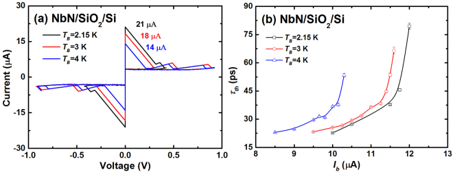
To investigate the temperature dependence of τth, we tuned the TB of SNSPDs from 2.15 K to 4 K using the resistive heater. Figure 2(a) shows the current-voltage (I-V) characteristics of Sample #6 at three different temperatures (2.15 K, 3 K, and 4 K). Isw decreased from 21 μA to 14 μA with increasing TB. As a result, the two-photon detection ranges changed to 10 μA ≤ Ib ≤ 12 μA (2.15 K), 9.5 μA ≤ Ib ≤ 11.6 μA (3 K), and 8.5 μA ≤ Ib ≤ 10.3 μA (4 K). Then, the Ib dependence of τth at different TB values were studied; τth gradually increased with Ib and presented a considerable change around the one-photon detection regime for all TB values (Fig. 2(b)). However, the τth-Ib curve shifted to a smaller Ib with increasing TB owing to the change of the two-photon detection range. In addition, at a fixed Ib, τth increased with TB; this could be attributed to the decreased electron relaxation rate at high TB. However, the (τth)min values at TB values of 2.15 K, 3 K, and 4 K were measured to be 22.7 ps ± 0.1 ps, 23.2 ps ± 0.1 ps, and 23.0 ps ± 0.1 ps, respectively. The results agree well with each other, and we saw a saturated trend of τth with decreasing the current, suggesting that the minimum hotspot relaxation time might be independent of TB. However, it is difficult to verify it by further decreasing the current since the device no longer worked in the two-photon detection regime anymore. This is different from the results reported for WSi SNSPDs, where (τth)min increased with TB from 250 mK to 2 K15. Possibly, the smaller Isw of WSi SNSPDs at higher temperatures restricts the ability to reach the lowest bias in the two-photon detection regime owing to the limit of the readout circuit. In other words, the (τth)min of WSi SNSPDs might be smaller at high temperatures as well.
Figure 2.
(a) I-V curves and (b) τth as a function of Ib measured at TB values of 2.15 K, 3 K, and 4 K, respectively, for Sample #6.
Dependence of τth on nanowire linewidth
It has been well established that the w of nanowire affects not only the optical absorption but also the intrinsic detection efficiency of SNSPD8,19. To understand the influence of w on τth, three different SNSPDs were fabricated on MgO substrate, wherein w was designed to be 90, 105, and 120 nm, respectively. The s value was set to 110 nm. The measured w/s is consistent with the design as the EBL and RIE processes were optimized. The other parameters are listed as Samples #1–3 in Table 1.
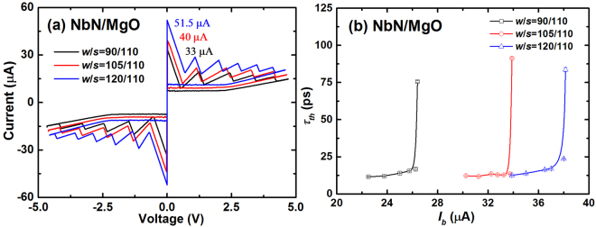
Figure 3(a) shows the I-V curves of the SNSPDs with three different w values (90, 105, and 120 nm) that give Isw values of 30, 40, and 51.5 μA respectively. Similarly, we obtained the relation of τth as a function of Ib for the three SNSPDs, as shown in Fig. 3(b). The trend displayed in the figure is comparable to that of Sample #6, and the curve shifts to the right with increasing w while maintaining the shape similarity. The (τth)min values for the three SNSPDs were determined to be 11.6 ps ± 0.1 ps, 12.2 ps ± 0.1 ps, and 12.6 ± 0.1 ps, respectively. The deviations were lesser than 1 ps. The slight increase in (τth)min with increasing w is explained by the presence of less thermal relaxation channels in the center of the nanowire as opposed to at the sides of the nanowire. In addition, the values are only half of those obtained for Sample #6; this is further discussed in the next section.
Figure 3.
(a) I-V curves and (b) τth as a function of Ib at TB values of 2.15 K for Sample #1–3 with different w/s.
Dependence of τth on the substrate
A variety of substrates, such as Si, MgO, MgF2, and Al2O3, have been used to grow ultrathin NbN films for fabricating SNSPDs20–22. To investigate the effect of substrates on τth, we measured and compared the τth of NbN SPSPDs on different substrates [Si (100), MgO (100), MgF2 (110), Al2O3 (0001), and SiO2/Si (100)].
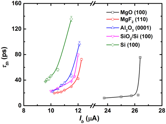
Figure 4 shows τth as a function of Ib at TB = 2.15 K for SNSPDs on various substrates. All SNSPDs showed a similar increase in τth with increasing Ib. The (τth)min values are summarized in Table 1 and follow the simple relation (τth)min(MgO) ≤ (τth)min(MgF2) ≤ (τth)min(Al2O3) ≤ (τth)min(SiO2/Si) ≤ (τth)min(Si). The maximum difference of (τth)min is roughly three times. The smallest (τth)min (11.6 ps ± 0.1 ps) was obtained from the MgO (100) substrate, and the largest (τth)min (34.5 ps ± 0.3 ps) was obtained from the Si (100) substrate. Typically, τth is controlled by carrier diffusion as well as by the recombination time of the quasiparticles, which is indeed related to the degrees of disorder of the materials15,23. In other words, a film with a lower disorder would have a smaller (τth)min.
Figure 4.

τth as a function of Ib at TB values of 2.15 K for various substrates.
Role of disorder on τth
The superconducting properties of NbN films are determined by many parameters of the films. One of the key parameters is actually the film crystal structure, which is strongly influenced by not only the substrate material but many other parameters including the deposition condition. Meanwhile, NbN is a conventional s-wave superconductor and the superconducting properties of the NbN ultrathin film can be partially characterized by its degrees of disorder, which is related to not only the substrate material, but also the film thickness. The degrees of disorder of NbN films on two substrates MgO and SiO2/Si were quantitatively analyzed. First, we measured the normalized resistance (RN) as a function of TB for the 5.5-nm-thick NbN/MgO and 7-nm-thick NbN/SiO2/Si substrates under different magnetic fields via transport measurements using a physical property measurement system. To accurately determine the resistivity (ρn), the electrical resistance (R) was measured on a 4 μm × 40 μm microbridge using the standard four-probe method. Figure 5(a) shows the RN-TB relations for the 5.5-nm-thick NbN/MgO sample. The upper critical field (Hc2) values at different temperatures were then determined at the point where RN becomes 50% of its normal value. The μ0Hc2-TB relations for the two samples are shown in Fig. 5(b). In the dirty limit of a superconductor film e.g., NbN, the mean electronic free path (l) is shorter than the Ginzburg–Landau coherence length (ξGL). Then, Hc2 (0), ξGL, and the electron diffusion coefficient (D) are obtained from , and , respectively, where Φ0 is the flux quantum, kB is the Boltzmann constant, and e is the electron charge24,25. For NbN films epitaxially grown on the MgO substrate, the obtained D value was 0.92 cm2/s, which is approximately twice the value of the film on the SiO2/Si substrate (0.47 cm2/s). The larger value of D suggests the presence of fewer defects and vacancies in the samples. The calculated ξGL values were 5.43 nm and 5.12 nm for the 5.5-nm-thick NbN/MgO and 7-nm-thick NbN/SiO2/Si samples, respectively.
Figure 5.
(a) RN as a function of TB for 5.5-nm-thick NbN on MgO (100) under different magnetic fields. (b) μ0Hc2 as a function of TB for 5.5-nm-thick NbN on MgO (100) and 7-nm-thick NbN on SiO2/Si (100). The solid lines are linear fits to the data. (c) ρxy as a function of B at 25 K for the two samples.
Next, Hall measurement was performed using a four-probe AC technique. Figure 5(c) shows the Hall resistivity (ρxy) as a function of B for the two samples measured at 25 K. The electron density (n) and the Ioffe–Regel parameter (kFl) are calculated from , and , respectively, where is determined from the slope shown in Fig. 5(c). All the obtained values are listed in Table 2. As expected, , indicating that all NbN films are in the dirty limit irrespective of which substrate is used. However, the kFl values described herein range from moderately disordered (5.31) to strongly disordered (1.53), suggesting that NbN grown on the MgO (100) substrates exhibits a less disordered structure. The film superconductivity decreases with increasing disorder. The Jsw values listed in Table 1 are also confirmed the above judgments to some extent.
Table 2.
Critical temperature (Tc), normal resistivity (ρn), upper critical field [Hc2(0)], Ginzburg–Landau coherence length (ξGL), electron diffusion coefficient (D), mean electronic free path (l), and Ioffe–Regel parameter (kFl) for the 5.5-nm-thick NbN/MgO (100) and 7-nm-thick NbN/SiO2/Si (100) samples.
| Substrate material | d (nm) | Tc (K) | ρn (μΩ cm) | μ0Hc2 (0) (T) | ξGL (nm) | D (cm2/s) | l (Å) | k F l |
|---|---|---|---|---|---|---|---|---|
| MgO (100) | 5.5 | 13.51 | 154 | 11.17 | 5.43 | 0.92 | 3.56 | 5.31 |
| SiO2/Si (100) | 7 | 7.71 | 562 | 12.56 | 5.12 | 0.47 | 1.08 | 1.53 |
Discussion
The minimum (τth)min measured in our NbN SNSPDs is about only one in seven of WSi SNSPDs’, indicating a significant difference in material properties. The direct method to decrease τth is to decrease the degrees of disorder of the materials. However, how to decrease τth together with optimizing other parameters such as intrinsic detection efficiency still remains an open question, which is very interesting for not only understanding the mechanism of SNSPD but also the practical SNSPD with high performance.
In summary, the τth of NbN SNSPDs on various substrates were investigated with a two-photon detection measurement technique. The experimental setup was based on pump-probe spectroscopy, wherein the laser pulses were separated by time delays tuned in a wide range. To realize two-photon detection in a reasonable bias range, the typically average photon number per pulse is set as 40. The high average photon number per pulse guarantee the data collection in a reasonable time (one dot per hour) compared with the correlated photon-pair source based on SPDC.
Our results show that the bath temperature and nanowire linewidth do not significantly affects (τth)min, i.e. they contribute less to the intrinsic upper limit of the count rate of SNSPD. However, (τth)min is strongly affected by the substrate because the degrees of disorder of NbN films are different. Epitaxially grown 5.5-nm-thick NbN on MgO (100) has a minimum (τth)min of 11.6 ps, and amorphous 7.5-nm-thick NbN on Si (100) has a maximum (τth)min of 34.5 ps. As a result, it is important to choose a suitable substrate if one wants to find lower τth. Besides, one may further study NbN films deposited on different substrates with different deposition conditions to further identify the minimum of τth. It may guide the further studies in the community on not only the physics of ultrathin superconducting films, but also the practical SNSPD with high count rate. Beyond that, the results are also interesting for the study of other superconducting sensors/detectors based on hot-electron effect, for example, hot-electron mixers.
Methods
Device fabrication
Ultrathin NbN films were synthesized through reactive DC magnetron sputtering of an 8-inch Nb target in an Ar + N2 gas mixture at room temperature. The typical background pressure was 9 × 10⁻⁶ Pa. The flow rates of Ar and N2 were set to 30 sccm and 4 sccm, respectively, under a total pressure of 0.27 Pa. The DC magnetron power was provided by current-regulated power supply set at 2.2 A with a resulting voltage of 270 V. The deposition rate was 6 Å/s, calibrated by X-ray reflectivity. The substrates used for film growth were Si (100) with its native oxide, MgO (100), MgF2 (110), Al2O3 (0001), and Si (100) with a 253-nm-thick buffering SiO2 layer. Prior to film deposition, all the substrates were cleaned by Argon ion-beam etching in a cleaning chamber. It is worth noting that the etching process removed native oxides of the Si (100) substrate. Because the MgO (100) and MgF2 (110) substrates allow the epitaxial growth of NbN films, NbN films on these substrates have higher critical temperature Tc than those on the Al2O3 (0001), SiO2/Si (100), and Si (100) substrates. To optimize the device performance, films with different thickness (d) values, i.e., 5.5, 4.5, 7, 7, and 7.5 nm were synthesized on the MgO (100), MgF2 (110), Al2O3 (0001), SiO2/Si (100), and Si (100) substrates, respectively. The films were structured into a conventional meandered nanowire structure, which covered a 15 μm-diameter area. All nanowire structures were patterned by electron-beam-lithography (EBL) with PMMA950-A2 as the e-beam resist. Electrode structures were formed by reactive ion etching (RIE) in the CF4 gas. Seven SNSPDs with different nanowire linewidth/space ratio (w/s) were fabricated. The detailed parameters of these SNSPDs are listed in Table 1.
Measurement setup
The experimental setup for τth measurement is shown in Fig. 6. The source of optical excitation is a 20-MHz mode-locked fiber laser with a 1550-nm wavelength, 500-fs pulse duration, and a typical maximum average output power of 2 mW. Laser pulses are attenuated and then divided by a 1:1 fiber beam splitter. One part of the pulse is sent to a fixed fiber, whereas the other part is sent to a motorized delay line. The time delay tD between the two pulses can be controlled by tuning the motorized delay line with a precision of ±0.01 ps. The two pulses are combined again on a 1:1 fiber beam splitter and then coupled to the SNSPDs. The measurements for the SNSPDs are performed in a Gifford–McMahon cryocooler at 2.15–5 K. A resistive heater is placed near the sample to tune the temperature of SNSPDs.
Figure 6.
Schematic of the experimental setup for the τth measurements. Two fiber beam splitters (BS) and a motorized delay line were used to control the tD between the two pulses.
Acknowledgements
This work was funded by National Key Research and Development Program of China (2017YFA0304000), National Natural Science Foundation of China (61671438) and Science and Technology Commission of Shanghai Municipality (STCSM) (16JC1400402).
Author Contributions
L.Z. and L.X.Y. designed the experiment, analyzed the data and wrote the paper. L.Z. and X.Y.Y. performed the measurements. L.Z. performed the thin film deposition and J.J.W. and W.J.Z. fabricated the SNSPD devices. C.L.L., Q.G. and H.L. provided assistance in the experiment. W.P., Z.W. and X.M.X. discussed the results. All authors reviewed the manuscript.
Competing Interests
The authors declare that they have no competing interests.
Footnotes
Publisher's note: Springer Nature remains neutral with regard to jurisdictional claims in published maps and institutional affiliations.
References
- 1.Dauler EA, et al. Review of superconducting nanowire single-photon detector system design options and demonstrated performance. Opt. Eng. 2014;53:081907. doi: 10.1117/1.OE.53.8.081907. [DOI] [Google Scholar]
- 2.Natarajan CM, Tanner MG, Hadfield RH. Superconducting nanowire single-photon detectors: physics and applications. Supercond. Sci. Technol. 2012;25:063001. doi: 10.1088/0953-2048/25/6/063001. [DOI] [Google Scholar]
- 3.Wang Z, Miki S, Fujiwara M. Superconducting Nanowire Single-Photon Detectors for Quantum Information and Communications. Ieee. J. Sel. Top. Quant. 2010;15:1741–1747. doi: 10.1109/JSTQE.2009.2034616. [DOI] [Google Scholar]
- 4.Hadfield RH. Single-photon detectors for optical quantum information applications. Nat. Photonics. 2007;3:696–705. doi: 10.1038/nphoton.2009.230. [DOI] [Google Scholar]
- 5.Renema JJ, et al. Experimental test of theories of the detection mechanism in a nanowire superconducting single photon detector. Phys. Rev. Lett. 2014;112:117604. doi: 10.1103/PhysRevLett.112.117604. [DOI] [PubMed] [Google Scholar]
- 6.Annunziata AJ, et al. Reset dynamics and latching in niobium superconducting nanowire single-photon detectors. J. Appl. Phys. 2010;108:084507. doi: 10.1063/1.3498809. [DOI] [Google Scholar]
- 7.Engel A, Renema JJ, Il’in K, Semenov A. Detection mechanism of superconducting nanowire single-photon detectors. Supercond. Sci. Technol. 2015;28:114003. doi: 10.1088/0953-2048/28/11/114003. [DOI] [Google Scholar]
- 8.Li H, et al. Large-sensitive-area superconducting nanowire single-photon detector at 850 nm with high detection efficiency. Opt. Express. 2015;23:17301–17308. doi: 10.1364/OE.23.017301. [DOI] [PubMed] [Google Scholar]
- 9.Marsili F, et al. Detecting single infrared photons with 93% system efficiency. Nat. Photonics. 2013;7:210–214. doi: 10.1038/nphoton.2013.13. [DOI] [Google Scholar]
- 10.Gol’tsman GN, et al. Picosecond superconducting single-photon optical detector. Appl. Phys. Lett. 2001;79:705. doi: 10.1063/1.1388868. [DOI] [Google Scholar]
- 11.Kerman AJ, Yang JKW, Molnar RJ, Dauler EA, Berggren KK. Electrothermal feedback in superconducting nanowire single-photon detectors. Phys. Rev. B. 2009;79:100509. doi: 10.1103/PhysRevB.79.100509. [DOI] [Google Scholar]
- 12.Shibata H, Takesue H, Honjo T, Akazaki T, Tokura Y. Single-photon detection using magnesium diboride superconducting nanowires. Appl. Phys. Lett. 2010;97:212504. doi: 10.1063/1.3518723. [DOI] [Google Scholar]
- 13.Heeres RW, Zwiller V. Superconducting detector dynamics studied by quantum pump-probe spectroscopy. Appl. Phys. Lett. 2012;101:112603. doi: 10.1063/1.4750139. [DOI] [Google Scholar]
- 14.Zhou Z, et al. Ultrasensitive N-photon interferometric autocorrelator. Phys. Rev. Lett. 2013;110:133605. doi: 10.1103/PhysRevLett.110.133605. [DOI] [PubMed] [Google Scholar]
- 15.Marsili F, et al. Hotspot relaxation dynamics in a current-carrying superconductor. Phys. Rev. B. 2016;93:094518. doi: 10.1103/PhysRevB.93.094518. [DOI] [Google Scholar]
- 16.Bitauld D, et al. Nanoscale optical detector with single-photon and multiphoton sensitivity. Nano. Lett. 2010;10:2977–2981. doi: 10.1021/nl101411h. [DOI] [PubMed] [Google Scholar]
- 17.Ferrari S, et al. Hot-spot relaxation time current dependence in niobium nitride waveguide-integrated superconducting nanowire single-photon detectors. Opt. Express. 2017;25:8739. doi: 10.1364/OE.25.008739. [DOI] [PubMed] [Google Scholar]
- 18.Kozorezov AG, et al. Quasiparticle recombination in hotspots in superconducting current-carrying nanowires. Phys. Rev. B. 2015;92:064504. doi: 10.1103/PhysRevB.92.064504. [DOI] [Google Scholar]
- 19.Guo Q, et al. Single photon detector with high polarization sensitivity. Sci. Rep. 2015;5:9616. doi: 10.1038/srep09616. [DOI] [PMC free article] [PubMed] [Google Scholar]
- 20.Yang X, et al. Superconducting nanowire single photon detector with on-chip bandpass filter. Opt. Express. 2014;22:16267–16272. doi: 10.1364/OE.22.016267. [DOI] [PubMed] [Google Scholar]
- 21.Wu JJ, et al. NbN superconducting nanowire single-photon detector fabricated on MgF2 substrate. Supercon. Sci. Technol. 2016;29:065011. doi: 10.1088/0953-2048/29/6/065011. [DOI] [Google Scholar]
- 22.Henrich D, et al. Broadening of hot-spot response spectrum of superconducting NbN nanowire single-photon detector with reduced nitrogen content. J. Appl. Phys. 2012;112:074511. doi: 10.1063/1.4757625. [DOI] [Google Scholar]
- 23.Leo A, et al. Quasiparticle scattering time in niobium superconducting films. Phys. Rev. B. 2011;84:014536. doi: 10.1103/PhysRevB.84.014536. [DOI] [Google Scholar]
- 24.Chockalingam SP, Chand M, Jesudasan J, Tripathi V, Raychaudhuri P. Superconducting properties and Hall effect of epitaxial NbN thin films. Phys. Rev. B. 2008;77:214503. doi: 10.1103/PhysRevB.77.214503. [DOI] [Google Scholar]
- 25.Lin SZ, et al. Characterization of thin-film NbN superconductor for single-photon detection by transport measurements. Phys. Rev. B. 2013;87:2450–2458. [Google Scholar]


