Abstract
Infantile myofibromatosis is a mesenchymal disorder characterized by the fibrous proliferation of the skin, bone, muscle and viscera. It is the most common fibrous tumor in childhood. We present a newborn with skin and bone disease without visceral involvement, who showed good response to vinblastine and methotrexate. Clinical features, etiology, diagnosis, and treatment are reviewed.
Keywords: Dermatology, Myofibromatosis, Neoplasms
INTRODUCTION
Infantile myofibromatosis (IM) is a mesenchymal disorder characterized by a fibrous proliferation of the skin, bone, muscle, and viscera. 1 Although rare, it is the most common fibrous tumor in childhood.1,2 Its name comes from the cells exhibiting distinct characteristics of fibroblasts and smooth muscle cells (myofibroblasts).3 IM usually appears as firm nodules, with purplish or normal skin, located in the subcutaneous tissue. 2 It can be solitary or multiple. Most cases are limited to the skin, but systemic involvement has also been described.4 The solitary type limited to the skin usually has a good prognosis with spontaneous regression. The visceral lesions are associated with high morbidity and mortality.3 Most cases are sporadic.4
CASE REPORT
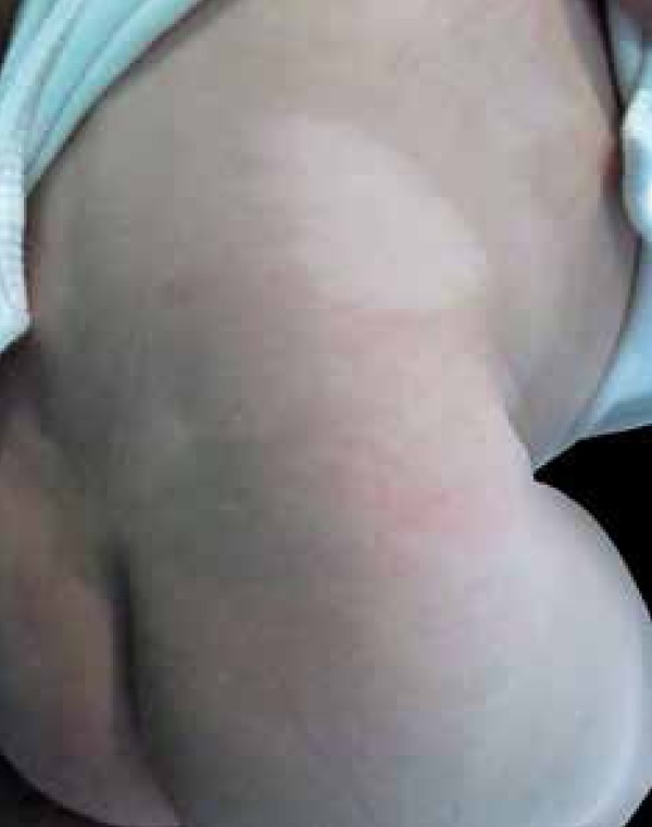
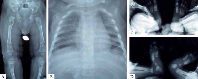
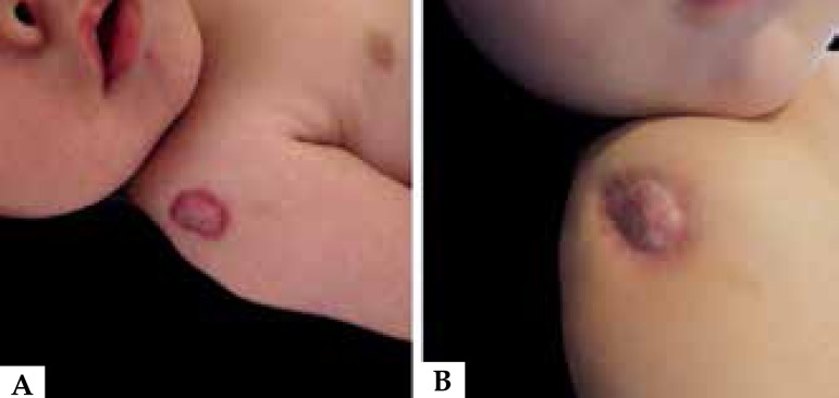
We present a male patient, full term, with no relevant medical history, which in the context of a routine hip x-ray control showed multiple osteolytic images in the sacrum and both femurs (Figure 1). Additional radiological images were performed in order to complete a full assessment of the child, involving most of the bony structures. Physical examination revealed a tumor consisting of an erythematous lesion with the central clearing of any atrophic appearance. Its edges were well delimited, irregular, and intensely colored. The lesion measured approximately 3 cm in diameter and was located on the right shoulder (Figure 2). He also presented a hypopigmented macule located on the right flank, as well as syndactyly of middle and ring fingers of his left hand, and third and fourth toes of his right foot (Figure 3).
Figure 1.
Multiple osteolytic images in A - Sacrum, femurs, and tibias B - Thorax C - and D - Tibias, fibula, and feet bones
Figure 2.
A: Erythematous plaque with central clearing of atrophic appearance with irregular, intensely colored, and well-delimited edges; B: Scar-like aspect of the same lesion after treatment.
Figure 3.

Hypopigmented macule on the right flank
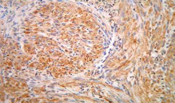
The pathology of the skin lesion posted a spindle cell proliferation in the reticular dermis, made up of elements with mild nuclear pleomorphism arranged in fascicles. The vasculature showed perycites-like characteristics (Figure 4). Immunohistochemistry was positive for vimentin (Figure 5), muscle specific actin (HHF35), and smooth muscle actin, while it was negative for desmin, S -100 protein, and cytokeratin AE1AE3 muscle actin. The Ki67 proliferative fraction was 5%.
Figure 4.
A - Spindle cell proliferation in reticular dermis made up of elements arranged in fascicles. (Hematoxylin & eosin, X10); B and C - Perycites like cells at the center of tapered cells lobes, (Hematoxylin & eosin, X40)
Figure 5.

Inmunohistochemistry showing inmunopositivity for vimentin, X40
With these results the presumption of infantile myofibromatosis was confirmed. Bone biopsies were consistent with the diagnosis. The patient showed no involvement of other structures. An oncology evaluation was requested. The boy was placed on a weekly treatment plan with vinblastine 1 mg/dose IV and methotrexate 7 mg/dose IV, with a progressive increase in dose until reaching 1.4 mg and 10 mg, respectively. The treatment lasted 12 months, achieving a complete resolution of bone and cutaneous lesions.
DISCUSSION
IM usually occurs before age two, but can be seen in older children and even adults. 1,2 IM is now considered within the spectrum of tumors with perivascular myoid differentiation, based on the involvement of myopericytoma, located in the vessel wall.2
There are three varieties: Solitary IM, multicentric IM without visceral involvement and multicentric IM with visceral compromise or generalized.1,4
When located in the skin, IM is heterogeneous. It usually appears as a subcutaneous nodule, but can also appear as an ulcer, pedunculated lesion, or similar to a hemangioma, as in the case of our patient. The most common locations are the head, neck, and trunk, while the involvement of the limbs is rare.5,6
The solitary form occurs predominantly in men and is typically seen in the dermis, subcutaneous tissues, and soft tissues; 50% of solitary forms and 90% of the multiple forms are congenital. 1,4 Bone involvement is rarely observed in the solitary form (5%), but it is common in the multicentric form (17-77%). 1
The etiology of IM is still quite unknown.1,3,4 Not long ago, it was established that the mutation in the receptor of the platelet-derived growth factor (PDGFRB) causes IM, and NOTCH3 was proposed as a candidate gene. 3,7 However, other genes could exist that determine IM, as well as other growth factors involved in its pathogenesis (basic fibroblast growth factor - bFGF). 3,8 Two patterns of inheritance - autosomal dominant and recessive - were described.1,3 The underlying mechanism of tumor regression and growth remains unknown, but it has been suggested to be related to angiogenic stimulation and regression, both triggered by bFGF.3,8 Urinary excretion of this factor increases in the active phase of the disease. 8
Histology shows well-circumscribed tapered cell lobes, resembling smooth muscle cells. At its center, perivascular round cells (hemangiopericitoides) are usually observed, giving a biphasic appearance. 6
Regarding immunohistochemistry, both vimentin and smooth muscle actin are positive.9
The characteristic image of IM is non-specific. In radiography, osteolytic lesions appear as defined areas with sclerotic rings. With ultrasound, the masses may show a hyperechoic or anechoic center with a surrounding ring. With CT, the tumor is observed as isodense or less dense than muscle, whereas bone involvement is seen as lytic lesions with sclerotic margins. MRI reveals low intensity on T1 and high on T2. 1
The prognosis of IM varies according to the type. Tumors without visceral involvement usually have an excellent prognosis with a spontaneous regression of lesions in one or two years.1,2 On the contrary, the visceral involvement type shows a severe compromise given by the gastrointestinal and cardiopulmonary complications with early morbidity and mortality. 1,10
Local complications are related to the mass effect and compression of the surrounding organs, such as the orbit, the larynx, the brachial plexus, and the vertebral cana.2,9 Bleeding from the tumor surface can be a fatal complication during the intrauterine period.5 There have also been reports associating IM with malformations. A probable role of bFGF in their appearance is therefore suggested. 8
The treatment of IM depends on its location. Although spontaneous regression often occurs, there have been reports of recurrence.2 Surgical excision is reserved for cases with compromised vital functions. The IM with visceral involvement may require surgery or chemotherapy (interferon alfa, vincristine-actinomycin D-cyclophosphamide, and vinblastine-methotrexate), although the results with both have been disappointing. 1,2
The focus of our work lies in the rarity of the disease and the good response to treatment. The dermatologist´s work stands out in its diagnosis.
Footnotes
Work performed at Hospital Alemán, Buenos Aires, Argentina
Financial support: none.
Conflict of interest: none.
REFERENCES
- 1.Wu W, Chen J, Cao X, Yang M, Zhu J, Zhao G. Solitary infantile myofibromatosis in the bones of the upper extremities: Two rare cases and a review of the literature. Oncol Lett. 2013;6:1406–1408. doi: 10.3892/ol.2013.1584. [DOI] [PMC free article] [PubMed] [Google Scholar]
- 2.Mashiah J, Hadj-Rabia S, Dompmartin A, Harroche A, Laloum-Grynberg E, Wolter M, et al. Infantile myofibromatosis: A series of 28 cases. J Am Acad Dermatol. 2014;71:264–270. doi: 10.1016/j.jaad.2014.03.035. [DOI] [PubMed] [Google Scholar]
- 3.Martignetti JA, Tian L, Li D, Ramirez MC, Camacho-Vanegas O, Camacho SC, et al. Mutations in PDGFRB Cause Autosomal-Dominant Infantile Myofibromatosis. Am J Hum Genet. 2013;92:1001–1007. doi: 10.1016/j.ajhg.2013.04.024. [DOI] [PMC free article] [PubMed] [Google Scholar]
- 4.Larralde M, Hoffner MV, Boggio P, Abad ME, Luna PC, Correa N. Infantile myofibromatosis: report of nine patients. Pediatr Dermatol. 2010;27:29–33. doi: 10.1111/j.1525-1470.2009.01073.x. [DOI] [PubMed] [Google Scholar]
- 5.Aye CY, Gould S, Akinsola SA. Congenital infantile myofibroma causing intrauterine death in a twin. BMJ Case Rep. 2011;2011 doi: 10.1136/bcr.09.2011.4851. [DOI] [PMC free article] [PubMed] [Google Scholar]
- 6.Kikuchi K, Abe R, Shinkuma S, Hamasaka E, Natsuga K, Hata H, et al. Spontaneous remission of solitary-type infantile myofibromatosis. Case Rep Dermatol. 2011;3:181–185. doi: 10.1159/000331325. [DOI] [PMC free article] [PubMed] [Google Scholar]
- 7.Cheung YH, Gayden T, Campeau PM, LeDuc CA, Russo D, Nguyen VH. A recurrent PDGFRB Mutation Causes Familial Infantile Myofibromatosis. Am J Hum Genet. 2013;92:996–1000. doi: 10.1016/j.ajhg.2013.04.026. [DOI] [PMC free article] [PubMed] [Google Scholar]
- 8.Inamadar AC, Palit A, Athanikar SB, Sampagavi VV, Deshmukh NS. Infantile myofibromatosis with multiple congenital anomalies. Pediatr Dermatol. 2005;22:281–282. doi: 10.1111/j.1525-1470.2005.22329.x. [DOI] [PubMed] [Google Scholar]
- 9.Mynatt CJ, Feldman KA, Thompson LD. Orbital infantile myofibroma: a case report and clinicopathologic review of 24 cases from the literature. Head Neck Pathol. 2011;5:205–215. doi: 10.1007/s12105-011-0260-4. [DOI] [PMC free article] [PubMed] [Google Scholar]
- 10.Okuda KV, Fitze G, Pablik J, Hahn G, Suttorp M, Vogelberg C. Infantile myofibromatosis as an unusual cause for unilateral atelectasis in an infant. Pediatr Blood Cancer. 2014;61:1158–1159. doi: 10.1002/pbc.24933. [DOI] [PubMed] [Google Scholar]


