Abstract
Salivary duct injury can be idiopathic, iatrogenic, or post-trauma and may result in sialocele or fistula. Most injuries regress spontaneously and botulinum toxin A is one of several therapeutic possibilities. We report a case of iatrogenic injury to the parotid duct after Mohs' micographic surgery for a squamous cell carcinoma excision in the left jaw region, treated by injection of botulinum toxin type A. Although the fistula by duct injury can be self-limiting, botulinum toxin injection by promoting the inactivity of the salivary gland allows rapid healing of the fistula.
Keywords: Botulinum toxins, type A; Mohs' surgery; Parotid region; Salivary gland fistula
INTRODUCTION
Salivary duct lesions may be idiopathic, iatrogenic, or post-traumatic, and may result in sialocele with painful facial edema or in the formation of skin fistulas.1 The onset of salivary fistula post-parotidectomy occurs in 4-14% of the cases, the majority of which are self-limiting and respond well to conservative treatment.2 Several techniques have been employed to treat this complication: percutaneous aspiration, compression bandage, anticholinergic drugs, radiation therapy, botulinum toxin, and surgical repair.1
This report presents a parotid duct iatrogenic lesion case, which occurred after Mohs' micrographic surgery (MMS) recommended for squamous cell carcinoma (SCC) excision in the left mandibular region, treated with the injection of type A botulinum toxin (BoNT-A).
CASE REPORT
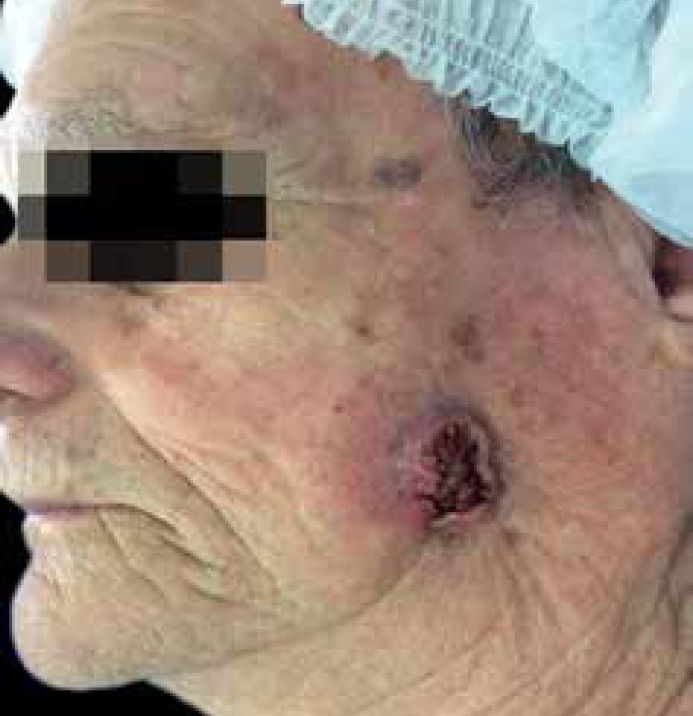
A 79-year-old male patient displayed a tumor measuring 2.5 x 2.3 cm, with an ulcerated center, in the left mandibular region, and with skin retraction in the area below the lesion (Figure 1).
Figure 1.
Clinical presentation of the lesion
The biopsy indicated SCC, and the pre-surgery ultrasound exam identified the muscular plane had been affected.
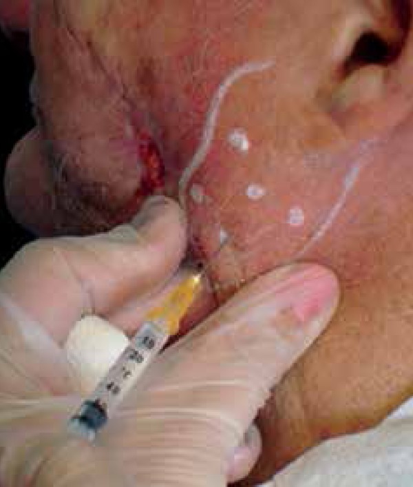
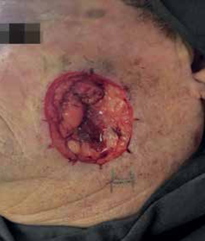
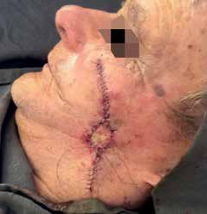
The MMS was performed up to the muscular plane, and two phases were required for full neoplasia resection. The final defect was 3.4 x 2.9 cm, repaired by detaching the side patches, associated to grafting (Figures 2 and 3). On the fifth day after surgery, the patient exhibited an edema and sensitivity on the left parotid gland. The hypothesis of parotitis was raised and treatment with Buscopan compound® every 8 hours for five days. On the 10th day after surgery, the patient indicated that secretion had been eliminated on the graft side during feeding, which was clinically confirmed. The eliminated secretion was collected and examined, revealing high amylase levels, confirming its salivary nature.1 Therefore, the patient was diagnosed with parotid duct lesion with skin fistula. On the 17th day after surgery, the patient received a BoNT-A (Botox®, Allergan, Inc.) injection. The vial was reconstituted with 2 ml of 0.9% saline solution. The patient received a 16U injection, divided among three points on the left parotid topography, using a 0.5 ml BD Ultrafine II syringe, with a 30G gauge, 8 mm long needle. The patient's condition persisted and, on the 24th day after surgery, the treatment was repeated, using the same dose, but using a 25G gauge, 40 mm long needle (Figure 4).
Figure 2.
Final defect
Figure 3.
Immediately after surgery
Figure 4.

Second application 24 days after surgery
After four days of the second application, the postprandial secretion was interrupted, which was maintained after 19 months. During this period, no secondary asymmetry related to the use of botulinum toxin was observed.
DISCUSSION
Knowledge of the facial anatomy is essential during surgery. The parotid gland is located on the preauricular region. The parotid duct (Stensen) originates on the anterosuperior portion of the gland, between the upper and middle thirds, 15 mm away from the zygomatic arch. From this point, it extends for 4 to 6 cm on the side surface of the masseter muscle to its anterior margin, where it forms a straight angle and perforates the buccinator muscle until it reaches the buccal mucosa, ending at the same height as the second upper molar.3 To locate this duct, two imaginary lines, one running from the tragus to the middle section of the upper lip, and the other one running from the external eye corner to the mental foramen, are used as references. The removal of invasive tumors in this area is very likely to result in parotid duct lesion, due to the duct being included in tumor block resection, as may have occurred in the reported case.4
The external parotid fistula usually occurs after one week, and the sialocele is manifested within 8 to 14 days post-trauma.5 In the case described above, the symptomatology of pain and inflammation started on the 5th day, but the saliva elimination was only observed after the 10th day.
When the fistula appears early on due to a sialocele complication, surgical exploration is the recommended treatment. If it appears late, the conservative treatment, with external pressure and anticholinergic drugs, is recommended.4 The way the fistula will be handled will depend on when it appears, on its complexity, on the area, and on the lesion's etiology.1
Botulinum toxin is known for releasing acetylcholine at the autonomic nervous system's cholinergic synapses. Because of this effect, it has been used for treating sialorrhea cases in Parkinson's disease, post-stroke, and in refractory post-parotidectomy parotid fistula cases.2,6 The BoNT-A doses vary between 10 and 100U.
Lim and Choi2 reported solving an acute fistula case after a parotid tumor resection with the use of 10U of BoNT-A.2
Two cases of BoNT-A application to treat parotid duct lesions caused by facial SCC resection are described in literature.
Hatzis and Finn described the case of a 58-year-old male patient who, after undergoing the excision of SCC on the left mandibular area, developed a salivary fistula on the 6th day after surgery. He was initially treated with 200U of BoNT-A (Botox®, Allergan, Inc.), followed by an additional 100U five days later. The fistula was fully resolved after a 21-day period.7
Krishnan et al. reported the case of a 62-year-old male patient who, after removal of right malar region SCC, exhibited the formation of persistent sialocele on the 5th day after surgery and was treated with ultrasound guided BoNT-A injections. Application was made at three parotid gland points, with a total dose of 23U, and the condition was solved after two weeks.8
In this study's case, the fistula was treated with 32U of BoNT-A in two applications. The first application of 16U with an 8 mm needle presented no improvement, and a second application of 16U with a 40mm needle was performed after seven days. The authors believe that the short needle did not reach the salivary gland.
In the second application, the longer needle reached the gland in an effective manner, resulting in a definitive interruption of the salivary secretion after four days, with no alteration of the facial muscle movements.
This fact draws attention to the importance of injecting at the appropriate depth, avoiding excessive doses. In the case reported above, 16U might have been sufficient. The use of long needles is recommended when ultrasound is not available to guide the application.
Footnotes
Work performed at the Dermatology Department, Hospital do Servidor Público Municipal de São Paulo (HSPM), São Paulo, SP, Brazil.
Financial support: none.
Conflict of interest: none.
REFERENCES
- 1.Kulyapina A, Lopez-de-Atalaya J, Ochandiano-Caicoya S, Navarro-Cuellar C, Navarro-Vila C. Iatrogenic salivar duct injury in head and neck câncer patients: report of four cases and review of the literature. J Clin Exp Dent. 2014;6:e291–e294. doi: 10.4317/jced.51438. [DOI] [PMC free article] [PubMed] [Google Scholar]
- 2.Lim YC, Choi EC. Treatment of an acute salivary fistula after parotid surgery: botulinum toxin type A injections primary treatment. Eur Arch Otorhinolaryngol. 2008;265:243–245. doi: 10.1007/s00405-007-0418-6. [DOI] [PubMed] [Google Scholar]
- 3.Oliveira MG. Manual de anatomia da cabeça e pescoço. 4. ed. Porto Alegre: Edipucrs; 2002. [Google Scholar]
- 4.Lazaridou M, Iliopoulos C, Antoniades K, Tilaveridis I, Dimitrakopoulos I, Lazaridis N. Salivary Gland Trauma: A Reviews of Diagnosis and Treatment. Craniomaxillofac Trauma Reconstr. 2012;5:189–196. doi: 10.1055/s-0032-1313356. [DOI] [PMC free article] [PubMed] [Google Scholar]
- 5.Parekh D, Glezerson G, Stewart M, Esser J, Lawson HH. Post-traumatic Parotid Fistulae and Sialoceles. Ann Surg. 1989;209:105–111. doi: 10.1097/00000658-198901000-00015. [DOI] [PMC free article] [PubMed] [Google Scholar]
- 6.Chow TL, Kwok SP. Use of botulinum toxin type A in a case of persistent parotid sialocele. Hong Kong Med J. 2003;9:293–294. [PubMed] [Google Scholar]
- 7.Hatzis GP, Finn R. Using Botox to Treat a Mohs Defect Repair Complicated by a Parotid Fistula. J Oral Maxillofac Surg. 2007;65:2357–2360. doi: 10.1016/j.joms.2006.10.017. [DOI] [PubMed] [Google Scholar]
- 8.Krishnan RS, Clark DP, Donnelly HB. The use of botulinum toxin in the treatment of a parotid duct injury during Mohs surgery and review of management options. Dermatol Surg. 2009;35:941–947. doi: 10.1111/j.1524-4725.2009.01159.x. [DOI] [PubMed] [Google Scholar]


