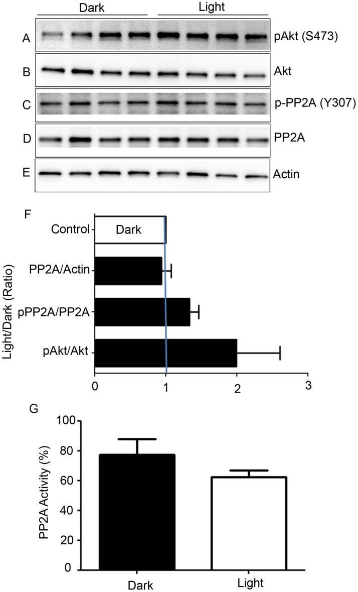
Figure 5. Biochemical characterization of PP2A, and phosphorylation state of PP2A-regulated protein.
Retinal lysates from dark- and light-adapted mice were subjected to immunoblot analysis with anti-pAkt (S473) A., anti-Akt B., p-PP2A (Y307) C., anti-PP2A D., and anti-actin E. antibodies. Densitometric analysis of pAkt, p-PP2A, and PP2A was performed in the linear range of detection, and absolute values were then normalized to Akt, PP2A, and actin F.. PP2A activity was measured from dark- and light-adapted mouse retinas G. as described in the Methods section. Data are mean + SEM, n = 4.

