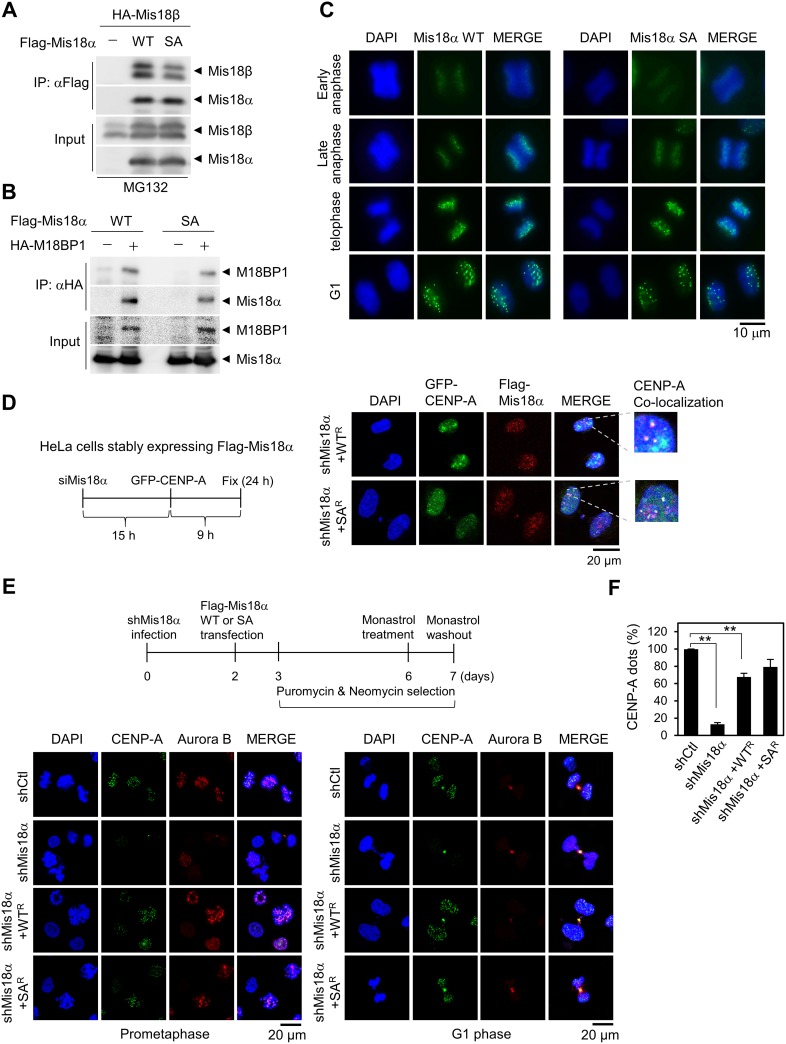
Figure 3. Mis18α phosphorylation is not required for CENP-A loading.
(A) HA-Mis18β with either Flag-Mis18α WT or Flag-Mis18α SA were transfected into 293T cells and the extracts were applied for co-IP assay. (B) The binding between Flag-Mis18α and HA-M18BP1 was analyzed as in A. (C) HeLa/Flag-Mis18α stable cells were synchronized by double-thymidine block and released into indicated phase. The cells were stained with anti-Flag antibody. The green dots indicate centromeric localization of Mis18α. Confocal image with 1,000x magnification. (D) Analysis scheme for the centromere recruitment of newly synthesized CENP-A (left). HeLa cells stably expressing siRNA-resistant form of Mis18α WT (WTR) or SA (SAR) were transfected sequentially with siRNA against Mis18α and with GFP-CENP-A (mimic newly synthesized CENP-A) as indicated in the scheme. Immunocytochemistry for Mis18α with anti-Flag antibody and GFP-CENP-A (right). (E) Scheme for the centromeric recruitment of CENP-A under prolonged Mis18α knockdown (upper). Knockdown of endogenous Mis18α was achieved by infecting lentivirus that is expressing shRNA against Mis18α. Lower left panel shows CENP-A dots in prometaphase cells and lower right panel represents G1 phase cells. (F) The number of CENP-A dot positive cells from E were calculated and expressed as a percentage of total cells. P value is calculated by t-test (**p < 0.01).

