Abstract
Medical images are essential in modern traumatology and orthopedic surgery. Access to images is often cumbersome due to a limited number of workstations. Moreover, due to the tremendous increase of data, the time to review or to communicate images has also become limited. One approach to overcome these problems is to make use of modern mobile devices, like tablet computers, to facilitate image access and associated workflows. Ten orthopedic surgeons were equipped with an Apple iPad mini 2 and specialized viewing software for medical images. The surgeons were able to send images from a workstation onto the tablets or to search for patient images directly. The software enabled the physicians to share images, annotated key slices, and messages instantly with their colleagues. The surgeons carried the tablets within or in the periphery of the hospital. The participants evaluated the software by means of daily questionnaires. Data was collected for a period of 9 months. Nearly 25 images were viewed in total by the surgeons per day. The tablet viewer was used for accessing approximately 30% of these images. On average, the surgeons were asked 1.7 times per day by a colleague for a second opinion. They used the tablets in approximately 29% of these cases. Furthermore, the mean time for accessing images was significantly lower using mobile software compared to conventional methods. Tablet computers can play a vital role for image access and communication in the daily routine of an orthopedic surgeon. Mobile image access is an important aspect for surgeons, especially in larger facilities, to facilitate and accelerate the clinical workflows.
Electronic supplementary material
The online version of this article (doi:10.1007/s10278-017-0011-5) contains supplementary material, which is available to authorized users.
Keywords: Tablet, Image, PACS, Orthopedics, Second opinion, DICOM
Introduction
Background
Digital radiological images are an essential tool in modern orthopedic surgery. They provide pre-operative planning, and intra-operative and post-operative control. While X-Ray images remain the most commonly used technique, tomographic image techniques, like computed tomography (CT) or magnetic resonance imaging (MRI), have become the gold-standard diagnostic for certain diseases.
Due to technical advancements of CT and MR imaging, acquisition times shortened and accuracy increased. Consequently, the amount of data being generated has also increased tremendously, while the number of medical professionals analyzing those images has remained the same or only slightly increased [1]. Nearly all EU Member States for which data are available showed a remarkable increase between 2008 and 2013 in the number of CT and MRI exams relative to the size of their population [2]. For the USA, the number of MR exams more than doubled between 2000 and 2013—the number of CT exams increased by 31% between 2004 and 2013 [3].
Having said that it is known that a second or more opinions on medical images increases the accuracy of the diagnosis for various diseases (e.g., in neuroradiology [4], for breast cancer diagnosis [5] or for reassessment of cervical spine CTs [6]), the disparity between the number of professionals and the amount of data has become a major challenge with respect to accurate and efficient reporting. Due to the sheer number of images and the lack of adequate tools, the time for reviewing radiological images has become less—not just for diagnostic reporting, but also for surgical preparation, for example. Moreover, the images are seldom shared with colleagues or other experts for a second opinion.
Evaluating new technologies to start coping with these problems is therefore a logical next step. Thanks to the digitalization of radiological images and technical advancements, modern smartphones and tablets are becoming increasingly important in clinical workflows in general and for managing medical image data in particular. In contrast to handhelds 10 or 15 years ago, modern mobile devices incorporate enough computing resources to work with high-resolution image data while providing high mobility.
It was an early finding that mobile devices may have a positive impact on efficiency and quality of care in the orthopedic field. For instance, Ricci et al. stated that “these systems also improve the comfort level of consulting orthopedic surgeons and potentially limit the risk of litigation for incorrect diagnosis” [7]. The evolution of mobile devices for clinical settings seems to be especially interesting for surgeons when considering various recent publications [8–10]. As a specific example which underlines the demand for mobile devices, Stahl et al. found that “evaluating […] CT scans transmitted to a smartphone is a readily accessible, simple, and inexpensive method” [11]. Although the topic is still in the focus of debates on data privacy and regulatory considerations, even the popular “WhatsApp” messenger was investigated by orthopedic surgeons as an “intradepartmental communication tool” [12]. However, the process and implementation of mobile techniques have just started and the real positive effects have scarcely been investigated.
Rationale
We expect mobile devices or tablets combined with specialized software to be a valuable tool to increase the efficiency of reading, interpreting, and communicating radiological images. We further expect that this is especially true for orthopedic surgeons or surgeons in general, since mobility plays a far more important role in their daily routine than for radiologists, for example.
There are numerous publications that mainly deal with the diagnostic quality on mobile devices and their comparability with the findings on a regular workstation (e.g., [13, 14]). The majority of these publications conclude that there is no major difference in the accuracy of reading while interaction with images could be more time-consuming on a mobile device. De Maio et al. summarized this with the conclusion “the diagnostic performance of interpreting MR images on a handheld mobile […] is similar to that of a conventional radiology workstation, however, requires a longer viewing time.”
We are, however, not aware of a study that has tried to evaluate tablets for image review and communication and their effects to workflow efficiency in the daily routine of surgeons. To answer these questions, we planned and conducted a study named “Ortho Mobile.” The study was approved by the local ethics committee in 2015. Results are presented in this article.
Material and Methods
Study Description
The “Ortho Mobile” was a prospective single-center study initiated at the end of 2015. A total of ten orthopedic surgeons took part. Data was collected for a period of 9 months. There were in total of six residents and four senior physicians.
The physicians were given an Apple iPad Mini 2 device, which has a suitable size and shape to be kept in the clinician’s white coat. The mini device assured both the permanent availability and a screen that is reasonably large enough for image reviewing. The term permanent availability refers to the fact that the tablet could be carried around, which is a decisive argument for employing tablet computers in a clinical setting. We did not expect tablets to be faster in terms of computing power or network connectivity (compared to conventional workstations), but the mobility of these devices guarantees immediate access everywhere. The study center is a supra-regional trauma hospital and has several associated facilities next to the main buildings, such as a rehabilitation center, an outpatient clinic, and a research and conference area nearby. It is not unusual that the physicians visit those buildings more than once per day. Wi-Fi access was available in the main buildings of the hospital and these surrounding structures, but complete coverage in every location could not be guaranteed. Thus, the selection of appropriate software was important. Please refer to the next section for a detailed description of the software and its integration.
The physicians received an initial training for the software and were then provided with the iPad devices. They were asked to use the device and the software according to their own needs and willingness so that there was no prior influence. The usage behavior was recorded by filling out daily online questionnaires. The online questionnaires were also installed as an “App” on the iPads for facilitating the process of data entry. The questionnaires were usually filled and collected at the end of the day. The questionnaire and the semantics of its questions were also presented during the initial participant training.
Please note that it was not possible to simply record the usage frequency using DICOM server logs. The physicians usually downloaded studies containing multiple series (or they were sent automatically before the ward round) out of which not all were necessary to review for, e.g., a second opinion. Thus, a software heuristic would have been needed that could tell whether single series were opened. This was not possible, in particular not for the existing workstation software.
Viewer Software
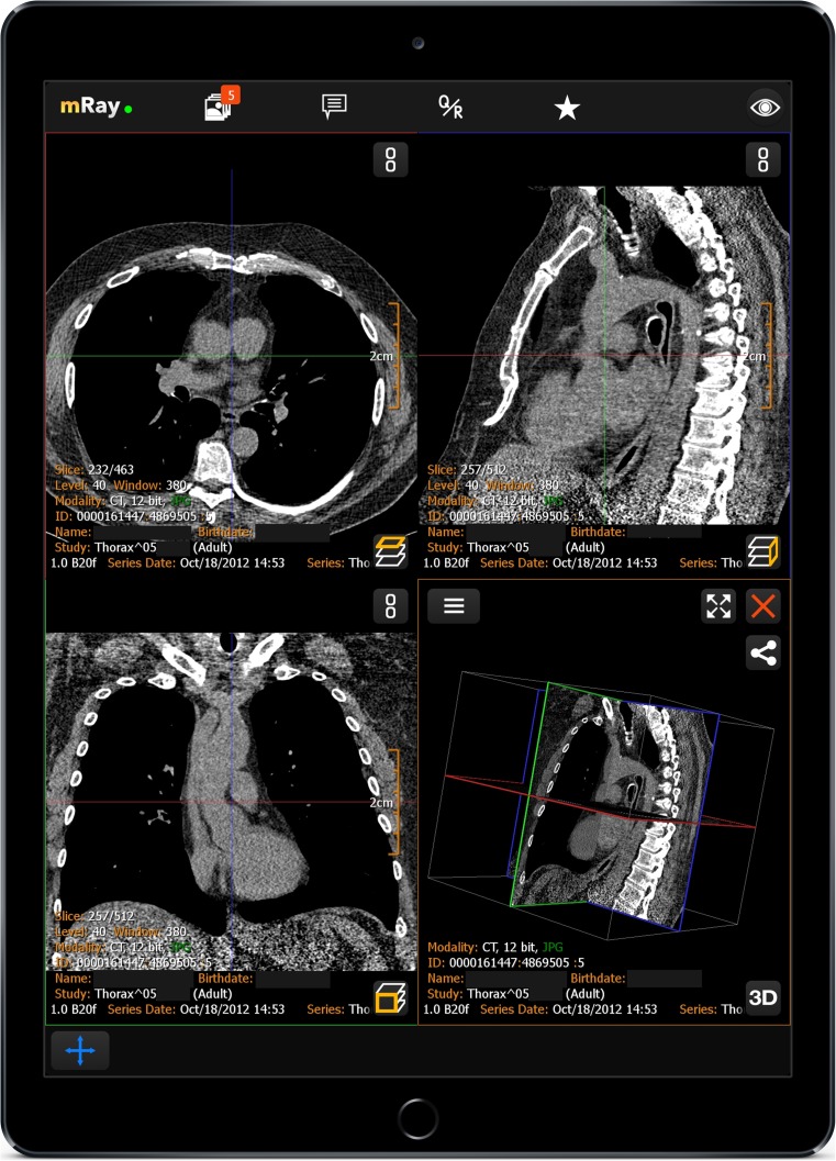
The choice of an appropriate software was important for conducting this study. While different vendors offer mobile extensions to their PACS (Picture Archiving and Communication System), it was of particular interest to have an offline-capable software which further incorporated means to exchange messages. We were able to utilize a mobile-only application called “mRay” [15]. The software is specifically designed for image display and analysis on mobile devices (cf. Fig. 1) and is independent of the underlying PACS. Data exchange and interoperability are realized using the DICOM standard (Digital Imaging and Communications in Medicine).
Fig. 1.
The medical image viewer used in this study runs on an Apple iPad device
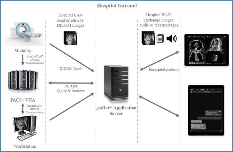
As shown in Fig. 2, the software is divided into two components. The client application (the “App”) enables image viewing on a tablet or smartphone. The application server, which takes the image data from the PACS, encrypts and compresses the images before distributing these to the mobile clients. An important aspect of the software is that the application does not require a permanent connection. Encrypted data is being downloaded and stored on the device temporarily. Due to this and by using individual logins, data privacy, and security was assured at all times.
Fig. 2.
Integration setup of the “mRay” software within the hospital: DICOM images can be sent from a workstation or the modality, whereas the PACS can be queried directly. The tablets receive these images, and the user can share parts of the images (key slices), text, and audio messages with other users of the software. Communication is managed by the application server
Apart from the image viewing functionality, the software has a communication platform that enables the sharing of images or key slices with annotations and for a quick communication by means of text and audio messaging.
This functionality is pretty similar to popular messenger applications except that sharing in this context means the sharing of radiological images, fixed viewing settings, or single key slices. The application was configured to have different ways of receiving data from the PACS: On the one hand, the physicians had the possibility to directly push image data from their workstations or even from a modality onto the tablets. On the other hand, it was also possible to search the PACS directly specifying different parameters. Moreover, an automated task served all patient image data of the last 24 h every morning before the ward round in the respective station.
Lastly, another important consideration was regulatory conformity of the software for an approval by the ethics committee. The software is a CE-certified medical device, so we were able to use it without further efforts.
Comparison of Workflows
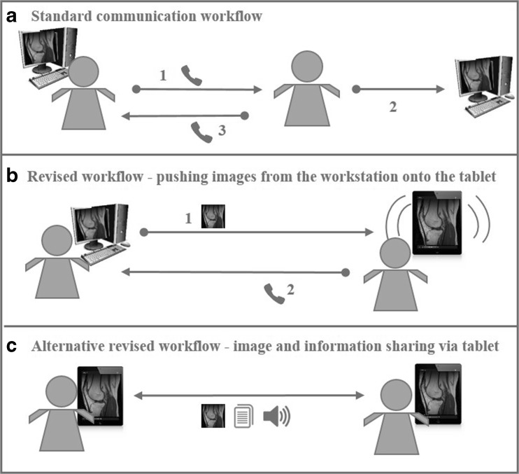
To give the reader a better understanding on how workflows were changed by the introduction of the tablets and which parts were subject to this investigation, the following diagram depicts the different possibilities.
The standard workflow A (Fig. 3) for providing a second opinion usually contains the following steps: A colleague is informed via phone to review a specific case (1). The called physician will then try to locate a workstation (if he is not already in the office) and to query the archive for the specific exam using the patient’s identification that was given to him verbally (2). As a side note and as an example for the difficulties of this workflow, the participating physicians reported that accessing images can take up to 10 min and more for locating an available workstation, potentially start it up, log on to the system, enter the patient’s data to search the dataset within the PACS (which can also be prone to errors) and to open the dataset on the workstation. The result of the review is normally also communicated back via phone (3).
Fig. 3.
The “traditional” workflow for second opinions (a) compared to the new possibilities when using tablets and specialized software (b, c)
Workflow B is the appropriate workflow when utilizing tablets as suggested in this manuscript. A physician who is sitting in front of a workstation and who conducts a reading is in need of a second opinion. He can then push studies or single series directly onto the tablet of a colleague who will be informed by a system notification telling him that new data is available (1). The dataset is automatically downloaded by the software so that a click on the notification brings up the ready-to-use image dataset. Results and other information are usually also communicated back via phone call.
In workflow C, the participants utilize solely their tablets and the software to exchange images, text, and/or audio messages concerning a specific case. Although phone calls remained the preferred way to contact someone directly, the messaging component enabled for an asynchronous exchange of a second opinion between the physicians.
Variables, Outcome Measures, Data Sources, and Bias
The daily questionnaire contained 14 questions, 12 of which represent countable measures that were analyzed in this study. Two questions could be answered with yes/no and gave the possibility of a free text entry. Please note that the term “image” does not refer to a complete study, but to a DICOM series, i.e., either a single X-Ray image or, for tomographic datasets, multiple slices. The physicians were instructed to use this term in this way, meaning as this was the common understanding of a radiological “image.” The list of questions is shown in Table 1.
Table 1.
Shows all questions of the daily issued questionnaire
| No. | Question |
|---|---|
| 1 | How often did you review X-ray images on a standard PC? |
| 2 | …X-ray images on your tablet? |
| 3 | …CT images on a standard PC? |
| 4 | …CT images on your tablet? |
| 5 | …MR images on a standard PC? |
| 6 | …MR images on your tablet? |
| 7 | How often did you use your tablet to show images to a patient? |
| 8 | How often have you been contacted for a second opinion today? |
| 9 | How often did you use your tablet for image retrieval and providing a second opinion? |
| 10 | How long did it take on average until image viewing was possible on a standard PC (in minutes)? |
| 11 | How long did it take on average until image viewing was possible on your tablet (in minutes)? |
| 12 | How often was it necessary to review images on a larger screen for better visibility? |
| 13 | Have you been unable to use your tablet due to technical reasons (e.g., Wi-Fi problems, software errors, or similar)? If yes please describe the problem(s). |
| 14 | Did you use your tablet in other places than the ambulance or the ward? If yes please state where. |
Questions 1–7 pertain to the general question of usage frequency, which was collected to evaluate the system in general. Questions 8 and 9 were asked for gathering information on the participants’ second opinion behavior.
Questions 10 and 11 are related to the efficiency of image access, which in turn relates to the study’s principal question as to whether tablets can accelerate the process of image retrieval. These questions created comparable variables between the two methods investigated in this study (tablet vs. traditional workstations), which was the prerequisite for a statistical test procedure. It is important to mention that the participants were instructed to estimate the complete time needed to access an image—starting from the initial request to do so until it was opened on the tablet or on a PC, respectively. We therefore had the intention to account for the daily routine of orthopedic surgeons in which the physician usually does not sit in front of a PC (see section “Comparison of Workflows,” workflow A).
The last three questions were gathered to record information on qualitative aspects of the tablet usage, i.e., technical problems and location of usage. A high interobserver variability was expected due to personal preferences. Values were therefore expected to be biased by this personal and subjective estimation.
Participant Selection
The selection of the physicians was made with the requirement of creating a heterogeneous group. The participant group of this study was nearly equally split in “experienced” senior physicians and “less-experienced” residents. However, there was only one woman in the study group. The work time spent in the field of traumatology and orthopedics ranged from less than 6 months to over 20 years. All participants had been using mobile devices in their private environment. None of them were inexperienced or had not used mobile devices before. Differing amounts of questionnaires were received from the participants: Six of the physicians returned between one and five questionnaires. The remaining four physicians filled out between 11 and 47 daily questionnaires in the course of this study (for detailed numbers please refer to Electronic Supplementary Material 1).
Statistical Analysis, Study Size, and Statistical Software
Survey participation of physicians and questionnaire results was first analyzed descriptively. The mean of all outcome variables of interest was estimated by linear random effects (i.e., variance components) models, and the mean difference in the duration to image access between standard PC and tablet was estimated by a linear mixed-effects model. For both corresponding 95%-Wald confidence intervals were computed. The individual physician was included as a random intercept to account for multiple observations per physician. Since graphical inspection did not suggest that time trends in outcomes existed and the number of observations per physician varied, no conclusive results on time trends could be expected and they were not considered in the models. If the confidence intervals excluded zero, estimates were considered as statistically significant. However, the study was exploratory in design and no adjustment for multiplicity was performed. Therefore, the results can only be interpreted descriptively. The collected data were complete, and thus, no methods to deal with missingness were applied. The analyses were performed in the R language and environment for statistical computing (R version 3.3.1).
Results
A total of 117 daily questionnaires were recorded. The estimated mean values of the answers after applying statistical analysis are listed in Table 2.
Table 2.
Statistical results of the survey on tablet usage for image review and communication
| No. | Question | Estimated mean [95% CI] |
|---|---|---|
| 1 | How often did you review X-ray images on a standard PC? | 10.1 [5.8; 14.4] |
| 2 | … X-ray images on your tablet? | 4.4 [0.8; 8.1] |
| 3 | … CT images on a standard PC? | 3.5 [2.6; 4.4] |
| 4 | … CT images on your tablet? | 1.5 [0.5; 2.5] |
| 5 | … MR images on a standard PC? | 3.5 [2.3; 4.7] |
| 6 | … MR images on your tablet? | 1.3 [0.4; 2.2] |
| 7 | How often did you use your tablet to show images to a patient? | 1.1 [0.4; 1.9] |
| 8 | How often have you been contacted for a second opinion today? | 1.7 [0; 3.4] |
| 9 | How often did you use your tablet for image retrieval and providing a second opinion? | 0.5 [0; 1.0] |
| Images in total viewed on your standard PC | 17.1 [11.4; 22.8] | |
| Images in total viewed on tablet | 7.1 [1.7; 12.5] | |
| 10 | How long did it take on average until image viewing was possible on a standard PC (in minutes)? | 2.2 [1.1; 3.3] mins |
| 11 | How long did it take on average until image viewing was possible on your tablet (in minutes)? | 1.0 [0.6; 1.4] mins |
| Time difference for accessing an image between PC and tablet (mean) | 1.1 [0.8; 1.4] mins | |
| 12 | How often was it necessary to review images on a larger screen for better visibility? | 0.2 [0; 0.5] |
Usage Statistics
The applied linear random effects model estimated a mean of 17 radiological images (X-ray 59%, CT 21%, MRI 20%) viewed per day using a standard PC, whereas the personal tablet was used to view 7.1 images per day on average (X-ray 62%, CT 20%, MRI 18%). This sums up to a mean overall value of nearly 25 images per day, i.e., the mobile software was used for accessing almost 30% of the images. Besides that, the surgeons used their tablet computers for bedside demonstration approximately 1.1 times per day.
Second Opinions
In total, the participants were asked 425 times by a colleague to review a patient dataset in the course of the study. After applying the random linear effects model, the estimated mean value for the frequency of second opinion inquiries was 1.7 times per day. In roughly 29% of these cases, the tablet was used to access images and give a second opinion.
Acceleration of Image Access
The participants were asked to estimate the average time for accessing images using a conventional PC and tablet computer, respectively. The estimated mean time for opening a specific patient image was 2.2 min for a regular PC and about 1 min when using the tablet. The mean difference in the duration to image access between the two methods was estimated by a linear mixed-effects model to be 1.1 min, i.e., the time to access images was estimated as being reduced when mobile image access through a tablet computer was used.
Other Relevant Findings
Images only needed to be reviewed on a stationary PC for a better visibility in just 0.2% of the cases. The participants reported several times that issues with the Wi-Fi connection were annoying. Besides on the ward or in the emergency room, the tablets were also used in the ambulatory, externa rehabilitation facility, the personal office, or in the operating theater.
Discussion
Background and Rationale
Radiological images are crucial for diagnosis and treatment in orthopedic and trauma surgery. Image data available for a single patient can, however, become overwhelming for physicians. New tools to facilitate image access, reading, and communication are vitally important, especially in the field of traumatology and orthopedics where image interpretation is just one part of the work.
The “Ortho Mobile” study targeted this specific challenge by investigating the benefits and drawbacks of modern tablet computers and special software for accessing and communicating images within a supra-regional trauma center. To our knowledge, no investigation has been published that attempted to analyze the benefits of mobile software tools on a quantitative basis and over a longer period of time. This study was therefore initiated to measure different parameters on a daily basis in order to explore the potential of this new technique.
Limitations
This study had several limitations and some of these were unavoidable. First of all, the study design was limited. It was inherently not possible to have separate “treatment” and “control” groups. Using tablet computers was only possible in conjunction with the existing infrastructure. They cannot be treated as a complete replacement, and this was not the intention of this investigation. Thus, there was no comparison between two independent procedures.
Second, the data presented in this study was collected using questionnaires. Questions asking for an estimation of an amount or a specific time are from their nature expected to be biased. Some of the participants invested more, some of them less time in filling out questionnaires. However, questionnaires were the only means to receive the data needed for to answer the fundamental questions of the study.
Last, the study was conducted at a single site. Specific peculiarities of the trauma center may have played a role. Although a multi-center study is always preferable from a statistical viewpoint, there is no major argument why the choice of the study location should have had an extraordinary influence on the results.
About Overall Usage and Acceptance
On average, the participating in this study needed to access about 25 images per day and almost 30% of these were accessed using the personal tablet. Nearly one out of three images viewed, reviewed, or communicated via the mobile software demonstrated its added value as felt and expressed by the participants. This total number gives a good overall judgment on the acceptance and the benefits of this new approach and met our expectations. This is in accordance with recent literature that even indicates a favorable “user experience” for mobile image viewers [16]. Boission et al. even suggested that “handheld devices could be a substitute for computer screens for teleconsultation by physicians working in emergency settings” [10].
The ratio of image types viewed on a standard PC and the tablets is nearly the same (approx. 60% of the images were x-rayed, 20% for CT, and MRI each), i.e., there is no indicator that a certain image type is favored for viewing on a mobile device. This is also in agreement with a large systematic review for radiological interpretation of images displayed on tablet computers [17], which indicated that mobile image viewing gives appropriate diagnostic results for these kinds of modalities.
Another encouraging result is the number of images discussed or shown to the patient (estimated mean, 1.1 per day). Although this aspect is rather subordinated finding, the participants emphasized the benefits of showing images to the patient with a tablet. It should be borne in mind that there was no systematic approach to interview the patients on this finding. A publication from 2014 revealed patients mostly reported that using tablets as a bedside information tool had no impact on their engagement [18]. We did not obtain data during our study that confirms or refutes this prior finding.
The participating physicians provided a varying number of questionnaires. This fact was statistically respected by applying a linear random effects model. However, it should be clearly stated that multiple questionnaires of the same person cannot be treated as an independent observation.
About the Influence of Mobile Image Access on the Workflow for Providing a Second Opinion
On average, the participating surgeons were asked 1.7 times per day by a colleague to review a patient dataset for a second opinion. They used the mobile software for this in approx. 29% of the cases. This is also a positive statement concerning the usage of mobile devices, although a better result was expected.
The software used in this study ([15]) incorporates a communication module similar to that in popular messenger applications but with a focus on radiological images. So besides just messaging, the software allows annotated key images or whole series to be shared through a secure channel (cf. section “Comparison of Workflows,” workflow C). Bearing in mind that “WhatsApp” was already a subject of investigation [12], we expected more usage of this functionality throughout the course of our study. However, only a few participants used it to ask for a second opinion. We therefore have no indication that the second opinion workflow is improved by using tablet computers and appropriate software. This could be because making a phone call is simply quicker. Although a collaborative communication platform is a non-blocking approach for information exchange, the fact that it is an entirely new workflow might be why our study participants neglected to use it.
The communication capabilities of a mobile image viewer may be more beneficial as soon as the software is implemented center-wide so that an inter-department communication is possible. The same applies to connecting referring physicians to the platform. More and specialized training on this functionality could also be beneficial.
About Workflow Acceleration
The estimation of access times probably has the greatest bias of all factors measured. It is difficult to estimate how long it took on average to access a certain dataset from the PACS using one means or the other. It is therefore also valid to say that this result is biased. Nevertheless, the numbers confirm the expectation that a mobile device can provide quicker access to clinical data and that this is also true for complex data like radiological images. The difference of 1.1 min is statistically significant.
To our knowledge, no other publication has attempted to capture the time parameter when comparing mobile applications to their desktop PC counterparts, although this is especially useful in emergency medicine. Surprisingly, even publications dealing with the usage of mobile devices for on-call emergency readings [14, 19], mainly discuss the assessment quality and do not provide findings on workflow acceleration even though this should be considered as a major improvement.
Other Relevant Findings
Images only needed to be reviewed on bigger screens for more clarity or visibility in just 0.2% of the cases. This in accordance with the literature in which the major part of the studies proves the comparability of image reading on stationary PCs and mobile devices [10, 13, 14]. Stable and center-wide Wi-Fi access continues to be an obstacle when implementing mobile solutions.
We further asked the participants if they used the software outside the ward or the ambulance. The trauma center that hosted this study has several external facilities the physicians may need to visit, sometimes multiple times a day. There is, e.g., a rehabilitation center, an outpatient clinic, and a research and conference area. As reported in the questionnaires, the physicians also made use of their tablets in this “periphery” and this was reported to be especially useful. Using mobile devices within larger institutions, like trauma centers or university hospitals, has more advantages than in smaller-sized facilities. The benefits of being mobile are especially important for orthopedic surgeons who are more frequently requested to move to different places within the hospital.
There are, of course, other mobile PACS viewers that could have been used. However, the selection of the software was crucial for the study because the impact of mobile technology is strongly tied to the software used and not just that a handheld device is used. This is why we described the application used in detail. We have chosen the software for the following reasons that other software solutions could not fully provide: First, the application can be used offline, i.e., images can be viewed without being connected to a network (access to the hospital Wi-Fi or a mobile network was not available everywhere). Second, the software enabled the streaming of images, which is especially important for large tomographic datasets. Third, it has a communication module, which allows images to be shared directly with a colleague so that he does not have to search for the images himself. The latter two points are important arguments for a quick accessibility of images. Nevertheless, we are convinced that some of the findings of this study could also have been discovered using other applications in the field.
Conclusions
The study presented in this paper was initiated to evaluate tablet computers and specialized software for image review and communication in the daily routine of orthopedic surgeons. Therefore, ten orthopedic surgeons documented the usage of this software on a daily basis.
A central finding of this study is that the mobile software was used in 30% of the cases when images were needed. This is a good indicator that tablets can be a valuable tool in the daily routine of a surgeon. There was also no indication that image viewing on a mobile device is less preferred or is lower in display quality compared to the normal stationary devices, this finding in accordance with the literature. Besides image reviewing, the participants of the study clearly emphasized the added value of using a personal tablet for image viewing when talking to the patient. The software was also judged as being useful for second opinions and as being quicker when images needed to be accessed.
Future research should investigate the effect of this new tool on a larger user base. With a limitation of only ten concurrent users, the possibilities to assess the positive effects of this collaborative technology were limited. We therefore intend to extend the implementation to more departments and external physicians—e.g., to external, abdominal surgeons that provide on-call services for the trauma center on a regular basis. Moreover, future investigations should also incorporate a review of how the age and the sex of the orthopedists influence the general acceptance for mobile devices.
To conclude, we see a high potential for mobile technology in the field of trauma surgery and orthopedics. Clinical orthopedic workflows and daily work of orthopedic surgeons will definitely benefit from these developments in terms of convenience, speed of decision making, and efficiency.
Electronic supplementary material
(PDF 6kb)
Acknowledgements
The study team wants to thank the IT department of the trauma center for supporting this study by implementing the infrastructural environment. Furthermore, we want to thank the local ethics committee for approving this study.
Compliance with Ethical Standards
Conflicts of Interest
Michael Müller is an employee of a company whose products are mentioned in the manuscript: mbits imaging GmbH.
Ethical Approval
All procedures performed in studies involving human participants were in accordance with the ethical standards of the institutional and/or national research committee and with the 1964 Helsinki declaration and its later amendments or comparable ethical standards. The study presented in this article was approved by the local ethics committee in 2015.
Informed Consent
Informed consent was obtained from all individual participants included in the study.
Footnotes
Electronic supplementary material
The online version of this article (doi:10.1007/s10278-017-0011-5) contains supplementary material, which is available to authorized users.
References
- 1.European Commission: Eurostat statistics database 2015. Healthcare personnel statistics - physicians. http://ec.europa.eu/eurostat/statistics-explained/index.php/Healthcare_personnel_statistics_-_physicians#Further_Eurostat_information. Accessed Nov 08, 2016
- 2.European Commission. : Eurostat statistics database 2015. Healthcare resource statistics - technical resources and medical technology.. http://ec.europa.eu/eurostat/statistics-explained/index.php/Healthcare_resource_statistics_-_technical_resources_and_medical_technology#Use_of_medical_technology. Accessed Nov 08, 2016
- 3.OECD: "Health care resources", OECD Health Statistics (database). doi:10.1787/data-00541-en. Accessed on 27 February 2017
- 4.Zan E, Yousem DM, Carone M, Lewin JS. Second-opinion consultations in neuroradiology. Radiology. 2010;255(1):135–141. doi: 10.1148/radiol.09090831. [DOI] [PubMed] [Google Scholar]
- 5.Mallory MA, Losk K, Lin NU, Sagara Y, Birdwell RL, Cutone L, Camuso K, Bunnell C, Aydogan F, Golshan M. The Influence of Radiology Image Consultation in the Surgical Management of Breast Cancer Patients. Ann Surg Oncol. 2015;22(10):3383–3388. doi: 10.1245/s10434-015-4663-4. [DOI] [PMC free article] [PubMed] [Google Scholar]
- 6.Khalilzadeh O, Rahimian M, Batchu V, Vadvala HV, Novelline RA, Choy G. Effectiveness of second-opinion radiology consultations to reassess the cervical spine CT scans: a study on trauma patients referred to a tertiary-care hospital. Diagn Interv Radiol. 2015;21(5):423–427. doi: 10.5152/dir.2015.15003. [DOI] [PMC free article] [PubMed] [Google Scholar]
- 7.Ricci WM, Borrelli J. Teleradiology in orthopaedics. Clin Orthop Relat Res. 2004;421:64–69. doi: 10.1097/01.blo.0000126751.94460.8d. [DOI] [PubMed] [Google Scholar]
- 8.Malan DF, van der Walt SJ, Raidou RG, van den Berg B, Stoel BC, Botha CP, Nelissen RGHH, Valstar ER. A fluoroscopy-based planning and guidance software tool for minimally invasive hip refixation by cement injection. Int J Comput Assist Radiol Surg. 2016;11(2):281–296. doi: 10.1007/s11548-015-1252-8. [DOI] [PMC free article] [PubMed] [Google Scholar]
- 9.Maruyama K, Kin T, Saito T, Suematsu S, Gomyo M, Noguchi A, Nagane M, Shiokawa Y. Neurosurgical simulation by interactive computer graphics on iPad. Int J Comput Assist Radiol Surg. 2014;9(6):1073–1078. doi: 10.1007/s11548-014-0993-0. [DOI] [PubMed] [Google Scholar]
- 10.Boissin C, Blom L, Wallis L, Laflamme L: Image-based teleconsultation using smartphones or tablets: qualitative assessment of medical experts. Emerg Med J. 2016 Oct 5. Publisher Item Identifier (PII): emermed-2015-205258 (Open Access) [DOI] [PMC free article] [PubMed]
- 11.Stahl I, Dreyfuss D, Ofir D, Merom L, Raichel M, Hous N, Norman D, Haddad E. Reliability of smartphone-based teleradiology for evaluating thoracolumbar spine fractures. Spine J. Publisher Item Identifier (PII) 2016;S1529-9430(16):30882–30888. doi: 10.1016/j.spinee.2016.08.021. [DOI] [PubMed] [Google Scholar]
- 12.Khanna V, Sambandam SN, Gul A. Mounasamy V: "WhatsApp"ening in orthopaedic care: a concise report from a 300-bedded tertiary care teaching center. Eur J Orthop Surg Traumatol. 2015;25(5):821–826. doi: 10.1007/s00590-015-1600-y. [DOI] [PubMed] [Google Scholar]
- 13.De Maio P, White LM, Bleakney R, Menezes RJ, Theodoropoulos J. Diagnostic accuracy of an iPhone DICOM viewer for the interpretation of magnetic resonance imaging of the knee. Clin J Sport Med. 2014;24(4):308–314. doi: 10.1097/JSM.0000000000000005. [DOI] [PubMed] [Google Scholar]
- 14.Tewes S, Rodt T, Marquardt S, Evangelidou E, Wacker FK, von Falck C. Evaluation of the use of a tablet computer with a high-resolution display for interpreting emergency CT scans. Rofo. 2013;185(11):1063–1069. doi: 10.1055/s-0033-1350155. [DOI] [PubMed] [Google Scholar]
- 15.mbits imaging GmbH, Heidelberg, Germany: mRay, Radiological image viewer for mobile devices with image processing functions. https://mbits.info/mray. Accessed on Nov 10, 2016
- 16.John S, Poh AC, Lim TC, Chan EH, Chong le R. The iPad tablet computer for mobile on-call radiology diagnosis? Auditing discrepancy in CT and MRI reporting. J Digit Imaging. 2012;25(5):628–634. doi: 10.1007/s10278-012-9485-3. [DOI] [PMC free article] [PubMed] [Google Scholar]
- 17.Caffery LJ, Armfield NR, Smith AC. Radiological interpretation of images displayed on tablet computers: a systematic review. Br J Radiol. 2015;88(1050):20150191. doi: 10.1259/bjr.20150191. [DOI] [PMC free article] [PubMed] [Google Scholar]
- 18.Baysari MT, Adams K, Lehnbom EC, Westbrook JI, Day RO. iPad use at the bedside: a tool for engaging patients in care processes during ward rounds? Intern Med J. 2014;44(10):986–990. doi: 10.1111/imj.12518. [DOI] [PubMed] [Google Scholar]
- 19.Park JH, Kim YK, Kim B, Kim J, Kwon H, Kim K, Choi SI, Chun EJ. Diagnostic performance of smartphone reading of the coronary CT angiography in patients with acute chest pain at ED. Am J Emerg Med. 2016;34(9):1794–1798. doi: 10.1016/j.ajem.2016.06.009. [DOI] [PubMed] [Google Scholar]
Associated Data
This section collects any data citations, data availability statements, or supplementary materials included in this article.
Supplementary Materials
(PDF 6kb)


