Factors that control skin inflammation are still being defined. Herro et al. demonstrate that the tumor necrosis factor superfamily protein LIGHT acts on keratinocytes via its receptor HVEM to promote characteristic features of atopic dermatitis, including epidermal hyperplasia and production of periostin.
Abstract
Dermatitis is often associated with an allergic reaction characterized by excessive type 2 responses leading to epidermal acanthosis, hyperkeratosis, and dermal inflammation. Although factors like IL-4, IL-13, and thymic stromal lymphopoietin (TSLP) are thought to be instrumental for the development of this type of skin disorder, other cytokines may be critical. Here, we show that the tumor necrosis factor (TNF) superfamily protein LIGHT (homologous to lymphotoxin, exhibits inducible expression, and competes with HSV glycoprotein D for binding to HVEM, a receptor expressed on T lymphocytes) is required for experimental atopic dermatitis, and LIGHT directly controls keratinocyte hyperplasia, and production of periostin, a matricellular protein that contributes to the clinical features of atopic dermatitis as well as other skin diseases such as scleroderma. Mice with a conditional deletion of the LIGHT receptor HVEM (herpesvirus entry mediator) in keratinocytes phenocopied LIGHT-deficient mice in exhibiting reduced epidermal thickening and dermal collagen deposition in a model of atopic dermatitis driven by house dust mite allergen. LIGHT signaling through HVEM in human epidermal keratinocytes directly induced proliferation and periostin expression, and both keratinocyte-specific deletion of HVEM or antibody blocking of LIGHT–HVEM interactions after disease onset prevented expression of periostin and limited atopic dermatitis symptoms. Developing reagents that neutralize LIGHT–HVEM signaling might be useful for therapeutic intervention in skin diseases where periostin is a central feature.
Introduction
There are several types of dermatitis or skin inflammation, but the most serious are those characteristic of allergen-driven atopic eczema and scleroderma. These diseases have several commonalities, not least of which is excessive keratinocyte activity. This results in the production of a range of inflammatory mediators that control the recruitment and cross talk with the immune system, and keratinocytes directly contribute to the pathological features of these diseases (Bieber, 2008; Yamamoto, 2009). In atopic dermatitis and scleroderma, molecules such as thymic stromal lymphopoietin (TSLP), which can be a product of keratinocytes, have been suggested to be central to disease development, contributing to a Th2-dominant environment that involves one or several cytokines such as IL-4, IL-13, TNF, and TGF-β (Yoo et al., 2005; Ziegler and Artis, 2010; Usategui et al., 2013). Through the activity of these molecules (and likely other inflammatory molecules), tissue-remodeling proteins such as collagen and periostin are overproduced, and epidermal hyperplasia is further promoted from the deregulated inflammatory signals received by keratinocytes.In particular, data suggest that the matricellular protein periostin is not only a feature of Type 2 skin inflammatory disease but also plays an essential role in the development of disease (Masuoka et al., 2012; Shiraishi et al., 2012; Yang et al., 2012; Taniguchi et al., 2014). It is primarily expressed by keratinocytes and fibroblasts in the skin, and it has been suggested to have several roles, such as controlling localization of inflammatory infiltrates, including dermal fibroblasts, and contributing to collagen production (Takayama et al., 2006; Sidhu et al., 2010; Ontsuka et al., 2012; Shiraishi et al., 2012; Taniguchi et al., 2014). In line with these activities, mice deficient in periostin are highly resistant to developing skin disease in models of atopic dermatitis and scleroderma (Masuoka et al., 2012; Yang et al., 2012). Moreover, periostin can also contribute to the speed of wound repair (Nishiyama et al., 2011; Ontsuka et al., 2012), further emphasizing its importance in the skin. Periostin is currently being used as a clinical marker of type 2 allergic inflammatory disease (Parulekar et al., 2014), and because IL-13 has been found to induce expression of periostin, it has been proposed that patients that highly express this protein will be responsive to therapies aimed at blocking IL-13 (Parulekar et al., 2014). However, it is not clear how many other molecules additionally contribute to the production of this inflammatory factor. Recently, we showed that TNF superfamily member 14 (TNFSF14), also known as LIGHT (homologous to lymphotoxin, exhibits inducible expression and competes with HSV glycoprotein D for binding to HVEM (herpesvirus entry mediator), a receptor expressed on T lymphocytes), had a strong role in driving development of skin inflammation in a model of scleroderma induced by the antibiotic bleomycin (Herro et al., 2015). LIGHT is primarily a T cell–derived cytokine, but other CD45+ hematopoietic cells such as neutrophils may also be a source, and LIGHT binds to two receptors: HVEM (TNFRSF14) and LTβR (TNFRSF3; Mauri et al., 1998; Scheu et al., 2002; Steinberg et al., 2009). Whether LIGHT signaling contributes to other types of skin inflammatory disease, such as atopic dermatitis, is unknown, and it is not clear which are the essential cell types that respond to LIGHT. Here, we show that neutralization of LIGHT–HVEM interactions, caused by a deficiency in LIGHT or therapeutic blocking of HVEM signaling, strongly reduced allergen-induced atopic dermatitis in mice. Furthermore, we demonstrate that HVEM expression in keratinocytes is critical for atopic dermatitis and that LIGHT signaling through HVEM specifically controlled keratinocyte hyperplasia and the expression of periostin.
Results and discussion
LIGHT deficiency limits house dust mite (HDM)–induced atopic dermatitis
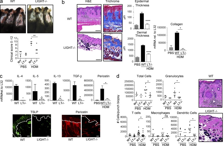
To test whether LIGHT controls the development of atopic dermatitis, we used a murine model (as previously described; Kawakami et al., 2007) induced by epicutaneous sensitization with HDM allergen combined with staphylococcal enterotoxin B, an exacerbating factor for human atopic dermatitis. Strikingly, LIGHT-deficient mice displayed minimal clinical symptoms characteristic of atopic dermatitis, including skin eruption, bleeding, redness, and scaling (Fig. 1 a); weak epidermal hyperplasia; and reduced dermal thickening, with less collagen deposited particularly in the adipose layers (Fig. 1 b). Additionally, all primary features of type 2 responses were lower in LIGHT-deficient mice, including TSLP and the Th2 cytokines, IL-4, IL-5, and IL-13 (Fig. 1 c). Moreover, TGF-β, which can contribute to collagen production, and periostin, which is deposited in the dermis and may also aid collagen expression among other activities, were almost absent in the skin of mice lacking LIGHT (Fig. 1 c). Lastly, overall inflammatory infiltrates were reduced in skin biopsy specimens from LIGHT-deficient hosts, with a trend toward lowered numbers of all cell types analyzed (Fig. 1 d). Thus, a genetic deficiency in LIGHT protected mice from developing allergen-driven atopic dermatitis, extending our prior finding that LIGHT was instrumental in the development of scleroderma-like skin disease induced by bleomycin.
Figure 1.
LIGHT deficiency limits HDM-induced atopic dermatitis. WT and LIGHT (LT)−/− mice were sensitized epicutaneously with HDM/SEB and analyzed on day 14. (a) Clinical symptoms, and scoring for skin eruption, scaling, bleeding, and redness from individual mice. (b) Skin sections stained with H&E or Masson’s trichrome blue and quantitated for epidermal and dermal thickness. Data represent means ± SEM from six mice. (c) mRNA expression of IL-4, IL-5, IL-13, and TGF-β in skin biopsy specimens and immunofluorescence (IF) staining of TSLP (green) and periostin (red). Dotted lines indicate epidermal-dermal border. Data are representative of (or means ± SEM from) 15–20 mice. (d) Quantitation of indicated cell populations in skin biopsy specimens by flow cytometry. Data are from individual mice. All results representative of (or from) four experiments. Bars, 200 µm. *, P < 0.05; **, P < 0.1; ****, P < 0.001. All statistical data were generated using the Mann–Whitney test.
HVEM-specific deletion in keratinocytes reduces atopic dermatitis
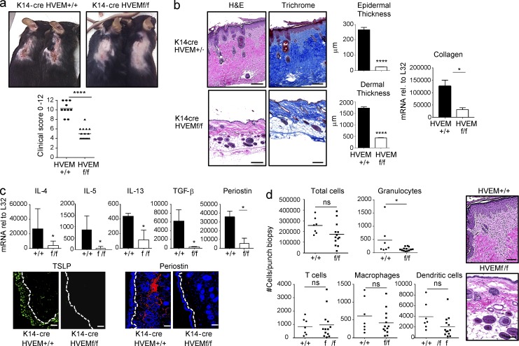
We previously found that both HVEM and LTβR are expressed in keratinocytes (Herro et al., 2015). To understand the importance of HVEM in these cells, we created conditional knockout mice in which HVEM was specifically deleted (K14-cre HVEMflox/flox). HVEM is expressed in many cells, including hematopoietic and nonhematopoietic cells. Showing these mice were not globally deficient in HVEM, flow analysis revealed a normal expression pattern of this molecule in the majority of splenic CD45+ and CD45− cells (not depicted) and normal expression of HVEM in skin fibroblasts, with a specific lack of expression in keratinocytes (Fig. S1). Importantly, mice lacking HVEM only in their keratinocytes to a large extent mimicked mice deficient in LIGHT and exhibited resistance to development of atopic dermatitis. Clinical atopic dermatitis symptoms (skin eruption, bleeding, redness, and scaling) were strongly reduced (Fig. 2 a), and minimal epidermal and dermal thickening was seen, along with weak deposition of collagen (Fig. 2 b) and generally poor expression of features of Th2 immunity (Fig. 2 c). Moreover, mice with a lack of HVEM in keratinocytes phenocopied LIGHT-deficient mice in exhibiting decreased expression of skin TGF-β and dermal periostin (Fig. 2 c). Interestingly, skin biopsy specimens still revealed significant infiltration of T cells, macrophages, and dendritic cells (Fig. 2 d), differing from the results with LIGHT-deficient mice (Fig. 1). Given that ILC2 have been proposed to contribute to atopic dermatitis (Salimi et al., 2013), and LIGHT might be active on ILC2 as these cells can express HVEM, it is possible that this may explain the difference in inflammatory infiltrates seen between mice lacking LIGHT and those lacking HVEM only in keratinocytes.
Figure 2.
Conditional knockout of HVEM in keratinocytes limits HDM-induced atopic dermatitis. K14-cre HVEMfl/fl mice (f/f) or K14-cre HVEM+/+ mice (+/+) were sensitized epicutaneously with HDM/SEB and analyzed on day 14. (a) Clinical symptoms and scoring for skin eruption, scaling, bleeding, and redness from individual mice. (b) Skin sections stained with H&E or Masson’s trichrome blue and quantitated for epidermal and dermal thickness. Data represent means ± SEM from 10 to 15 mice. (c) mRNA expression of IL-4, IL-5, IL-13, and TGF-β in skin biopsy specimens, and IF staining of TSLP (green) and periostin (red; DAPI blue). Dotted lines indicate the epidermal–dermal border. Data are representative of (or means ± SEM from) 10 to 15 mice. (d) Quantitation of indicated cell populations in skin biopsy specimens by flow cytometry. Data are from individual mice. All results are representative of two experiments. Bars, 200 µm. *, P < 0.05; ****, P < 0.001. All statistical data were generated using the Mann–Whitney test.
LIGHT–HVEM signaling promotes hyperplasia and periostin expression in keratinocytes
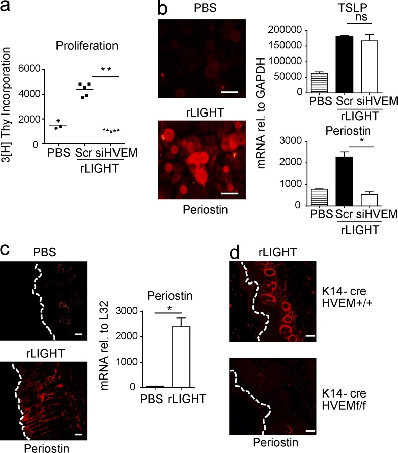
To understand what might be the main activity of HVEM in keratinocytes, we first assessed the action of LIGHT in vitro on normal human epidermal keratinocytes. There were two primary effects that we hypothesized could explain the dramatic effect on atopic dermatitis symptoms when HVEM could not be expressed in these cells. First, LIGHT–HVEM signaling might have regulated division of keratinocytes, driving epidermal hyperplasia. Indeed, in vitro, LIGHT promoted proliferation of keratinocytes, and this was completely prevented by siRNA knockdown of HVEM (Fig. 3 a).
Figure 3.
HVEM regulates proliferation and periostin expression in keratinocytes. (a and b) Human epidermal keratinocytes were cultured with PBS or rLIGHT for 72 h with or without siRNA knockdown of HVEM. (a) Proliferation (tritiated thymidine incorporation). Data are from three to five individual replicate cultures. (b) Periostin expression by IF stain (red) and mRNA expression of TSLP and periostin. Scr, scrambled siRNA. Data are representative of (or means ± SEM from) three replicate cultures. (c) WT mice were injected s.c. with rLIGHT for 2 d. Periostin IF stain on skin tissue or mRNA in biopsy specimens by quantitative PCR on day 3 (means ± SEM). (d) K14-cre HVEM+/+ or K14-cre HVEMfl/fl mice were injected s.c. with rLIGHT for 2 d. Periostin IF stain on skin tissue on day 3. All results are representative of three experiments. Bars, 200 µm. *, P < 0.05; **, P < 0.1. All statistical data were generated using the Mann–Whitney test.
Second, because both TSLP and periostin have been shown to play a strong role in development of skin inflammation in HDM-driven models (Masuoka et al., 2012; Ando et al., 2014), HVEM signals might have directly controlled the production of either or both proteins. We previously showed in vitro with human keratinocytes that LIGHT could up-regulate TSLP expression (Herro et al., 2015), but TSLP expression was not affected by siRNA knockdown of HVEM in keratinocytes (Fig. 3 b). Interestingly, differing from the in vitro results, TSLP was reduced in vivo in the skin in the HVEM conditional knockout animals (Fig. 2). The latter may then have been a consequence of the lack of HVEM signaling preventing the hyperplasia of keratinocytes necessary for increased TSLP to be expressed.
Most significantly, we found that LIGHT also strongly induced periostin production in keratinocytes at both the mRNA and protein levels and that knockdown of HVEM completely prevented LIGHT activity in this regard (Fig. 3 b). Previous studies have shown that periostin is highly expressed in the skin of patients with atopic dermatitis and scleroderma (Zhou et al., 2010; Yang et al., 2012), but exactly how it contributes to tissue inflammation is not clear. Periostin has been described to possess several activities, including binding to extracellular matrix proteins like collagen that could enhance structural rigidity in tissues; binding to several αV integrins (β1/β3/β5) and inducing chemokine production that combined could facilitate localization of T cells, mast cells, and eosinophils with epithelial cells such as keratinocytes; and synergizing with TGF-β to enhance epithelial-mesenchymal transition and collagen deposition that could contribute to tissue remodeling.
To further the conclusion that LIGHT-HVEM interaction is a primary regulator of periostin production by keratinocytes, we treated naive mice subcutaneously with recombinant LIGHT (rLIGHT) given on 2 successive days. Periostin expression was strongly up-regulated in the epidermis and dermis at the transcript and protein levels (Fig. 3 c) at a magnitude only marginally lower than seen in animals epicutaneously primed with house dust mite allergen (see Fig. 1). Moreover, mice lacking HVEM in their keratinocytes did not express significant amounts of skin periostin when injected with rLIGHT (Fig. 3 d). This further illustrates the role of LIGHT signaling through HVEM in these cells and suggests that LIGHT is a novel mediator of periostin production.
As well as keratinocytes/epithelial cells, periostin is a product of fibroblasts and has been primarily thought to be controlled by IL-13 and TGF-β based on studies showing that either cytokine can induce this protein in vitro in these cell types (Takayama et al., 2006; Sidhu et al., 2010; Zhou et al., 2010). It was then a possibility that LIGHT-HVEM signaling might have up-regulated periostin indirectly via these cytokines. However, IL-13 is not a known product of keratinocytes, and blocking this cytokine or TGF-β, that can be produced by keratinocytes, had no effect on LIGHT induction of periostin in vitro (Fig. S2 a). Importantly, we also found that LIGHT was as effective as IL-13 in driving periostin expression, but interestingly no additive or synergistic activity was seen when LIGHT was combined with IL-13 (Fig. S2 b). This implies that both cytokines may target the same intracellular pathway that leads to transcription of the periostin gene. We also injected rLIGHT into the skin of IL-4Rα-deficient mice, and found the overall skin reaction was strongly reduced, although not completely, with still some evidence of epidermal thickening and some periostin (Fig. S2 c). At present, it is not clear how to interpret this result. Given that keratinocyte-specific deletion of HVEM (Figs. 2 and 3 d) results in weak periostin expression with rLIGHT or allergen exposure, the data with IL-4Rα−/− mice might suggest that IL-13 does synergize with LIGHT in vivo. An alternative possibility is that there is cooperation between HVEM and IL-4Rα that might be independent of IL-13 or IL-4. Further studies will be required to understand these results. Nevertheless, it is likely that LIGHT acts together with both IL-13 and TGF-β in vivo.
Another question for future studies is whether LIGHT is a primary regulator of periostin production in dermal fibroblasts. These cells express both HVEM and LTβR, and preliminary experiments in vitro show that LIGHT can up-regulate periostin expression (not depicted). Therefore, it will be of interest to determine whether fibroblast-specific deletion of HVEM additionally impacts development of atopic dermatitis.
Therapeutic blocking of LIGHT–HVEM interactions suppresses atopic dermatitis
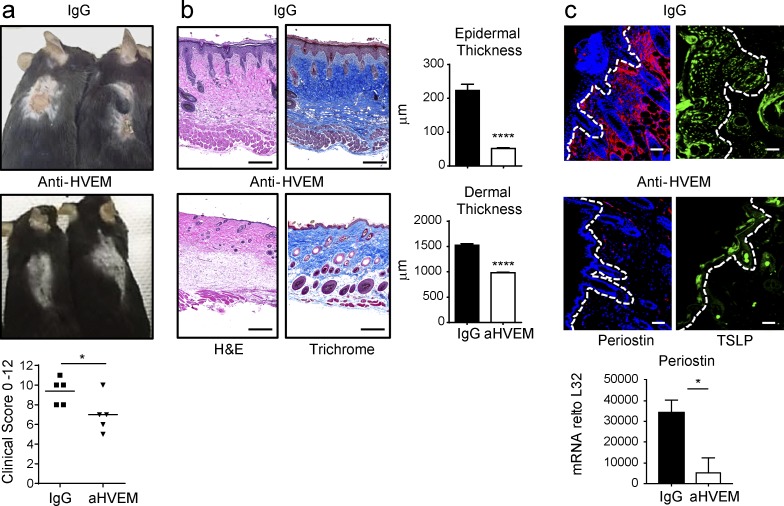
To extend our results, we lastly assessed whether blocking LIGHT–HVEM interactions therapeutically with an anti–HVEM antibody (which neutralizes LIGHT–HVEM, but not LIGHT–LTβR) might reduce allergen-induced atopic dermatitis symptoms and periostin expression. Because we observed full-onset atopic dermatitis by day 7, we treated mice previously sensitized with HDM at this time, with the antibody given every other day until the end of the experiment (Fig. 4). Importantly, atopic dermatitis clinical scores were significantly abrogated in anti-HVEM treated mice (Fig. 4 a), along with a strong reduction in epidermal thickening and a moderate albeit significant decrease in dermal thickness (Fig. 4 b). Most significantly, periostin expression in the dermis was almost absent after blocking HVEM activity, and less epidermal TSLP expression was observed (Fig. 4 c). Inflammatory immune infiltrates were largely unaffected, and there were variable effects on Th2 cytokines and TGF-β (Fig. S3).
Figure 4.
Therapeutic blocking of LIGHT–HVEM reduces HDM-driven atopic dermatitis. (a–c) WT mice were sensitized epicutaneously with HDM/SEB and treated with anti-HVEM or IgG starting on day 7 and analyzed on day 14. (a) Clinical symptoms and scoring for skin eruption, scaling, bleeding, and redness from individual mice. (b) Skin sections stained with H&E or Masson’s trichrome blue and quantitated for epidermal and dermal thickness. Data represent means ± SEM from five mice. (c) IF staining of TSLP (green) and periostin (red). Dotted lines indicate epidermal-dermal border. mRNA expression of periostin in skin biopsy (means ± SEM). Data representative of 15 mice, with similar data in three experiments. Bars, 200 µm. *, P < 0.05; ****, P < 0.001. All statistical data were generated using the Mann–Whitney test.
In summary, our results reveal a novel role for LIGHT–HVEM activity in keratinocytes that is essential for dermatitis in a model of eczema and show that blocking LIGHT–HVEM interactions after disease onset suppresses symptoms of skin inflammatory disease. HVEM is emerging as an important regulator of immune and inflammatory responses, acting in a variety of sites and different cell types, including lymphocytes, myeloid cells, and epithelial cells, but this is the first study that it directly controls immune reactions in the skin and the first showing that it is essential in keratinocytes in vivo. We further delineate a new pathway dependent on LIGHT–HVEM that controls periostin expression in the skin.
Although our studies highlight the role of HVEM in atopic dermatitis, this does not rule out a contribution of LTβR, the other receptor for LIGHT. We knocked down LTβR in human keratinocytes with siRNA and found a significant reduction in proliferation similar to knockdown of HVEM and a reduction in TSLP, but we observed no change in periostin expression (unpublished data). Together with our previous data in a scleroderma model, where blocking LTβR significantly reduced several aspects of skin inflammation (Herro et al., 2015), it is therefore likely that LTβR is also involved in the pathogenesis of atopic dermatitis. However, future studies with specific deletion of this receptor in vivo will be required to understand its overall role in keratinocytes.
Importantly, human studies have found that LIGHT is up-regulated in the serum of atopic dermatitis patients (Kotani et al., 2012; Morimura et al., 2012), suggesting it could be highly active in human disease. Interestingly, polymorphisms linked to decoy receptor 3, a soluble binding partner and presumed antagonist of FasL, TL1A, and LIGHT, also associate with susceptibility to atopic dermatitis (Sun et al., 2011; Ellinghaus et al., 2013), and decoy receptor 3 is up-regulated in human atopic dermatitis samples (Chen et al., 2004; Ellinghaus et al., 2013). The significance of these latter observations is not clear, but the polymorphisms and enhanced expression could lead to defective function of this protein, and subsequently, a bias in the balance between FasL, TL1A, and LIGHT may exist, which might further augment the action of LIGHT in the skin. Together with our results, these findings suggest that reagents that either target LIGHT–HVEM interactions alone or LIGHT interactions with both of its receptors may be beneficial for therapies aiming to halt and potentially abrogate atopic dermatitis in humans.
Materials and methods
Mice
6–8-wk-old male WT, LIGHT−/− (Scheu et al., 2002), and HVEMflox/flox mice crossed to K14-cre mice (Jackson Labs) were bred in-house on the C57BL/6 background. HVEMfl/fl mice were generated at the La Jolla Institute. A frt site-flanked LacZ reporter cassette was inserted into intron 2 of the Tnfrsf14 (HVEM) gene and a variant frt site (F3)-flanked Neor cassette, for drug selection, was inserted into intron 6 of the Tnfrsf14 gene by recombineering. Germline transmitted Tnfrsf14flox-neo/flox-neo mice were crossed with mice expressing the FLPe variant of the Saccharomyces cerevisiae FLP1 recombinase mice that deleted the LacZ reporter and the Neo cassette to generate HVEMflox/flox mice. In some experiments, WT BALB/c and IL-4R alpha-deficient mice (Jackson Labs) were used. All experiments were in compliance with the regulations of the La Jolla Institute for Allergy and Immunology Animal Care Committee.
Skin inflammation protocols
Atopic dermatitis–like disease was induced with two cycles of epicutaneous sensitization with HDM extract (10 µg/mouse; Greer Labs) and Staphylococcal enterotoxin B (SEB; 500 ng/mouse; Toxin Technology) applied directly on the shaved and abraded (using a Dremel for 10 s) back of mice at days 0 and 8, before sacrificing the mice at day 14. After the first and second epicutaneous application of the allergen, the backs of the mice were covered with a gauze pad for 3 d, and then this was removed for 4 d to allow mice to scratch. Clinical symptoms were evident at day 4 and peaked around day 7, with the disease fully established and persisting throughout the second cycle. Clinical symptoms were scored for skin eruption, scaling, bleeding, and redness on a scale from 0 to 3 each, with a cumulative maximum of 12. For neutralization of LIGHT–HVEM interactions, mice were administered 200 µg anti–HVEM mAb (LH1; Anand et al., 2006) given i.v. on day 7 1 d before the second cycle of epicutaneous sensitization with HDM/SEB and every other day i.p. until the end of the experiment.
Collagen, skin thickness, and H&E measurements
Formalin (4% in PBS)–fixed skin sections were stained with Masson’s trichrome blue to evaluate collagen, and H&E to evaluate inflammation. An image analysis system and software (Image-Pro Plus; Media Cybernetics) were used to quantitate epidermal and dermal thickening.
Immunohistochemical staining
Skin samples were deparaffinized by sequential placement in xylene and ethanol. Sections were treated with Fc block in 10% donkey serum (in PBS) and stained with (1) rabbit polyclonal antibody to periostin (clone ab14041; Abcam) at 1:200 concentration followed by Rhodamine Red-X AffiniPure donkey anti–rabbit (711295152; Jackson ImmunoResearch) at 1:500 or (2) goat polyclonal antibody to TSLP (clone L18; Santa Cruz Biotechnology) at 1:200 followed by donkey anti–goat IgG-FITC (sc-2024; Santa Cruz Biotechnology) at 1:500. Slides were read on a Nikon 80i microscope and analyzed by LSM-Image software.
Analysis of skin immune cell infiltrates
Skin, including both dermis and epidermis, was minced in the presence of trypsin/dispase, and cellular infiltration was assessed by flow in single-cell suspensions using CD45.2-BV711 (clone 104; BD), CD3e-BV650 (clone 145-2C11; BD), Mac3–Alexa Fluor 647 (clone M4/84; BioLegend), Ly6-G/Ly6-G–Alexa Fluor 700 (Gr-1, clone RB6-8C5; BioLegend), SiglecF-PE-CF594 (clone E50-2440; BD), CD11b-BB515 (clone M1/70; BD), and CD11c-Brilliant Violet 785 (clone N418; BioLegend). Macrophages were gated as CD45+, autofluorescent high, Mac3+, CD11c+, SiglecF+; dendritic cells as CD45+, CD11c+, CD11b+; neutrophils as CD45+, GR1+, CD11b+, SiglecF−; and T cells as CD45+, CD3+.
Real-time quantitative PCR
Total RNA was isolated using TRIzol (Invitrogen) and RNeasy Fibrous Tissue mini kit (74704; Qiagen). Single-strand cDNA was prepared by reverse transcribing 5 µg total RNA using the Transcriptor First Strand cDNA kit (Roche). Samples were amplified in IQ SYRB Green Supermix (Bio-Rad Laboratories) using the following primer pairs: TGF-β forward, 5′-CCCTATATTTGGAGCCTGGA-3′; reverse, 5′-GGAAGCTTCGGGATTTATGG-3′; huTSLP forward, 5′-TATGAGTGGGACCAAAAGTACCG-3′; reverse, 5′-GGGATTGAAGGTTAGGCTCTGG-3′; muPeriostin forward, 5′-CCCTCCAGCAAATTCTGGGCACCA-3′; reverse, 5′-GAGACTCACGTTTTCTTCCCGCAG A-3′; huPeriostin forward, 5′-GACTCAAGATGATTCCCTTT-3′; reverse, 5′-GGTGCAAAGTAAGTGAAGG-3′; mIL13 forward, 5′-CTTGCCTTGGTGGTCTCG-3′; reverse, 5′-CGTTGCACAGGGGAGTCT-3′; mIL-4 forward, 5′-AGATCATCGGCATTTTGAACG-3′; reverse, 5′-TTTGGCACATCCATCTCCG-3′; mIL-5 forward, 5′-CGCTCACCGAGCTCTGTTG-3′; reverse, 5′-CCAATGCATAGCTGGTGATTTTT-3′; and mCollagen forward, 5′-GAGCCCTCGCTTCCGTACTC-3′; reverse, 5′-TGTTCCCTACTCAGCCGTCTGT-3′. All samples were run in triplicate, and the mean values were used for quantification. Data are expression relative to ribosomal protein L32 or GAPDH.
Stimulation of keratinocytes and transfection of siRNA
Human epidermal keratinocytes from neonates (HEK) or mouse epidermal keratinocytes (PAM212 cell line) were stimulated with 20 ng/ml recombinant human LIGHT (664-LI/CF; R&D), mouse LIGHT (1794-LT-025/CF; R&D), or mouse IL-13 (413-ML-005; R&D) for 72 h in Epilife media (Life Technologies). In some cases, 30 µg/ml anti–TGF-β mAb (1D11; BioXCell) or anti–IL-13 (MAB413-SP; R&D) or isotype control were added into culture. Periostin was measured by immunostaining using anti–hPeriostin mAb (clone ABT292; Millipore) and quantitative PCR analyses. ON-TARGETplus siRNAs were purchased from Dharmacon. A 5–25 nM concentration range of control scrambled or HVEM siRNA was transfected into HEK cells using HiPerFect transfection reagent (Qiagen). Efficiency of HVEM knockdown was monitored over time by both mRNA and surface protein levels and was stable during the 24- to 72-h culture period. HVEM was visualized using anti-HVEM (BioLegend).
Statistical analyses
Statistical analysis was performed using GraphPad Prism software. A nonparametric Mann–Whitney test was used where indicated. A p-value <0.05 was considered statistically significant.
Online supplemental material
Fig. S1 shows HVEM expression in skin cells from K14-cre HVEMfl/fl mice. Fig. S2 shows LIGHT regulation of periostin expression independently of IL-13 and TGF-β. Fig. S3 shows cellular infiltration and Th2 cytokines in WT mice sensitized epicutaneously with HDM/SEB and treated with anti-HVEM or IgG.
Supplementary Material
Acknowledgments
We thank Klaus Pfeffer for originally making available mice deficient in LIGHT.
This work was supported by the National Institutes of Health (grants AI070535 and AI100905 to M. Croft, AI061516 to M. Kronenberg, and F32 DK082249 to J-W. Shui).
The authors declare no competing financial interests.
Author contributions: R. Herro performed experiments. J-W. Shui, S. Zahner, K. Tamada, and M. Kronenberg contributed reagents or mice. R. Herro, Y. Kawakami, T. Kawakami, and D. Sidler contributed experimental protocols and advice. R. Herro and M. Croft conceived the project, directed the studies, analyzed the data, and wrote the manuscript. All authors discussed the results and commented on the manuscript.
References
- Anand S., Wang P., Yoshimura K., Choi I.H., Hilliard A., Chen Y.H., Wang C.R., Schulick R., Flies A.S., Flies D.B., et al. . 2006. Essential role of TNF family molecule LIGHT as a cytokine in the pathogenesis of hepatitis. J. Clin. Invest. 116:1045–1051. 10.1172/JCI27083 [DOI] [PMC free article] [PubMed] [Google Scholar]
- Ando T., Xiao W., Gao P., Namiranian S., Matsumoto K., Tomimori Y., Hong H., Yamashita H., Kimura M., Kashiwakura J., et al. . 2014. Critical role for mast cell Stat5 activity in skin inflammation. Cell Reports. 6:366–376. 10.1016/j.celrep.2013.12.029 [DOI] [PMC free article] [PubMed] [Google Scholar]
- Bieber T. 2008. Atopic dermatitis. N. Engl. J. Med. 358:1483–1494. 10.1056/NEJMra074081 [DOI] [PubMed] [Google Scholar]
- Chen C.C., Yang Y.H., Lin Y.T., Hsieh S.L., and Chiang B.L.. 2004. Soluble decoy receptor 3: increased levels in atopic patients. J. Allergy Clin. Immunol. 114:195–197. 10.1016/j.jaci.2004.02.048 [DOI] [PubMed] [Google Scholar]
- Ellinghaus D., Baurecht H., Esparza-Gordillo J., Rodríguez E., Matanovic A., Marenholz I., Hübner N., Schaarschmidt H., Novak N., Michel S., et al. . 2013. High-density genotyping study identifies four new susceptibility loci for atopic dermatitis. Nat. Genet. 45:808–812. 10.1038/ng.2642 [DOI] [PMC free article] [PubMed] [Google Scholar]
- Herro R., Antunes R.D.S., Aguilera A.R., Tamada K., and Croft M.. 2015. The Tumor Necrosis Factor Superfamily Molecule LIGHT Promotes Keratinocyte Activity and Skin Fibrosis. J. Invest. Dermatol. 135:2109–2118. 10.1038/jid.2015.110 [DOI] [PMC free article] [PubMed] [Google Scholar]
- Kawakami Y., Yumoto K., and Kawakami T.. 2007. An improved mouse model of atopic dermatitis and suppression of skin lesions by an inhibitor of Tec family kinases. Allergol. Int. 56:403–409. 10.2332/allergolint.O-07-486 [DOI] [PubMed] [Google Scholar]
- Kotani H., Masuda K., Tamagawa-Mineoka R., Nomiyama T., Soga F., Nin M., Asai J., Kishimoto S., and Katoh N.. 2012. Increased plasma LIGHT levels in patients with atopic dermatitis. Clin. Exp. Immunol. 168:318–324. 10.1111/j.1365-2249.2012.04576.x [DOI] [PMC free article] [PubMed] [Google Scholar]
- Masuoka M., Shiraishi H., Ohta S., Suzuki S., Arima K., Aoki S., Toda S., Inagaki N., Kurihara Y., Hayashida S., et al. . 2012. Periostin promotes chronic allergic inflammation in response to Th2 cytokines. J. Clin. Invest. 122:2590–2600. 10.1172/JCI58978 [DOI] [PMC free article] [PubMed] [Google Scholar]
- Mauri D.N., Ebner R., Montgomery R.I., Kochel K.D., Cheung T.C., Yu G.L., Ruben S., Murphy M., Eisenberg R.J., Cohen G.H., et al. . 1998. LIGHT, a new member of the TNF superfamily, and lymphotoxin alpha are ligands for herpesvirus entry mediator. Immunity. 8:21–30. 10.1016/S1074-7613(00)80455-0 [DOI] [PubMed] [Google Scholar]
- Morimura S., Sugaya M., Kai H., Kato T., Miyagaki T., Ohmatsu H., Kagami S., Asano Y., Mitsui H., Tada Y., et al. . 2012. High levels of LIGHT and low levels of soluble herpesvirus entry mediator in sera of patients with atopic dermatitis. Clin. Exp. Dermatol. 37:181–182. 10.1111/j.1365-2230.2011.04079.x [DOI] [PubMed] [Google Scholar]
- Nishiyama T., Kii I., Kashima T.G., Kikuchi Y., Ohazama A., Shimazaki M., Fukayama M., and Kudo A.. 2011. Delayed re-epithelialization in periostin-deficient mice during cutaneous wound healing. PLoS One. 6:e18410 10.1371/journal.pone.0018410 [DOI] [PMC free article] [PubMed] [Google Scholar]
- Ontsuka K., Kotobuki Y., Shiraishi H., Serada S., Ohta S., Tanemura A., Yang L., Fujimoto M., Arima K., Suzuki S., et al. . 2012. Periostin, a matricellular protein, accelerates cutaneous wound repair by activating dermal fibroblasts. Exp. Dermatol. 21:331–336. 10.1111/j.1600-0625.2012.01454.x [DOI] [PubMed] [Google Scholar]
- Parulekar A.D., Atik M.A., and Hanania N.A.. 2014. Periostin, a novel biomarker of TH2-driven asthma. Curr. Opin. Pulm. Med. 20:60–65. 10.1097/MCP.0000000000000005 [DOI] [PubMed] [Google Scholar]
- Salimi M., Barlow J.L., Saunders S.P., Xue L., Gutowska-Owsiak D., Wang X., Huang L.C., Johnson D., Scanlon S.T., McKenzie A.N., et al. . 2013. A role for IL-25 and IL-33–driven type-2 innate lymphoid cells in atopic dermatitis. J. Exp. Med. 210:2939–2950. 10.1084/jem.20130351 [DOI] [PMC free article] [PubMed] [Google Scholar]
- Scheu S., Alferink J., Pötzel T., Barchet W., Kalinke U., and Pfeffer K.. 2002. Targeted disruption of LIGHT causes defects in costimulatory T cell activation and reveals cooperation with lymphotoxin β in mesenteric lymph node genesis. J. Exp. Med. 195:1613–1624. 10.1084/jem.20020215 [DOI] [PMC free article] [PubMed] [Google Scholar]
- Shiraishi H., Masuoka M., Ohta S., Suzuki S., Arima K., Taniguchi K., Aoki S., Toda S., Yoshimoto T., Inagaki N., et al. . 2012. Periostin contributes to the pathogenesis of atopic dermatitis by inducing TSLP production from keratinocytes. Allergol. Int. 61:563–572. 10.2332/allergolint.10-OA-0297 [DOI] [PubMed] [Google Scholar]
- Sidhu S.S., Yuan S., Innes A.L., Kerr S., Woodruff P.G., Hou L., Muller S.J., and Fahy J.V.. 2010. Roles of epithelial cell-derived periostin in TGF-beta activation, collagen production, and collagen gel elasticity in asthma. Proc. Natl. Acad. Sci. USA. 107:14170–14175. 10.1073/pnas.1009426107 [DOI] [PMC free article] [PubMed] [Google Scholar]
- Steinberg M.W., Shui J.W., Ware C.F., and Kronenberg M.. 2009. Regulating the mucosal immune system: the contrasting roles of LIGHT, HVEM, and their various partners. Semin. Immunopathol. 31:207–221. 10.1007/s00281-009-0157-4 [DOI] [PMC free article] [PubMed] [Google Scholar]
- Sun L.D., Xiao F.L., Li Y., Zhou W.M., Tang H.Y., Tang X.F., Zhang H., Schaarschmidt H., Zuo X.B., Foelster-Holst R., et al. . 2011. Genome-wide association study identifies two new susceptibility loci for atopic dermatitis in the Chinese Han population. Nat. Genet. 43:690–694. 10.1038/ng.851 [DOI] [PubMed] [Google Scholar]
- Takayama G., Arima K., Kanaji T., Toda S., Tanaka H., Shoji S., McKenzie A.N., Nagai H., Hotokebuchi T., and Izuhara K.. 2006. Periostin: a novel component of subepithelial fibrosis of bronchial asthma downstream of IL-4 and IL-13 signals. J. Allergy Clin. Immunol. 118:98–104. 10.1016/j.jaci.2006.02.046 [DOI] [PubMed] [Google Scholar]
- Taniguchi K., Arima K., Masuoka M., Ohta S., Shiraishi H., Ontsuka K., Suzuki S., Inamitsu M., Yamamoto K.I., Simmons O., et al. . 2014. Periostin controls keratinocyte proliferation and differentiation by interacting with the paracrine IL-1α/IL-6 loop. J. Invest. Dermatol. 134:1295–1304. 10.1038/jid.2013.500 [DOI] [PubMed] [Google Scholar]
- Usategui A., Criado G., Izquierdo E., Del Rey M.J., Carreira P.E., Ortiz P., Leonard W.J., and Pablos J.L.. 2013. A profibrotic role for thymic stromal lymphopoietin in systemic sclerosis. Ann. Rheum. Dis. 72:2018–2023. 10.1136/annrheumdis-2012-202279 [DOI] [PMC free article] [PubMed] [Google Scholar]
- Yamamoto T. 2009. Scleroderma--pathophysiology. Eur. J. Dermatol. 19:14–24. [DOI] [PubMed] [Google Scholar]
- Yang L., Serada S., Fujimoto M., Terao M., Kotobuki Y., Kitaba S., Matsui S., Kudo A., Naka T., Murota H., and Katayama I.. 2012. Periostin facilitates skin sclerosis via PI3K/Akt dependent mechanism in a mouse model of scleroderma. PLoS One. 7:e41994 10.1371/journal.pone.0041994 [DOI] [PMC free article] [PubMed] [Google Scholar]
- Yoo J., Omori M., Gyarmati D., Zhou B., Aye T., Brewer A., Comeau M.R., Campbell D.J., and Ziegler S.F.. 2005. Spontaneous atopic dermatitis in mice expressing an inducible thymic stromal lymphopoietin transgene specifically in the skin. J. Exp. Med. 202:541–549. 10.1084/jem.20041503 [DOI] [PMC free article] [PubMed] [Google Scholar]
- Zhou H.M., Wang J., Elliott C., Wen W., Hamilton D.W., and Conway S.J.. 2010. Spatiotemporal expression of periostin during skin development and incisional wound healing: lessons for human fibrotic scar formation. J. Cell Commun. Signal. 4:99–107. 10.1007/s12079-010-0090-2 [DOI] [PMC free article] [PubMed] [Google Scholar]
- Ziegler S.F., and Artis D.. 2010. Sensing the outside world: TSLP regulates barrier immunity. Nat. Immunol. 11:289–293. 10.1038/ni.1852 [DOI] [PMC free article] [PubMed] [Google Scholar]
Associated Data
This section collects any data citations, data availability statements, or supplementary materials included in this article.


