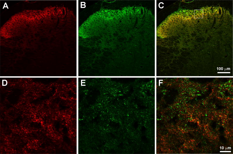
Figure 4. Mu opioid receptor and mGluR5 are differentially expressed in spinal cord.
Representative sections of mouse spinal cord perfused eight weeks after spared nerve-injury. A) MOR-ir, red B) mGluR5-ir, green C) merged image suggesting expression of the two receptors in different structures. Boxed image represents the section magnified in D-F. D) higher magnification of the MOR-ir, red E) higher magnification of mGluR5-ir, green E) higher magnification of the merged image of both receptors confirming predominantly differential expression of the two receptors.

