Abstract
The advent of multicellularity was a watershed event in the history of life, yet the transition from unicellularity to multicellularity is not well understood. Multicellularity opens up opportunities for innovations in intercellular communication, cooperation, and specialization, which can provide selective advantages under certain ecological conditions. The unicellular alga Chlamydomonas reinhardtii has never had a multicellular ancestor yet it is closely related to the volvocine algae, a clade containing taxa that range from simple unicells to large, specialized multicellular colonies. Simple multicellular structures have been observed to evolve in C. reinhardtii in response to predation or to settling rate-based selection. Structures formed in response to predation consist of individual cells confined within a shared transparent extracellular matrix. Evolved isolates form such structures obligately under culture conditions in which their wild type ancestors do not, indicating that newly-evolved multicellularity is heritable. C. reinhardtii is capable of photosynthesis, and possesses an eyespot and two flagella with which it moves towards or away from light in order to optimize input of radiant energy. Motility contributes to C. reinhardtii fitness because it allows cells or colonies to achieve this optimum. Utilizing phototaxis to assay motility, we determined that newly evolved multicellular strains do not exhibit significant directional movement, even though the flagellae of their constituent unicells are present and active. In C. reinhardtii the first steps towards multicellularity in response to predation appear to result in a trade-off between motility and differential survivorship, a trade-off that must be overcome by further genetic change to ensure long-term success of the new multicellular organism.
Introduction
The evolutionary transition from unicellularity to multicellularity represents a major increase in complexity during the history of life. Higher cell number opens the door to cellular differentiation, enabling diverse processes to be spatially and temporally segregated and to be made more efficient via cellular division of labor. In the volvocine algae, size correlates with level of cellular differentiation. Large-colony members of this clade always contain both somatic and reproductive cells, while small-colony taxa are typically composed of undifferentiated cells that perform both functions. In large colonies, somatic cells are specialized for colony movement and energy production, whereas germ line cells are well-provisioned with resources and dedicated to colony reproduction; this arrangement potentially increases both metabolic efficiency and reproductive potential of the colony as a whole [1]. The unicell- to multicell- transition is not an easy one to make, however, with complex life often requiring specific structural and physiological adaptations to overcome challenges associated with increased size. For example, the diminished ratio of external surface area to internal volume in a large versus a small organism puts a premium on effective nutrient transport and metabolic waste removal. Increased size associated with the transition to multicellularity is therefore subject to biophysical constraints that can result in trade-offs. These trade-offs may be so severe that the evolutionary success of the new multicellular organism depends on further innovation.
While an integral part of life’s history, the transition from unicellular to multicellular organisms is not yet well understood. Phylogenetic analyses indicate that multicellularity has arisen multiple times in at least 25 independent lineages [2]. However, the comparative approach gives only a partial view into the genetic changes that led to the first steps along the path towards multicellularity. The comparative approach uses present instead of ancestral relatives due to limited knowledge of ancestral populations, and requires multiple assumptions, including the assumption of a simple and repeatable evolutionary history. Model systems allow us to expand our view of how multicellularity might arise by simulating its evolution in the laboratory. By experimentally studying this innovation, considered one of life’s major transitions [3], we can gain insight into the evolutionary processes and environmental constraints that may have led to the advent of complex, multicellular organisms.
Given their morphological diversity and the varying levels of differentiation observed among closely related species, the volvocine algae provide an especially useful model system to study the evolution of complexity and multicellularity. Chlamydomonas reinhardtii, a unicellular, photosynthetic green alga in the Chlamydomonadaceae, has never had a multicellular ancestor yet is closely related to the volvocine algae, which express multicellularity in colonies of up to 50,000 cells [4]. Ranging from small, undifferentiated multicellular clusters to large, well-differentiated spherical colonies, the volvocine algae show many ways in which multicellularity can be organized. The more complex volvocine algae such as Volvox carteri exhibit elaborate structures in which individual haploid cells are anchored and oriented via a shared extra-cellular matrix that also serves as a major interface between cells and their environment. This higher level of organization distinguishes volvocine evolution from the evolution of the much simpler Tetrasporales, in which haploid, thin-walled, nonmotile spores are formed during reproduction [5, 6]. Species of volvocine algae contain organized groups of C. reinhardtii-like cells with varying levels of complexity, indicating that a simple colonial structure composed of unicellular C. reinhardtii may be possible.
Two forms of selection have been used to produce simple multicellularity in C. reinhardtii. Predation has long been thought to favor multicellularity as it would lead to survivorship among clusters too large to be ingested [7, 8], and indeed Herron et al. have successfully used this form of selection to experimentally evolve simple multicellular structures [9]. Ratcliff et al. reported the evolution of such forms in response to settling rate selection, which favors algae having larger mass [10]. Significantly, de novo multicellular lineages that arise in response to either form of selection obligately produce clusters for successive generations in cultures subjected to neither centrifugation nor predation, indicating the new trait is heritable. Two distinct cluster types have been observed in these two types of experiments: clumped, irregular clusters of cells arose in response to centrifugation, while clusters that evolved in response to predators consist of cells that remain within the parental cell wall, forming clusters as a result of their failure to separate after division [9, 10]. Structure in the latter appears more orderly, as de novo multicell number is frequently a power of two (21–24). Because many of these structures are reminiscent of extant volvocine species, the predation-evolved isolates were chosen for the present study.
Like all other Volvocales, C. reinhardtii is a freshwater organism that is capable of photosynthesis for energy production. C. reinhardtii are negatively buoyant, and sink if no effort is expended to maintain their position in the water column [11]. Because maintaining an optimal light environment contributes to its ecological success, it is perhaps unsurprising that C. reinhardtii is flagellated and capable of taxis in response to a number of different stimuli, including light intensity. Equipped with two flagella and a single eyespot, C. reinhardtii detect differing light levels and use a coordinated breast stroke-like movement to change their position relative to optical stimulus [12]. While C. reinhardtii is heterotrophic and capable of utilizing environmentally sourced carbon, photosynthesis offers not only additional energy production with carbon uptake, but also an alternative source if environmental carbon is limited [13]. As has been demonstrated with Chlamydomonas humicola, a close relative of C. reinhardtii in the Chlamydomonas genus, mixotrophic cultures have a growth rate >2-times higher than heterotrophic cultures and a rate ~8 times higher than autotrophic cultures [14]. Clearly, the capacity to utilize multiple carbon sourcing pathways plays a major role in setting algal growth rates. Without the ability to undergo phototaxis, C. reinhardtii in nature would almost certainly have a diminished capacity to generate energy. Motility is therefore likely to be an important component of fitness in this species.
Multicellular relatives of C. reinhardtii such as Gonium, Eudorina and Volvox also rely on flagellar movement. Regardless of their colony complexity, Volvocine algae have been observed to have exteriorly oriented flagella regardless of colony complexity. These negatively buoyant multicellular species rely on flagellar movement not only to respond to changes in light environment but also to increase nutrient flow around the colony, facilitating nutrient transport and waste removal [11].
Here we explore the capacity of de novo multicellular strains of C. reinhardtii evolved under predation to undergo phototaxis. Our goal is to evaluate the impact of this selective process on motility—a determinant of fitness in this freshwater phototroph—in order to understand the role that predative selection may have played in the first steps of this major evolutionary transition. By evaluating the capacity of multicellular C. reinhardtii to move in response to a key environmental stimulus, we can gauge its potential viability in nature and better understand what further evolutionary innovations and selection pressures would be needed to evolve differentiated multicellularity in the laboratory.
Methods
Strain construction and culture conditions
Details of the evolution experiment that gave rise to the experimental strains are provided in Herron et al. 2018. Briefly, experimental populations of C. reinhardtii were generated by crossing parental strains CC1690 (mating type +) and CC2290 (mating type -), resulting in a genetically diverse pool of F2 progeny. Crosses were performed using the protocol described by Harris [15], modified to a volume of 10 mL for growth of vegetative cells and gametogenesis. F1 progeny from the initial CC1690 x CC2290 cross were crossed in bulk to produce the F2 pool used for the experiment. Subsamples of this outcrossed population were then used for all treatment and control groups.
C. reinhardtii in the treatment groups were co-cultured with the predator Paramecium tetraurelia, while those in the control groups were cultured axenically. Experimental and control populations initially included ~2 × 10⁵ C. reinhardtii cells in 1.5 mL of COMBO medium [16]; experimental populations additionally included ~2 × 10⁴ P. tetraurelia. Both experimental and control populations were grown in 24-well plates at 22.5°C under cool white light (4100K); 0.1 mL of each replicate culture was transferred to fresh medium weekly for 50 weeks (~300 C. reinhardtii generations).
The strains used in the motility assays were isolated from experimental and control populations through three rounds of serial plating. Eight colonies were picked at random from each population in the first round of plating. Each colony was diluted, re-plated, and a single colony picked in the second and third rounds to ensure that each isolate was uniclonal. Isolates were then cultured in 3 mL TAP media [17] and stored on TAP + 1.5% agar plates. Strains were regrown in 5 mL WC [18] at a temperature of 22.5°C under cool white illumination at 4100 K. Cultures provided upon request.
Assay of motility in experimentally evolved algae
Motility in response to light was evaluated on cell lines that displayed varying degrees of multicellularity, as well as on a unicellular control population. Three populations of C. reinhardtii were grown until dense enough to see with the naked eye, a density of about 2 x10⁵ cells/mL. Eight strains were isolated from each of two experimental populations and one control population. The control population, K1, contained eight unicellular strains, experimental population B2 contained three unicellular strains and five multicellular strains, and experimental population B5 contained eight multicellular strains (Fig 1). Once cultured to a density of ~2 x10⁵ cells/mL, cultures were diluted to the same absorbance of 0.1 using a spectrophotometer set at λ = 800 nm. The 24-well tissue culture plates used had their longitudinal textured edges removed to reduce light scattering and to expose wells directly to the light source. Strains were randomized in such a way as to ensure one replicate of each strain per plate, at least one multicellular and one unicellular strain per plate, and varying replicate position between plates to reduce positioning effects. Using this randomized order, strains were placed into the six wells along the long side of the modified plate to ensure as equal light distribution among wells as possible.
Fig 1. Multicellular and unicellular phenotypes.
Cells from the B2 population (a), B5 population (b) and K1 population (c). Scale bars = 20 μm (images by Josh Ming Borin).
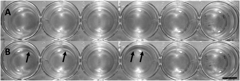
To measure phototaxis, a copy stand, designed to hold camera positioning and light level steady, was mounted with a Canon Rebel T3 Powershot camera equipped with a Canon EF-S 18-55mm f/3.5–5.6 IS II lens. Modified tissue culture plates were placed on the platform of the copy stand with the algal wells facing a cool white (4100 K) directional incubator light. Light level at the plate was fixed at 170 lux. An image of the plate was taken before the directional light was switched on using the neutral light provided by the copy stand and a cable release to minimize camera movement. Then, the copy stand lights were turned off, the directional light was turned on, and plates were exposed from one edge for a period of five minutes. Light from all other sides was blocked. Once the five-minute period had elapsed, the directional light was switched off, copy stand lights switched on, and a final image was taken. Because cultures were grown to a density that could be seen with the naked eye, density changes in the well due to algal phototaxis were immediately visible during and after light exposure. The images taken recorded these visual changes for later analysis. This process was repeated with all plates; ultimately six sets of plates were analyzed and six replicates per strain were obtained (Fig 2). Each replicate was assayed one time, and separate cultures were used.
Fig 2. Experimental design.
Illustration of the experimental design of motility assays.
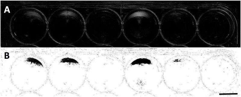
To compare levels of phototaxis between unicellular and multicellular strains, images were loaded into ImageJ photo-analysis software [19]. For each set of “before” and “after” images, density changes were analyzed by comparing the pixel density changes over the time-course of light exposure. Because camera position and lighting were held constant between the images, changes in pixel density could be attributed to algal cell movement within the well in response to light. In order to determine net change in pixel density, we subtracted one image from the other. This procedure creates a map of change in algal density over the five-minute exposure time. To further improve our analysis, the color threshold on this map could be set to display only green-spectrum pixel changes. This helps to reduce any possible effects due to shadowing or slight movements of the plate or camera, and specifies our analysis for green cells and clusters of C. reinhardtii. For each well in each plate a plot profile was obtained to determine where the highest density changes of algae occurred and in which strains. The difference between the average density of the lighter half of the well (closer to the directional light source) and that of the darker half was then taken to obtain the direction of movement, and to indicate no directional movement if this difference proved insignificant. In this way, a relative magnitude and direction of phototaxis was measured in each strain (Figs 3 and 4).
Fig 3. Well plate before and after light exposure.
Longitudinal wells in a modified 24-well plate before and after light exposure. Image A shows the plate before exposure, while image B was taken after light exposure. Black arrows indicate changes in algal density due to directional light exposure. Scale bar = 8mm.
Fig 4. Well plate analysis in ImageJ.
Images from Fig 3 analyzed to indicate density differences in the plate before and after directional light exposure. Image A shows the ImageJ analysis showing the changes in density of green pixels between the before and after photographs, showing algal movement due to light exposure. Image B shows this same analysis image with a color threshold set to determine relative magnitude of algal movement. Strains analyzed here with their corresponding magnitude of pixel density change in grayscale units (from left to right) are: K107, 75.5; K108, 73.7; B505, 1.5; K105, 96.4; B501, 40.3; B207, 0.2. Scale bar = 8mm.
Microscopy
To further explore why movement is limited in multicellular C. reinhardtii, strains were observed under a light microscope to determine whether structural features peculiar to multicellular colonies might limit motility. To preserve colony integrity and viability no stains or fixing agents were used. Microscopy was performed at 80x magnification on a Zeiss Axio Vert.A1 inverted microscope and images recorded using a Zeiss Axiocam 105 Color camera.
Results
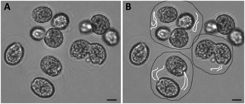
Microscopy indicates that in the multicellular form, unicellular flagellated C. reinhardtii appear to be encased within an external wall with few protrusions into the surrounding media. (Fig 5). We hypothesize that in the case of predation-selected lines, multicellularity evolved by failure of daughter cells to separate after cell division. If so, then each colony consists of a group of daughter cells trapped within the maternal cell wall. Importantly, each individual algal cell within the confines of the group remains flagellated and can be observed moving these flagella within the colony (S1 File). These individual cells can also move themselves around within the colony walls, changing position with one another but unable to escape the confines of their shared, external barrier. In some cases—a handful out of about 1000 colonies observed—a few flagella were observed to be moving outside of the colony wall. Phototactic assays were performed to determine whether these flagella and those inside the colony can generate movement despite being largely confined within the nascent colony.
Fig 5. Microscopy of multicellular colony.
Multicellular colonies from population B2 appear to remain within parental cell wall and retain visible flagella, which were observed to be capable of movement. In (b) black lines have been drawn to better indicate potential colony walls, while white lines indicate flagella. Scale bar = 10μm.
In phototactic assays multicellular strains of C. reinhardtii exhibited very limited—if any—phototaxis (Fig 6). Because direct microscopy indicates that the multicellular stages are largely immobile (S1 File), we hypothesize that the low levels of phototaxis observed in multicellular strains (Figs 6–8) were due to the presence of two phenotypes in these cultures: multicellular colonies and motile unicellular propagules released during the colonial reproductive cycle.
Fig 6. Density changes per strain.
Change in algal density distributions in unicellular and multicellular strains due to light exposure.
Fig 8. Directionality of phototaxis.
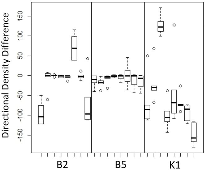
Analysis of algal movement due to light exposure where positive values indicate accumulation in the half of the well toward the light source and negative values indicate accumulation in the other half.
Transmission microscopy revealed that multicellular cultures contain a small number of these motile unicellular propagules, which are released during multicellular strains’ reproductive cycle when a colony reaches its characteristic burst size of 16 cells. These propagules were observed to have functional flagella and appear to remain phototactic, as indicated by low levels of algal accumulation observed in multicellular cultures with a small population of these propagules (S1 Table). Importantly, time-lapse microscopy reveals that these unicellular propagules obligately develop into multicellular colonies after their release from the parent colony, even in the absence of a predator [9]. It is this non-plastic, obligate colony formation in particular that distinguishes these strains from palmelloid colonies, the formation of which has also been observed in C. reinhardtii in the presence of certain environmental factors [10, 20]. Some strains, when viewed under a microscope at 80x magnification, had a small number of these unicellular propagules swimming among the multicellular clusters regardless of when strains were analyzed, so a slight response is expected due to these motile propagules.
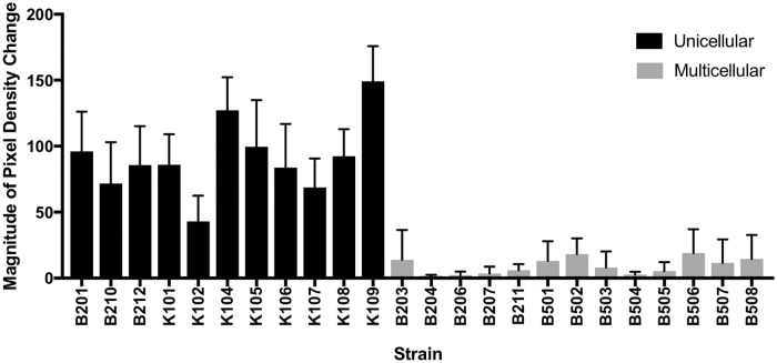
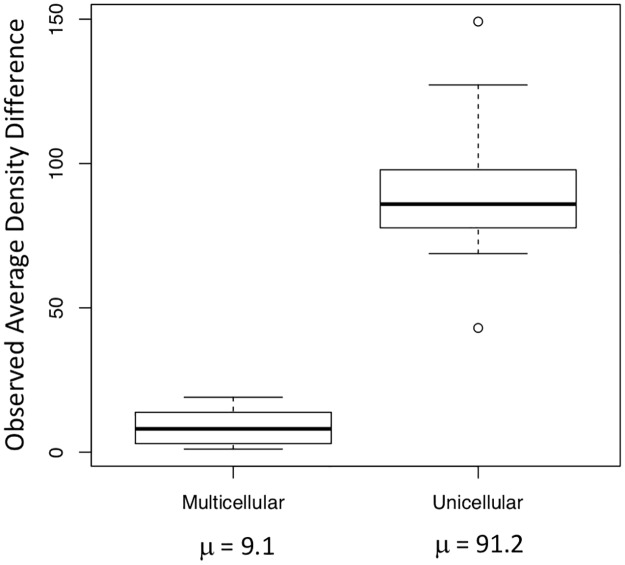
Despite this small margin of error, levels of phototaxis in unicellular strains were ten times higher on average than levels of phototaxis in multicellular strains (S1 Table). When average density changes were compared using ANOVA, there was an order of magnitude difference between average density changes of multicellular versus unicellular strains (p < 0.0001) (Fig 7). Thus, cells within multicellular units that evolve from unicellular ancestors under predation appear to lose the capacity to be motile when in a colony, at least in the earliest stages of this transition. While individual cells remain capable of movement within the colony, cells can neither move themselves out of the colony nor move the colony as a whole because too few flagella extend past the cell wall to exert force against the surrounding fluid. From this, we can conclude that the advent of multicellularity in C. reinhardtii under predation selection alone severely limits motility in comparison to unicellular ancestors.
Fig 7. Magnitude of algal accumulation.
Analysis of the relative magnitude of algal movement due to light exposure between multicellular and unicellular strains.
All unicellular strains observed in this experiment exhibited significant directional movement. The direction and magnitude of movement differed among strains, though this is not necessarily an unusual result when analyzing genetically diverse populations. Direction and level of phototaxis have been shown to be influenced by the presence of reactive oxygen species, by varying CO2 concentrations as well as by a number of other environmental factors [12, 21]. Two strains exhibited strong negative phototaxis at the same light intensity that all other strains exhibited positive phototaxis; moreover, among unicells values for positive phototaxis varied over an order of magnitude (Fig 8). The two negatively phototactic strains came from different populations: these strains were B210 from the B2 experimental population and K104 from the K1 control population. These results indicate differential light sensitivity in unicellular strains. Nevertheless, despite these variable responses, all genetically unicellular strains were phototactic in contrast to the genetically multicellular strains, which were not, indicating that the absence of motility in the latter is not attributable to some environmental cause or simply to differential phototaxis. In no instance did we observe flagellar defects or other motility-limiting factors in either genetically unicellular strains or in unicellular propagules from multicellular colonies, indicating that predation selection did not impact the structures that confer motility in the single cells of this species.
Discussion
In multicellular C. reinhardtii strains evolved under predation, structure limits motility. With over 99% of flagella internalized, no amount of beating can alter the position of the colony. In other volvocine species externally oriented flagella are a key characteristic of the algal colony, and elegant mechanisms have evolved to regulate and coordinate their movement [5, 11]. We see this in species as simple as the regularly clumped Gonium to species as complex as Volvox, whose colonies contain thousands of cells. The use of these flagella is critical and their purposes are multifaceted, indicating that a loss of function would likely be detrimental. Flagella facilitate boundary layer mixing and nutrient transport as well as motility, and all three functions contribute to algal viability [11, 22]. In our experiments, populations were evolved in illuminated incubators with ample nutrients obviating the need for photo- or chemotaxis. This process therefore allows multicellular colonies to settle and survive without selection for motility. Thus, while our evolved multicells thrive in a laboratory environment, their immobility would likely place them at a strong fitness disadvantage in nature. For this reason, additional or alternative selection pressures will be required to study how motile multicellular colonies might evolve from unicells in natural environments. These selective measures might include the use of a chemical gradient or a light gradient in conjunction with predation, as described below.
Cells within de novo multicellular C. reinhardtii colonies remain capable of individual flagellar movement and are often able to move themselves within the colony (S1 File). In addition, when colonies break apart during their life cycle unicellular propagules remain capable of phototaxis. Thus, de novo multicellular strains appear not to have lost photo-sensing ability or flagellar function under selection for large size. This characteristic differentiates these nonmotile colonies from simple tetrasporic groups, as a shared cell wall is present but cells within the wall remain flagellated and motile [6]. We also observe that despite consistent treatment through the selection experiments, levels and direction of phototaxis vary widely among strains that remain unicellular. This is not an unexpected result, as various factors may affect levels of phototactic response in genetically diverse populations [12, 21].
While structural barriers are the most obvious factors hindering motility in de novo multicellular algae, the lack of cellular coordination may be a significant hindrance as well. Colonies cannot easily accomplish directional movement if all cells are acting independently. In larger volvocine colonies, cells must synchronize their activities to create a pattern of flagellar movement that can move the group [11]. This coordination is made possible via a highly structured extra-cellular matrix that anchors cells in the correct orientation relative to one another [5]. We cannot observe enough free (external) flagellar movement in our multicellular strains to ascertain whether their movement is in any way coordinated, however we did not observe any structural organization offered by the external matrix.
In conclusion, experimental laboratory evolution can be used not only to explore metabolic adaptation [23, 24] and life history evolution [25], but also to identify the genetic pathways and ecological pressures that promote major evolutionary transitions [10]. By examining the motility phenotype of newly evolved multicellular Chlamydomonas we have uncovered a trade-off that arises from their novel morphology. However, because flagellar structure and function are unchanged, and unicellular propagules remain phototactic, barriers to the subsequent evolution of externalized flagella and coordinated movement in multicells may not be too high to overcome in the laboratory. Multiple, and possibly sequential, selective pressures are likely required to experimentally evolve naturally viable multicellular algal clusters. Identifying the nature of these selection pressures, as well the order and intensity required to produce differentiated multicellularity promises to yield insight into mechanisms underlying a major evolutionary transition in the history of life on Earth.
Supporting information
Individual cells within the colony are capable of using their flagella to move themselves within the colony, but are not capable of moving the colony as a whole.
(MOV)
Values of the pixel density difference between the front and back half of each well for each strain.
(TIFF)
Acknowledgments
Funding for this work was provided by the NASA Astrobiology Institute (NNA17BB05A), the National Science Foundation (DEB-1457701) and the John Templeton Foundation (43285).
Data Availability
All relevant files are available through the Open Science Framework at osf.io/qfvt4.
Funding Statement
Funding for this work was provided by the National Aeronautics and Space Administration Astrobiology Institute (NNA17BB05A) FR, the National Science Foundation (DEB-1457701) MDH & FR, and the John Templeton Foundation (43285) MDH. The funders had no role in study design, data collection and analysis, decision to publish, or preparation of the manuscript.
References
- 1.Bonner JT. On the origin of differentiation [Internet]. Vol. 28, Journal of Biosciences. 2003. [cited 2015 Nov 15]. 523–528 p. Available from: http://link.springer.com/10.1007/BF02705126 [DOI] [PubMed] [Google Scholar]
- 2.Grosberg R, Strathmann R. The evolution of multicellularity: a minor major transition? Annu Rev Ecol Evol Syst [Internet]. 2007;38:621–654. Available from: http://www-eve.ucdavis.edu/grosberg/Grosbergpdfpapers/2007Grosberg&Strathmann.AREES.pdf [Google Scholar]
- 3.Szathmáry E, Maynard Smith JM. The major evolutionary transitions. Nature [Internet]. 1995. March 16 [cited 2015 Nov 7];374(6519):227–32. Available from: http://dx.doi.org/10.1038/374227a0 [DOI] [PubMed] [Google Scholar]
- 4.Herron MD, Hackett JD, Aylward FO, Michod RE. Triassic origin and early radiation of multicellular volvocine algae. Proc Natl Acad Sci U S A [Internet]. 2009. March 3 [cited 2015 Nov 10];106(9):3254–8. Available from: http://www.pnas.org/content/106/9/3254.full [DOI] [PMC free article] [PubMed] [Google Scholar]
- 5.Hallmann A. Evolution of reproductive development in the volvocine algae. Sex Plant Reprod [Internet]. Springer-Verlag; 2011. June 21 [cited 2017 Sep 29];24(2):97–112. Available from: http://link.springer.com/10.1007/s00497-010-0158-4 [DOI] [PMC free article] [PubMed] [Google Scholar]
- 6.Sharma OP. Textbook of algae [Internet]. Tata McGraw-Hill; 1986. [cited 2017 Sep 29]. 396 p. https://www.alibris.com/Textbook-of-Algae-O-P-Sharma/book/13862042 [Google Scholar]
- 7.Bell G. The origin and evolution of germ cells as illustrated by the Volvocales. In: The Origin and Evolution of Sex [Internet]. 1985 [cited 2015 Nov 10]. http://www.researchgate.net/publication/247936930_The_origin_and_evolution_of_sex_chap._The_origin_and_evolution_of_germ_cells_as_illustrated_by_the_Volvocales
- 8.Boraas M, Seale D, Boxhorn J. Phagotrophy by a flagellate selects for colonial prey: a possible origin of multicellularity. Evol Ecol. 1998;12(2):153–164. [Google Scholar]
- 9.Herron MD, Borin JM, Boswell JC, Walker J, Knox CA, Boyd M, et al. De novo origin of multicellularity in response to predation bioRxiv [Internet]. Cold Spring Harbor Laboratory; 2018. January 12 [cited 2018 Jan 14];247361 https://www.biorxiv.org/content/early/2018/01/12/247361 [Google Scholar]
- 10.Ratcliff WC, Herron MD, Howell K, Pentz JT, Rosenzweig F, Travisano M. Experimental evolution of an alternating uni- and multicellular life cycle in Chlamydomonas reinhardtii. Nat Commun [Internet]. 2013;4:2742 Available from: http://www.nature.com/ncomms/2013/131106/ncomms3742/full/ncomms3742.html [DOI] [PMC free article] [PubMed] [Google Scholar]
- 11.Solari CA, Kessler JO, Goldstein RE. Motility, mixing, and multicellularity. Genet Progr Evolvable Mach [Internet]. 2007. [cited 2015 Nov 10];8(2):115–29. Available from: http://www.damtp.cam.ac.uk/user/gold/pdfs/gpem.pdf [Google Scholar]
- 12.Wakabayashi K, Misawa Y, Mochiji S, Kamiya R. Reduction-oxidation poise regulates the sign of phototaxis in Chlamydomonas reinhardtii. Proc Natl Acad Sci U S A [Internet]. 2011. July 5 [cited 2016 May 22];108(27):11280–4. Available from: http://www.pnas.org/cgi/doi/10.1073/pnas.1100592108 [DOI] [PMC free article] [PubMed] [Google Scholar]
- 13.Stavis RL. Phototaxis in Chlamydomonas Reinhardtii. J Cell Biol [Internet]. 1973. November 1 [cited 2016 May 22];59(2):367–77. Available from: http://jcb.rupress.org/cgi/content/long/59/2/367 [DOI] [PMC free article] [PubMed] [Google Scholar]
- 14.Lalibertè G, de la Noüie J. Auto-, hetero-, and mixotrophic growth of Chlamydomonas humicola (cmloroimiyckak) on acetate. J Phycol [Internet]. 1993. October [cited 2017 Aug 15];29(5):612–20. Available from: http://doi.wiley.com/10.1111/j.0022-3646.1993.00612.x [Google Scholar]
- 15.Hoober JK. The Chlamydomonas Sourcebook. A Comprehensive Guide to Biology and Laboratory Use. Elizabeth H. Harris. Academic Press, San Diego, CA, 1989. xiv, 780 pp., illus. $145. Science (80-) [Internet]. 1989 Dec 15 [cited 2018 Jan 10];246(4936):1503–4. http://www.ncbi.nlm.nih.gov/pubmed/17756009 [DOI] [PubMed]
- 16.Kilham SS, Kreeger DA, Lynn SG, Goulden CE, Herrera L. COMBO: a defined freshwater culture medium for algae and zooplankton. Hydrobiologia [Internet]. Kluwer Academic Publishers; 1998. [cited 2018 Jan 10];377(1/3):147–59. Available from: http://link.springer.com/10.1023/A:1003231628456 [Google Scholar]
- 17.Chlamydomonas Resource Center. TAP and Tris-minimal [Internet]. 2017 [cited 2017 Jul 9]. http://www.chlamycollection.org/methods/media-recipes/tap-and-tris-minimal/
- 18.UTEX Culture Collection of Algae. WC Medium [Internet]. 2017 [cited 2017 Jul 9]. https://utex.org/products/wc-medium
- 19.Rasband WS. ImageJ [Internet]. Bethesda, Maryland, USA: U. S. National Institutes of Health; 2016. http://imagej.nih.gov/ij/ [Google Scholar]
- 20.Lurling M, Beekman W. Palmelloids formation in Chlamydomonas reinhardtii: defence against rotifer predators? Ann Limnol—Int J Limnol [Internet]. EDP Sciences; 2006. June 15 [cited 2017 Sep 29];42(2):65–72. Available from: http://www.limnology-journal.org/10.1051/limn/2006010 [Google Scholar]
- 21.Fischer BB, Wiesendanger M, Eggen RIL. Growth condition-dependent sensitivity, photodamage and stress response of Chlamydomonas reinhardtii exposed to high light conditions. Plant Cell Physiol [Internet]. Oxford University Press; 2006. August [cited 2016 Jul 19];47(8):1135–45. Available from: http://www.ncbi.nlm.nih.gov/pubmed/16857695 [DOI] [PubMed] [Google Scholar]
- 22.Goldstein RE. Green algae as model organisms for biological fluid dynamics. Annu Rev Fluid Mech. 2015;47:343–377. doi: 10.1146/annurev-fluid-010313-141426 [DOI] [PMC free article] [PubMed] [Google Scholar]
- 23.Leiby N, Marx CJ. Metabolic Erosion Primarily Through Mutation Accumulation, and Not Tradeoffs, Drives Limited Evolution of Substrate Specificity in Escherichia coli. Moran NA, editor. PLoS Biol [Internet]. Public Library of Science; 2014. February 18 [cited 2017 Sep 29];12(2):e1001789 Available from: http://dx.plos.org/10.1371/journal.pbio.1001789 [DOI] [PMC free article] [PubMed] [Google Scholar]
- 24.Rosenzweig F, Sherlock G. Experimental evolution: prospects and challenges. Genomics [Internet]. 2014. December [cited 2017 Aug 15];104(6 Pt A):v–vi. Available from: http://www.ncbi.nlm.nih.gov/pubmed/25496938 [DOI] [PMC free article] [PubMed] [Google Scholar]
- 25.Burke MK, King EG, Shahrestani P, Rose MR, Long AD. Genome-wide association study of extreme longevity in Drosophila melanogaster. Genome Biol Evol [Internet]. 2014. January [cited 2017 Aug 15];6(1):1–11. Available from: https://academic.oup.com/gbe/article-lookup/doi/10.1093/gbe/evt180 [DOI] [PMC free article] [PubMed] [Google Scholar]
Associated Data
This section collects any data citations, data availability statements, or supplementary materials included in this article.
Supplementary Materials
Individual cells within the colony are capable of using their flagella to move themselves within the colony, but are not capable of moving the colony as a whole.
(MOV)
Values of the pixel density difference between the front and back half of each well for each strain.
(TIFF)
Data Availability Statement
All relevant files are available through the Open Science Framework at osf.io/qfvt4.


