ABSTRACT
To study the influenza virus determinants of pathogenicity, we characterized two highly pathogenic avian H5N1 influenza viruses isolated in Vietnam in 2012 (A/duck/Vietnam/QT1480/2012 [QT1480]) and 2013 (A/duck/Vietnam/QT1728/2013 [QT1728]) and found that the activity of their polymerase complexes differed significantly, even though both viruses were highly pathogenic in mice. Further studies revealed that the PA-S343A/E347D (PA with the S-to-A change at position 343 and the E-to-D change at position 347) mutations reduced viral polymerase activity and mouse virulence when tested in the genetic background of QT1728 virus. In contrast, the PA-343S/347E mutations increased the polymerase activity of QT1480 and the virulence of a low-pathogenic H5N1 influenza virus. The PA-343S residue (which alone increased viral polymerase activity and mouse virulence significantly relative to viral replication complexes encoding PA-343A) is frequently found in H5N1 influenza viruses of several subclades; infection with a virus possessing this amino acid may pose an increased risk to humans.
IMPORTANCE H5N1 influenza viruses cause severe infections in humans with a case fatality rate that exceeds 50%. The factors that determine the high virulence of these viruses in humans are not fully understood. Here, we identified two amino acid changes in the viral polymerase PA protein that affect the activity of the viral polymerase complex and virulence in mice. Infection with viruses possessing these amino acid changes may pose an increased risk to humans.
KEYWORDS: influenza, PA, polymerase activity, virulence
INTRODUCTION
Highly pathogenic avian H5N1 influenza viruses are now enzootic in poultry populations in several Asian and Middle Eastern countries, with occasional outbreaks being reported in Africa and Europe. Human contact with infected birds likely caused most of the 859 reported human infections with these viruses since 2003, which have resulted in 453 deaths as of 25 July 2017 (http://www.who.int/influenza/human_animal_interface/2017_07_25_tableH5N1.pdf?ua=1). In Vietnam, highly pathogenic avian H5N1 influenza viruses were first detected in 2001 and have caused numerous outbreaks in poultry (1–10). In 2004 to 2005, 90 human infections with these viruses were reported in Vietnam, resulting in 39 deaths (http://www.who.int/influenza/human_animal_interface/2017_07_25_tableH5N1.pdf?ua=1). The number of human H5N1 cases has declined considerably in Vietnam in recent years; in fact, no human infections with H5N1 influenza viruses have been reported there since 2014. Nonetheless, the circulation of H5N1 viruses in poultry in Vietnam poses a continuing risk to humans.
Influenza virulence and pathogenicity are determined by several viral proteins, most prominently the hemagglutinin (HA) protein, which is essential for virus binding to cellular receptors and fusion of the viral envelope with the endosomal membrane of the cell. In addition, the components of the viral polymerase complex, that is, the polymerase proteins PB2, PB1, and PA, affect influenza pathogenicity (11–39). The PB2 protein, which binds to the cap structure of cellular mRNAs and recruits them for the initiation of viral replication, contains several residues that are associated with the pathogenicity of H5N1 influenza viruses in mammals, including PB2-627K (PB2 with K at position 627) (21–23), PB2-701N (PB2 with N at position 701) (11, 12), PB2-591R/K (PB2 with R or K at position 591) (13, 15, 19), PB2-271A (PB2 with A at position 271) (18, 19), PB2-158G (PB2 with G at position 158) (20), and PB2-147T/339T/588T (PB2 with T at positions 147, 339, and 588) (24). The viral RNA-dependent RNA polymerase PB1 also encodes amino acids that affect the polymerase activity and/or transmissibility of H5N1 influenza viruses in mammals, including PB1-207R (PB1 with R at position 207) (25) and PB1-99Y/368V (PB1 with Y at position 99 and V at position 368) (26).
The third subunit of the influenza virus polymerase complex, PA, encodes an endonuclease that cleaves the cap structure and subsequent nucleotides from cellular mRNA to generate primers for influenza viral transcription (reviewed in reference 40). As for PB1 and PB2, several studies have identified amino acid positions in PA (including PA-44, PA-101, PA-127, PA-185, PA-224, PA-237, PA-241, PA-343, PA-347, PA-353, PA-383, and PA-573) that affected replication and/or virulence (27–38, 41–44).
Previously, we conducted surveillance activities in Vietnam and isolated several H5N1 viruses. Here, we characterized two of these viruses, isolated in 2012 and 2013, that displayed similar virulence in mice but differed significantly in their viral polymerase activities. This phenotypic difference was traced to two amino acids in the viral PA protein that have not previously been identified as markers of H5 virulence.
RESULTS
Virulence of H5N1 viruses in BALB/c mice.
Previously, we collected tissue samples and/or swabs from apparently healthy, sick, or dead chickens, ducks, and wild birds in Vietnam from 2008 to 2013. From these surveillance studies, we isolated 53 H5N1 influenza viruses belonging to clades 1.1.2, 2.3.2.1a, 2.3.2.1c, and 2.3.4.1 (all of these viruses possess multiple basic amino acids at their HA cleavage site). Twenty-three viruses representing these clades were tested for their pathogenicity in mice by intranasally infecting groups of three 6-week-old BALB/c mice (Jackson Laboratory) with 104 PFU/50 μl of virus. Mortality and body weight were monitored daily for 2 weeks (Fig. 1). Mice infected with viruses of clades 2.3.2.1a, 2.3.2.1c, and 2.3.4.1 lost body weight rapidly and died or had to be euthanized due to their body weight loss within 3.0 to 6.7 days of infection, whereas mice infected with viruses of clade 1.1.2 showed comparatively less weight loss and died or had to be euthanized due to their body weight loss after 7.3 to 9.0 days of infection.
FIG 1.
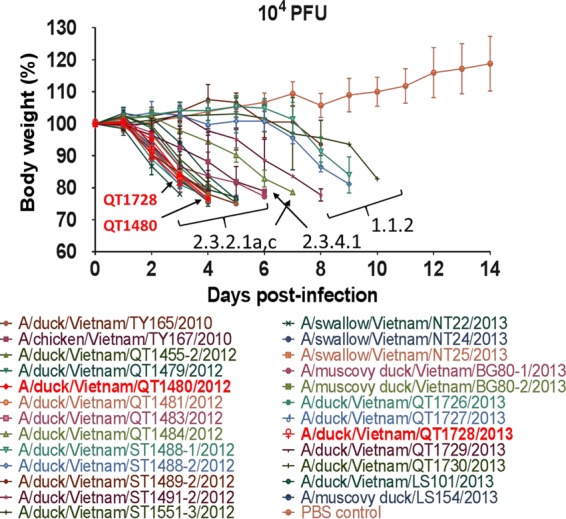
Virulence of H5N1 influenza viruses in BALB/c mice. Three mice per group were inoculated intranasally with 104 PFU of the indicated virus and observed daily for 14 days for body weight loss and lethality. The results shown are mean values ± standard deviations (error bars) from three individual mice.
We selected two viruses, A/duck/Vietnam/QT1480/2012 (QT1480) and A/duck/Vietnam/QT1728/2013 (QT1728), as representatives of the currently circulating clades 2.3.2.1a and 2.3.2.1c. QT1480 and QT1728 viruses generated by reverse genetics displayed 50% mouse lethal doses (MLD50s) of 100.7 and 101.5 PFU, respectively (Fig. 2A to D). We also intranasally inoculated six mice per group with 103 PFU/50 μl of QT1480 and QT1728 viruses. On days 3 and 6 postinfection, three mice per group were euthanized, and virus titers in different mouse organs were assessed by means of plaque assays in MDCK cells. Both viruses replicated to high titers in the lungs of infected animals and spread systemically, resulting in virus replication in the nasal turbinates, spleen, kidney, and brain on day 3 and/or 6 postinfection (Fig. 2E and F). Collectively, these results demonstrated the high virulence of QT1480 and QT1728 in mice.
FIG 2.
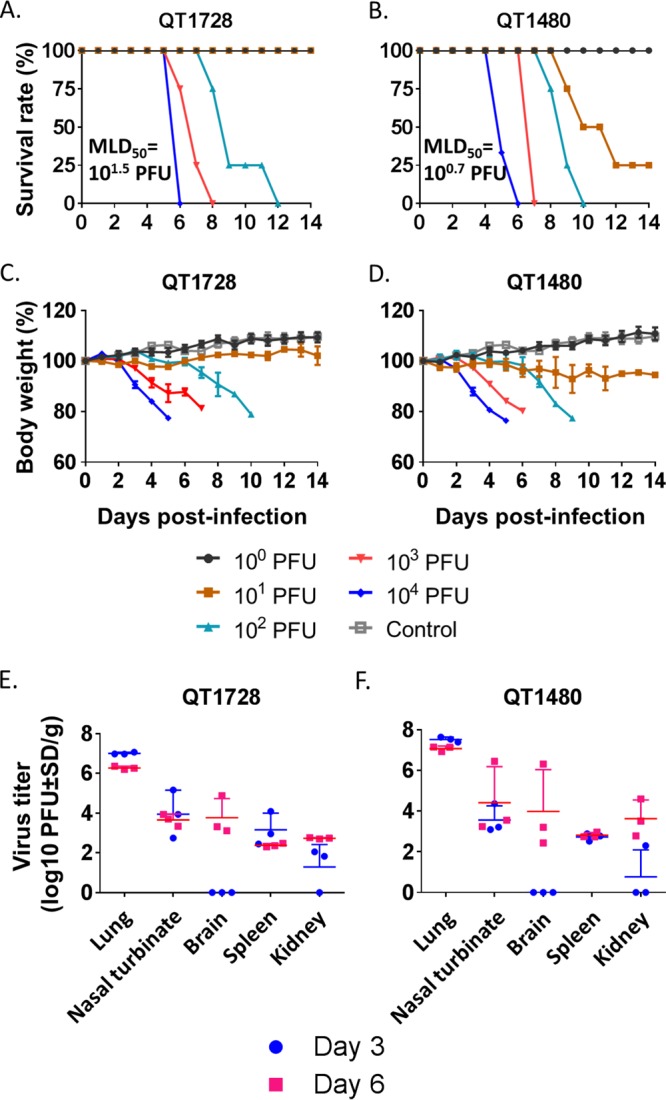
Virulence of QT1728 and QT1480 viruses in BALB/c mice. (A to D) Three or four mice per group were inoculated intranasally with the indicated doses of QT1728 or QT1480 virus and monitored for survival (A and B) and weight changes (C and D) for 14 days. The results shown are mean values ± standard deviations (error bars) from three or four individual mice. (E and F) To assess virus titers in mouse organs, six mice per group were inoculated intranasally with 103 PFU of virus. Three mice per group were euthanized on day 3 and day 6 postinfection, respectively, and lungs, nasal turbinates, brains, spleens, and kidneys were collected for virus titration in MDCK cells. Each symbol represents the value for an individual mouse. The data shown are mean values (long horizontal lines) plus standard deviations (SD) (short horizontal lines).
QT1480 and QT1728 viruses have different polymerase activity in mammalian cells.
Next, we tested the polymerase activity of the QT1480 and QT1728 viral polymerase complexes in minireplicon assays in mammalian cells at 37°C and 33°C (i.e., the temperatures of the lower and upper respiratory tracts of humans). Human 293T cells were transfected with plasmids expressing the components of the viral replication complex (i.e., PB2, PB1, PA, and nucleoprotein [NP]) and with a plasmid directing the transcription of a virus-like RNA that encodes the reporter protein luciferase. In addition, cells were transfected with an internal transfection control encoding Renilla luciferase. Twenty-four hours later, luciferase activity was measured as a surrogate for the activity of the viral polymerase complex. Although the QT1480 and QT1728 viruses caused similar virulence in mice, their polymerase activities were markedly different (Fig. 3A). At 37°C, the QT1728 polymerase activity was 9-fold higher than that of QT1480; at 33°C, this phenotype was even more pronounced with an 809-fold difference in polymerase activity between QT1480 and QT1728.
FIG 3.
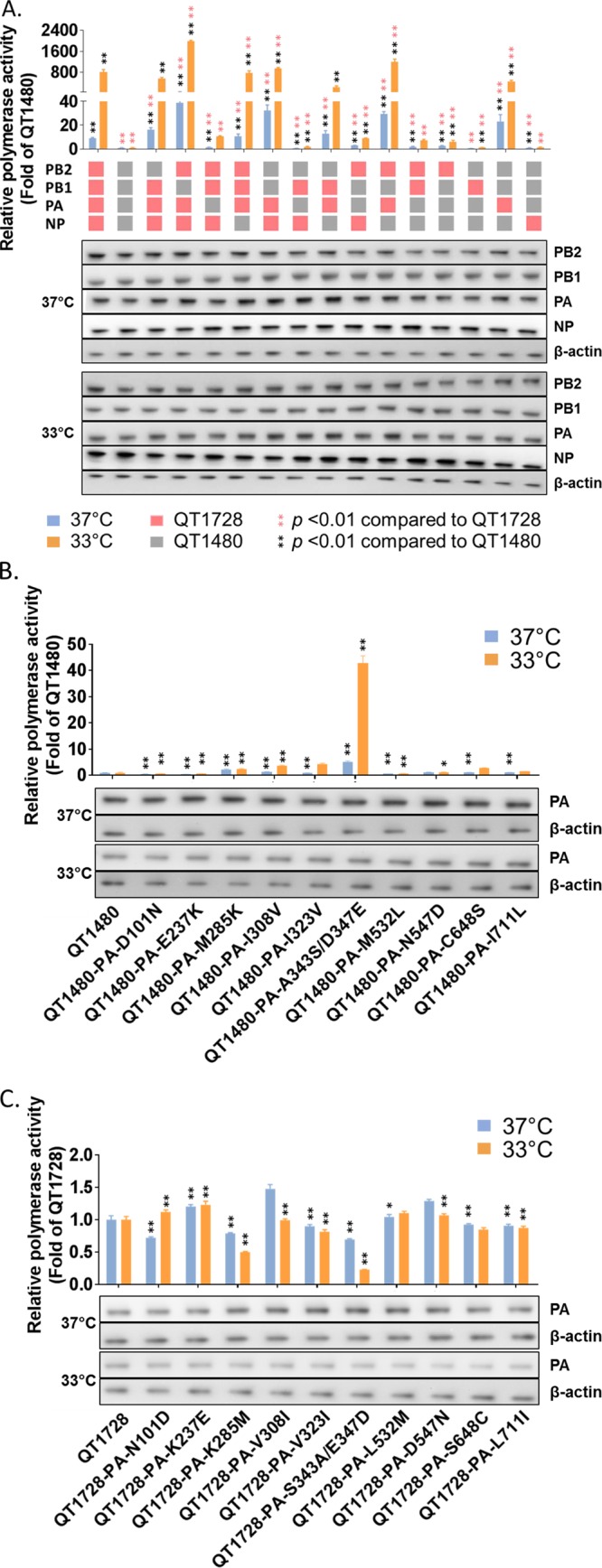
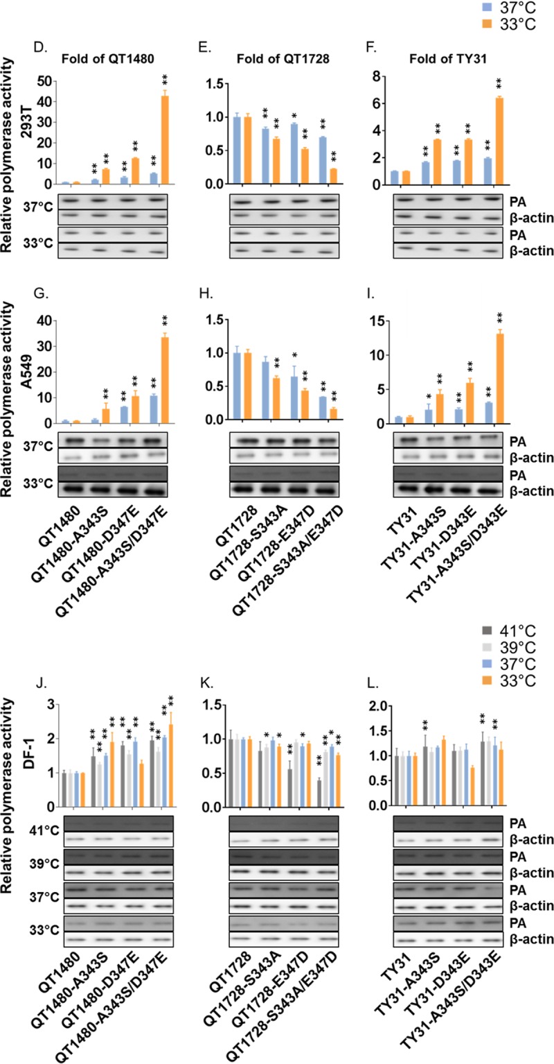
Identification of viral genes and amino acids that contribute to the difference in the polymerase activities of QT1728 and QT1480 viruses. (A) 293T cells were transfected with four protein expression plasmids for PB2, PB1, NP, and wild-type or mutant PA proteins, with a plasmid for the expression of a virus-like RNA encoding the firefly luciferase gene, and with a control plasmid encoding Renilla luciferase, and assayed after a 24-h incubation at 33°C and 37°C. (B and C) Minireplicon assays as described in panel A were carried out with the indicated mutant QT1480 (B) and QT1728 (C) PA proteins. (D to L) Minireplicon assays of QT1480, QT1728, and TY31 PA proteins possessing mutations at positions 343 and/or 347 in 293T (D to F), A549 (G to I), and DF-1 (J to L) cells. Data shown are mean values plus standard deviations for the results of three independent experiments. P values were calculated by one-way ANOVA, followed by Dunnett's test (*, P < 0.05; **, P < 0.01). In experiments carried out in parallel, cells were transfected as described above and processed for Western blot analysis.
The PA proteins of QT1480 and QT1728 viruses are critical for the differences in viral polymerase activity in minireplicons in mammalian cells.
To identify the component of the viral polymerase complex responsible for the difference in the polymerase activity between these two viruses, we tested minireplicons in which individual or multiple components of the replication complex were exchanged between QT1480 and QT1728 (Fig. 3A). Introduction of various (but not all) combinations of the QT1728 polymerase and NP proteins into the QT1480 viral replication complex increased the QT1480 polymerase activity significantly at 33°C and/or 37°C (Fig. 3A; see Table S1 in the supplemental material). The greatest increase in polymerase activity was detected upon introduction of the QT1728 PA protein into the QT1480 replication complex. Conversely, the QT1728 polymerase activity was reduced by the introduction of several (but not all) combinations of the QT1480 polymerase and/or NP proteins (Fig. 3A; Table S1). In general, viral minireplicons with the QT1728 PA protein displayed high polymerase activity, whereas those with the QT1480 PA protein showed low polymerase activity (Fig. 3A). These data demonstrate that the QT1480 and QT1728 PA proteins are primarily responsible for the low or high polymerase activity of the respective viral replication complexes.
Two amino acid residues in PA are critical for the differences in the polymerase activities of QT1480 and QT1728 viruses.
The PA proteins of QT1480 and QT1728 differ by 11 amino acids (Table 1). To identify the amino acid residues in the QT1480 and QT1728 PA proteins responsible for the difference in polymerase activity, we tested mutant QT1428 PA proteins in which single amino acids were replaced with the amino acid residue encoded by QT1480 at the respective position and vice versa; due to their close proximity, the residues at positions 343 and 347 were tested together. Several amino acid changes significantly increased the viral polymerase activity of QT1480 in minireplicon assays in 293T cells (Fig. 3B and C; Table S1); among them, the introduction of the PA-A343S/D347E (PA with the A-to-S change at position 343 and the D-to-E change at position 347) mutations into the QT1480 polymerase complex had the greatest effect, increasing the viral polymerase activity by 5-fold at 37°C and 43-fold at 33°C, respectively (Fig. 3B). Conversely, the PA-S343A/E347D mutations caused the greatest reductions in the polymerase activity of the QT1728 replication complex (Fig. 3C; Table S1).
TABLE 1.
Amino acid differences between the PA proteins of QT1728 and QT1480 viruses
| Virus | Clade | Positions at which the amino acids differ between the PA proteins of QT1728 and QT1480a |
||||||||||
|---|---|---|---|---|---|---|---|---|---|---|---|---|
| 101 | 237 | 285 | 308 | 323 | 343 | 347 | 532 | 547 | 648 | 711 | ||
| QT1728 | 2.3.2.1c | N | K | K | V | V | S | E | L | D | S | L |
| QT1480 | 2.3.2.1a | D | E | M | I | I | A | D | M | N | C | I |
| TY31 | 2.3.2 | E | E | M | I | V | A | D | L | D | N | L |
TY31 is listed for reference purposes.
We then tested the PA-A343S and PA-D347E mutations individually. Both mutations increased the QT1480 polymerase activity in 293T cells significantly when tested individually and simultaneously (Fig. 3D; Table S1). Introduction of the reciprocal mutations (PA-S343A and PA-E347D) into the QT1728 polymerase resulted in small, but statistically significant, reductions in polymerase activity (Fig. 3E; Table S1). Similar data were obtained in human lung adenocarcinoma (A549) cells, although the expression levels of PA were low at 33°C, and the effect of the amino acid at position 343 was not statistically significant at 37°C (Fig. 3G and H; Table S1).
To determine whether the effects of the PA-343A/S and PA-347D/E mutations were specific to mammalian cells, we also performed minireplicon assays in chicken DF-1 cells at 33°C and 37°C and at 39°C and 41°C. The latter two temperatures were chosen because the body temperature of birds ranges from 38 to 42°C (the body temperature of an active adult chicken is ∼41°C, whereas that of a resting adult or young chicken is slightly lower); at 39°C and 41°C, the expression levels of PA were low (Fig. 3J to L). Largely, the introduction of PA-A343S/D347E into the replication complex of QT1480 increased the QT1480 polymerase activity, whereas the introduction of PA-S343A/E347D into the replication complex of QT1728 reduced its polymerase activity, although not all comparisons were statistically significant (Fig. 3J and K; Table S1).
Together, these data demonstrate that the amino acids at positions 343 and 347 of PA affect the polymerase activity of QT1480 and QT1728 in minireplicon assays in cultured cells and that this effect is stronger in mammalian cells than in a chicken cell line.
The amino acids at positions 343 and 347 of PA affect H5N1 virulence in mice.
To determine the significance of PA-343A/S and PA-347D/E to the virulence of QT1428 and QT1728 in mice, mutant viruses possessing the respective amino acid changes should be generated and tested in mice. However, the introduction of the PA-A343S/D347E mutations into QT1480 was considered by the U.S. National Institutes of Health (U.S. NIH) to be a potential “gain-of-function” experiment and was not approved. The introduction of PA-S343A/E347D mutations into QT1728 was, however, approved because it was expected to result in a “loss-of-function.”
To test the effects of the PA-A343S/D347E mutations on H5N1 virulence, we used a surrogate H5N1 influenza virus that has low pathogenicity in mice (A/chicken/Vietnam/TY31/2005 [TY31]; clade 2.3.2) (34). This virus encodes multiple basic amino acids at its HA cleavage site but does not cause lethal infection when inoculated into mice at 104 or 105 PFU (34); TY31 possesses the QT1480-like residues PA-343A and PA-347D (Table 1). Due to its low virulence in mice, the U.S. NIH and the University of Wisconsin Institutional Biosafety Committee granted us permission to generate and characterize TY31 virus with the PA-A343S and PA-D347E mutations.
First, we tested whether the PA-A343S/D347E mutations affected the polymerase activity of the TY31 replication complex in minireplicon assays in 293T (Fig. 3F; Table S1), A549 (Fig. 3I; Table S1), and DF-1 (Fig. 3L; Table S1) cells. In mammalian cells, TY31 minireplicons with the PA-A343S and/or PA-D347E mutations replicated more efficiently than wild-type TY31 minireplicons did, although the increase in replication efficiency was less pronounced than the increase detected with the QT1480 polymerase complex. In chicken DF-1 cells, the introduction of the PA-A343S and/or PA-D347E mutations did not consistently increase the polymerase activity of the TY31 polymerase complex.
Next, mice were inoculated with wild-type QT1728 and TY31 viruses or with single and double mutants possessing the respective mutations at positions 343 and/or 347 of PA. Body weight loss and lethality were monitored for 14 days. The amino acid changes PA-S343A and/or PA-E347D mildly to moderately attenuated QT1728 virus (Fig. 4), with MLD50 values of 101.5 PFU for QT1728, 102.3 PFU for QT1728-PA-S343A, 101.8 PFU for QT1728-E347D, and 102.5 PFU for QT1728-PA-S343A/E347D. The introduction of PA-D347E into TY31 did not affect mouse virulence (Fig. 5). In contrast, TY31 virus encoding PA-A343S caused body weight loss and lethality in mice infected with high doses of this virus, and the MLD50 value of TY31-PA-A343S (103.8 PFU) was considerably higher than that of TY31 (MLD50 > 105.5 PFU). TY31 virus possessing the PA-A343S/D347E double mutation was even more virulent in mice (MLD50 of 102.8 PFU; Fig. 5).
FIG 4.
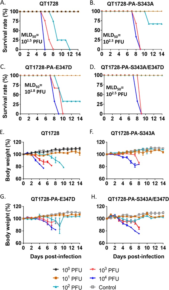
Effects of mutations in PA at positions 343 and 347 on virulence of QT1728 virus in mice. (A to H) Three mice per group were inoculated intranasally with the indicated dose of viruses and monitored for survival (A to D) and weight changes (E to H) for 14 days. The body weight loss data shown in panel A are identical to those in Fig. 2A; they are presented again for a better comparison of wild-type and mutant viruses; likewise, the body weight loss data presented in panel E are identical to those in Fig. 2C.
FIG 5.

Effects of PA mutations at positions 343 and 347 on the virulence of TY31 in mice. (A to H) Three mice per group were inoculated intranasally with the indicated dose of viruses and monitored for survival (A to D) and weight changes (E to H) for 14 days.
Infection of mice with 103 PFU of QT1728 caused systemic infection, with virus replication in the respiratory tract, spleen, kidney, and brain (Fig. 6A), whereas the viruses bearing single or double mutations in PA were restricted to the lungs of infected animals (with the exception of QT1728-S343A/E347D, which was isolated from the nasal turbinates of one infected mouse) (Fig. 6B to D). After infection of mice with 103 PFU of TY31, no virus was detected in any of the organs tested on day 3 or 6 after inoculation (Fig. 6E), consistent with the known low virulence of this virus. The TY31-PA-D347E mutant was detected in the nasal turbinates of one mouse, but not in any of the other organs tested (Fig. 6G). The PA-A343S mutation in the TY31 genetic background resulted in moderate virus titers in the lungs of most infected animals, and virus was isolated from the nasal turbinates of one mouse (Fig. 6F). TY31 viruses encoding both mutations in PA (i.e., PA-A343S/D347E) replicated efficiently in the lungs of infected animals, and virus was isolated from the nasal turbinates of one animal (Fig. 6H). Thus, the PA-A343S and PA-D347E mutations individually increased the mouse virulence of an H5 virus, although the effect of the PA-A343S mutation was greater than that of the PA-D347E mutation. The combination of these mutations resulted in a further increase in the virulence and pathogenicity of TY31 virus.
FIG 6.
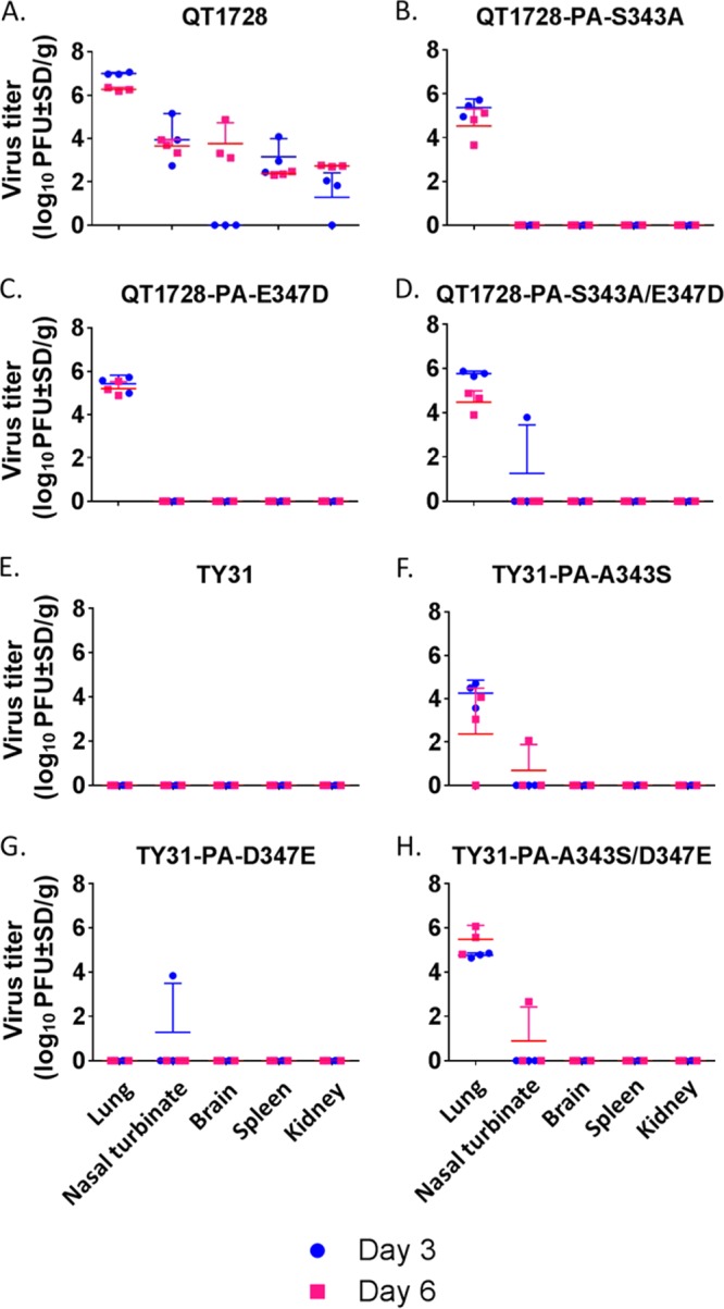
Effects of PA mutations at positions 343 and 347 on virus titers in the organs of mice infected with QT1728 or TY31 virus. (A to H) Six mice per group were inoculated intranasally with 103 PFU of wild-type or mutant QT1728 (A to D) or TY31 (E to H) viruses. Three mice per group were euthanized on day 3 and day 6 postinfection, and lungs, nasal turbinates, brains, spleens, and kidneys were collected for virus titration in MDCK cells. Each symbol represents the value for an individual mouse. Weight change and virus titer data are presented as mean values with standard deviations. The virus titer data shown in panel A are identical to those in Fig. 2E; they are presented again to aid in the comparison of the wild-type and mutant viruses.
Prevalence of PA-343A/S and PA-347D/E among influenza viruses.
Our data indicated that the PA-A343S and PA-D347E mutations increase the virulence of H5N1 influenza viruses in mice. Therefore, we assessed the prevalence of these amino acid residues among influenza A viruses. At position 347, PA-347D (i.e., the “low-virulence type”) is highly conserved among influenza A viruses (Table 2). At position 343, human H3N2 viruses encode primarily PA-343S (i.e., the “high-virulence type”), whereas human H2N2, human seasonal H1N1, and currently circulating human H1N1 viruses (which possess a PA gene of North American avian influenza virus origin) predominantly encode PA-343A (Table 2). Most avian influenza viruses encode PA-343A, but the prevalence of PA-343S has increased noticeably among H5, H7, H9, and H10 influenza viruses isolated since 2011, 2014, 2013, and 2013, respectively (Table 2). Further inspection of H5 influenza viruses revealed PA-343S among viruses of clades 1.1.2, 2.3.2.1a, 2.3.2.1b, and 2.3.2.1c (Table 3), all of which have been isolated in Vietnam. In contrast, PA-343S is not highly prevalent among viruses of clade 2.3.4.4 (Table 3), which have undergone multiple reassortment events to form viruses of the H5N2, H5N3, H5N5, H5N6, and H5N8 subtypes, some of which have spread to North America and Europe.
TABLE 2.
Prevalence of PA-343A/S and PA-347D/E
| Host and subtype | No. of isolates analyzed | Prevalence (%) of the indicated amino acid at the following position in PA protein: |
|||
|---|---|---|---|---|---|
| 343 |
347 |
||||
| A | S | D | E | ||
| Human isolates | |||||
| Seasonal H1N1 | 1,650 | 20.1a | 0.2 | 99.5 | 0.06 |
| H1N1pdm | 7,578 | 93.2 | 0.05 | 99.9 | 0.01 |
| H2N2 | 105 | 100 | 0 | 100 | 0 |
| H2N2 | 9,714 | 7.4 | 92.6 | 99.9 | 0.04 |
| Swine isolates | |||||
| H1 | 3,293 | 95.1 | 0.9 | 95.7 | 3.6 |
| H3 | 1,577 | 94.0 | 2.3 | 94.9 | 3.2 |
| Other | 88 | 94.3 | 1.1 | 100 | 0 |
| Equine isolates | |||||
| H3N8 | 128 | 60.1b | 0 | 99.2 | 0 |
| H7N7 | 12 | 58.3 | 41.7 | 100 | 0 |
| Avian isolates | |||||
| H1 | 822 | 99.4 | 0 | 100 | 0 |
| H2 | 527 | 99.8 | 0 | 100 | 0 |
| H3 | 2,140 | 99.2 | 0.8 | 99.95 | 0 |
| H4 | 1,962 | 98.1 | 0.3 | 99.8 | 0 |
| H5c | 3,972 | 89.0 | 8.4d | 99.7 | 0.2 |
| H6 | 1,622 | 94.3 | 0.7 | 99.9 | 0.06 |
| H7e | 2,165 | 96.9 | 2.9f | 99.9 | 0 |
| H8 | 170 | 100 | 0 | 100 | 0 |
| H9 | 2,164 | 89.7 | 9.1g | 99.8 | 0.05 |
| H10 | 1,099 | 87.2 | 12.6h | 99.8 | 0.09 |
| H11 | 653 | 99.4 | 0.3 | 100 | 0 |
| H12 | 231 | 87.0 | 1.3 | 100 | 0 |
| H13 | 395 | 94.7 | 5.3 | 100 | 0 |
| H14 | 32 | 100 | 0 | 100 | 0 |
| H15 | 17 | 100 | 0 | 100 | 0 |
| H16 | 229 | 100 | 0 | 100 | 0 |
| Avian and human H5 | 3,972 | 89.0 | 8.4d | 99.7 | 0.2 |
| Avian H5 | 3,717 | 89.0 | 8.6i | 99.7 | 0.2 |
| Human H5 | 255 | 88.6 | 5.5j | 100 | 0 |
| Avian and human H7N9 | 616 | 90.3 | 9.3 | 99.8 | 0 |
| Avian H7N9 | 518 | 89.8 | 10.0 | 99.8 | 0 |
| Human H7N9 | 98 | 92.9 | 5.1 | 100 | 0 |
79.6% of seasonal human H1N1 viruses encode E at this position.
39.8% of equine H3N8 viruses encode E at this position.
Avian and human isolates.
Until 2010, 0.04%; since 2011, 21.5% (mostly from Vietnam and Cambodia).
Avian H7 viruses only; for human H7N9 viruses, see below.
Until 2013, 0.2%; since 2014, 10.4% (almost all are H7N9 viruses).
Until 2012, 1.5%; since 2013, 26.7% (mostly H9N2 viruses from China).
Until 2012, 0.3%; since 2013, 40.5%.
Until 2010, 0.04%; since 2011, 23.1%.
Until 2010, 0%; since 2011, 43.8% (all are H5N1 viruses obtained from Cambodia in 2013).
TABLE 3.
Prevalence of PA-343A/S and PA-347D/E in selected subclades of H5 viruses
| Clade | No. of sequences | Amino acid(s) (no. of isolates with the respective amino acid) at the following position in PA protein: |
|
|---|---|---|---|
| 343 | 347 | ||
| 1.1 | 60 | A (60) | D (60) |
| 1.1.1 | 10 | A (10) | D (10) |
| 1.1.2 | 90 | A (57), S (33) | D (90) |
| 2.1.3.2 | 87 | A (87) | D (87) |
| 2.2.1 | 48 | A (48) | D (48) |
| 2.2.1.1 | 28 | A (28) | D (28) |
| 2.2.1.2 | 40 | A (40) | D (40) |
| 2.3.2.1 | 40 | A (40) | D (40) |
| 2.3.2.1a | 217 | A (181), S (36) | D (216), N (1) |
| 2.3.2.1b | 28 | A (21), S (7) | D (28) |
| 2.3.2.1c | 407 | A (136), S (260), T (11) | D (398), N (2), E (7) |
| 2.3.4.4 | 524 | A (515), E (1), S (5), V (3) | D (523), N (1) |
DISCUSSION
Here, we report that the PA-A343S/D347E mutations increase the polymerase activity and virulence of an avian H5N1 influenza virus in mice. Both mutations contribute to polymerase activity and virulence individually, with the PA-A343S mutation having a greater effect than the PA-D347E mutation. However, the greatest effect on virulence was conferred by the double mutant, suggesting a functional relationship between the amino acids at positions 343 and 347 of PA.
The amino acids at positions 343 and 347 of PA had been reported to affect influenza virus replication (34, 41–44), but these studies did not assess the particular amino acid changes tested here and did not evaluate the combined effect of PA-343/347 on influenza virus replication and pathogenicity. Previously, we reported that the PA gene of a human H5N1 influenza virus (A/Vietnam/UT36285/2010) increased the polymerase activity and mouse virulence of an avian H5N1 influenza virus (A/chicken/Vietnam/TY31/2005) (34). Further studies identified five amino acid residues in PA, among them PA-343T, that contributed to the increased replication and virulence of the reassortant virus (34). Comparative testing of human pandemic H1N1 viruses revealed an isolate (A/Sichuan/1/2009) that replicated more efficiently in epithelial cells and caused higher mouse virulence than other human pandemic H1N1 viruses tested (41). A/Sichuan/1/2009 possessed five unique amino acid changes in four viral proteins, including PA-A343T, but the authors did not test the individual contributions of these amino acid changes to the polymerase activity and mouse virulence of A/Sichuan/1/2009 (41). In another study, serial passage of an H5N6 influenza virus in mice resulted in a more virulent variant that carried a PA-A343T mutation together with other amino acid changes in four other viral proteins; however, the significance of these amino acid changes for increased mouse virulence was not tested (42).
Treanor et al. (43) reported that a PA-D347N mutation suppressed the temperature sensitivity (ts) and attenuation (att) phenotypes of a reassortant virus possessing the PB2 gene of the cold-adapted A/Ann Arbor/1/60 virus in the background of human A/Korea/82 (H3N2) virus, suggesting a functional relationship between PA-347N and PB2-265S (which confer the ts and att phenotypes). In another study, a PA-D347N mutation and several other mutations were detected after serial passages of an H9N2 influenza virus in mice, but these amino acid changes were not tested individually (44). Nevertheless, collectively, these studies demonstrate a role for the amino acids at positions 343 and 347 of PA in influenza virus replication and virulence.
Our analysis of influenza virus sequences revealed that PA-347E is very rare among influenza A viruses (Table 2). PA-343S is commonly found in human H3N2 influenza viruses, but not human H2N2, human seasonal H1N1, or human pandemic 2009 H1N1 influenza viruses (in which the PA gene is derived from a North American avian influenza virus). Among avian influenza viruses, PA-343S was not commonly found until 2010, but its prevalence has increased appreciably in certain subtypes in recent years (Table 2). These subtypes include H5, H7, and H9, which occasionally infect humans and play an important role in influenza virus ecology because viruses of these subtypes are enzootic in poultry populations where they have the opportunity to reassort frequently. A detailed assessment of major H5 virus subclades revealed PA-343S in viruses of clades 1.1.2, 2.3.2.1a, 2.3.2.1b, and 2.3.2.1c (Table 3), which circulate in Vietnam and have undergone reassortment; this may explain the broader prevalence of PA-343S in H5 viruses in Vietnam. In fact, viruses of clade 2.3.2.1c (the currently dominant subclade of H5 viruses in Vietnam) encode predominantly PA-343S.
Currently, the mechanisms through which the amino acids at positions 343 and 347 of PA affect virus replication and pathogenicity are not understood. These positions are located in the so-called PA-C domain of the polymerase complex (Fig. 7) (45) but are not part of the two phosphoserine-5 binding sites (45, 46), which are essential for the interaction of PA with the cellular RNA polymerase II C-terminal domain. The amino acids at positions 343 and 347 of PA are located at the edge of an α-helix and are exposed to the surface; interestingly, amino acid 336 of PA, which affects the viral polymerase activity and pathogenicity of recombinant influenza viruses (28), is located on the same α-helix as residues 343 and 347. Therefore, we speculate that these residues interact with host factors and that it is the intensity of these interactions that affects the viral polymerase activity.
FIG 7.
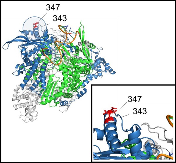
Localization of amino acids PA-343 and PA-347 on the three-dimensional (3D) structure of influenza A polymerase (PDB 4WSB). The PB2, PB1, and PA subunits are shown in gray, green, and blue, respectively.
In conclusion, we identified two novel virulence markers in the influenza H5N1 virus PA protein, (i.e., PA-343S and PA-347E) that together significantly increase influenza virus polymerase activity and virulence in mice. The detection of these amino acids in H5 influenza viruses may signal an increased risk for severe infection in mammals.
MATERIALS AND METHODS
Cells.
All cell lines used in this study were purchased from the American Type Culture Collection (ATCC). Madin-Darby canine kidney (MDCK) cells were maintained in minimal essential medium (MEM) containing 5% newborn calf serum, essential amino acids, vitamins, and antibiotics. Human embryonic kidney (293T) cells, human lung adenocarcinoma (A549) cells, and chicken fibroblast (DF-1) cells were maintained in Dulbecco's modified Eagle's medium with 10% fetal bovine serum and antibiotics. All cells were incubated at 37°C with 5% CO2, unless otherwise noted.
Viruses.
A/duck/Vietnam/QT1480/2012 (QT1480) (H5N1) and A/duck/Vietnam/QT1728/2013 (QT1728) (H5N1) viruses were isolated from avian species in Vietnam and regenerated by reverse genetics as described by Neumann et al. (47). A/chicken/Vietnam/TY31/2005 (TY31) (H5N1) has been described previously (34). Mutant variants of QT1480, QT1728, and TY31 viruses were generated by using reverse genetics. All viruses were amplified in 9- to 11-day-old embryonated chicken eggs and sequenced before their experimental characterization.
Mouse experiments.
Six-week-old female BALB/c mice (Jackson Laboratory, Bar Harbor, ME) were anesthetized with isofluorane and inoculated intranasally with the indicated doses of H5N1 viruses in a volume of 50 μl. Body weight and mortality were monitored daily for 2 weeks. Infected mice that lost more than 25% of their initial weight were euthanized humanely. The dose lethal to 50% of infected mice (MLD50) was calculated by the method of Reed and Muench.
To assess virus replication in the organs of infected mice, groups of six animals were anesthetized and infected intranasally with the indicated dose of virus. On days 3 and 6 postinfection, three mice per group were euthanized humanely, and organs (lungs, brains, nasal turbinates, spleens, and kidneys) were collected for virus titration in MDCK cells. The animal experiments were reviewed and approved by the Institutional Animal Care and Use Committee at the University of Wisconsin—Madison before the start of the experiments.
Minireplicon assay.
To determine the relative polymerase activities of H5N1 viruses, the PB1, PB2, NP, and wild-type and mutant PA genes were inserted into the pCAGGS/BsmBI protein expression vector. All constructs were sequenced to ensure the absence of unwanted mutations.
Human 293T and A549 cells and avian DF-1 cells were transfected with four protein expression plasmids for PB2, PB1, NP, and wild-type or mutant PA proteins, with a plasmid for the expression of a virus-like RNA encoding the firefly luciferase gene under the control of the human (for 293T and A549 cells) or avian (for DF-1 cells) RNA polymerase I promoter and with a control plasmid encoding Renilla luciferase by using TransIT-LT1 (Mirus, Madison, WI, USA) for 293T cells, xfect (Clontech, Mountain View, CA, USA) for A549 cells, or Lipofectamine 3000 (Thermo Fisher, Waltham, MA, USA) for DF-1 cells. Cells were incubated for 24 h at 33°C and 37°C for 293T cells, for 48 h at 33°C and 37°C for A549 cells and for 48 h at 33°C, 37°C, 39°C, and 41°C for DF-1 cells. The cells were then lysed, and the relative luciferase activity was measured by using a dual-luciferase reporter assay kit (Promega, Madison, WI, USA). Data shown are the mean values with standard deviations for the results of three independent experiments.
Western blot assay.
Cells were lysed with passive lysis buffer (Promega, Madison, WI, USA). Total protein was quantified by using the BCA assay (Thermo Fisher, Waltham, MA, USA), mixed with Laemmli sample buffer (Bio-Rad, Hercules, CA, USA), and heated at 95°C for 5 min. Samples were run on NuPAG 4%–12% Bis-Tris protein gels (Thermo Fisher, Waltham, MA, USA), and proteins were transferred onto iBlot 2 transfer stacks polyvinylidene difluoride (PVDF) membrane by using the iBlot 2 dry transfer system (Thermo Fisher, Waltham, MA, USA). The membranes were blocked with 5% nonfat milk powder (Bio-Rad, Hercules, CA, USA) in Tris-buffered saline containing 0.1% Tween 20 (TBS-T) for 1 h at room temperature. For primary antibodies, we used mouse anti-PB2 monoclonal antibody 18/1 (48), mouse anti-PB1 monoclonal antibody 136/1, mouse anti-PA monoclonal antibody 65/4 (49) (all of which were gifts from H. Kida, Hokkaido University, Sapporo, Japan), rabbit anti-NP antibody (catalog no. PA5-32242; Thermo Fisher, Waltham, MA, USA), and mouse anti-β-actin antibody (ab8224; Abcam, Cambridge, MA, USA). The primary antibodies were incubated overnight at 4°C, followed by incubation with the horseradish peroxidase (HRP)-conjugated secondary antibodies (i.e., HRP-conjugated goat anti-mouse [catalog no. G-21040] and anti-rabbit [catalog no. G-21234] [both catalog numbers for Thermo Fisher, Waltham, MA, USA]. Reactions were visualized with SuperSignal West Dura extended-duration substrate (Thermo Fisher, Waltham, MA, USA). Western blot images were acquired by using the FluorChem HD2 system (ProteinSimple, San Jose, CA, USA).
Statistical analysis.
The minireplicon data were analyzed by using the R statistical package (www.r-project.org) and the Multcomp package (50). We performed the one-way analysis of variance (ANOVA) test, followed by Dunnett's posthoc test to compare each mutant group to the wild-type viruses. P values less than 0.05 were considered significant (*, P < 0.05; **, P < 0.01).
Biosafety.
This study was assessed and approved by the U.S. NIH and the University of Wisconsin Institutional Biosafety Committee prior to the start of the experiments. All experiments with infectious H5N1 influenza viruses were performed in a biosafety level 3 (BSL3) containment laboratory.
Accession number(s).
The sequences of these viruses have been deposited in GenBank under the following accession numbers: KX513118, KX513212, KX513283, KX513287, KX513316, KX513336, KX513361, and KX644102 for QT1728 and KX513120, KX513186, KX513203, KX513215, KX513235, KX513262, KX513308, and KX644123 for QT1480.
Supplementary Material
ACKNOWLEDGMENTS
We thank Susan Watson for scientific editing and H. Kida (Hokkaido University, Sapporo, Japan) for mouse anti-PB2 monoclonal antibody 18/1 (48), mouse anti-PB1 monoclonal antibody 136/1, and mouse anti-PA monoclonal antibody 65/4 (49). We thank the CRIP sequencing core at the Icahn School of Medicine at Mt. Sinai (New York, NY) for sequencing and assembly of the viral genomes of QT1480 and QT1728.
This work was supported by the NIAID-funded Center for Research on Influenza Pathogenesis (CRIP) (HHSN272201400008C), by the Japan Initiative for Global Research Network on Infectious Diseases (J-GRID) from the Ministry of Education, Culture, Science, Sports, and Technology (MEXT) of Japan, and by the Leading Advanced Projects for medical innovation (LEAP) from the Japan Agency for Medical Research and Development (AMED). Research reported in this study was supported by the Office of Research Infrastructure of the National Institutes of Health under award number S10OD018522.
The content is solely the responsibility of the authors and does not necessarily represent the official views of the National Institutes of Health.
Footnotes
Supplemental material for this article may be found at https://doi.org/10.1128/JVI.01557-17.
REFERENCES
- 1.Nguyen DC, Uyeki TM, Jadhao S, Maines T, Shaw M, Matsuoka Y, Smith C, Rowe T, Lu X, Hall H, Xu X, Balish A, Klimov A, Tumpey TM, Swayne DE, Huynh LP, Nghiem HK, Nguyen HH, Hoang LT, Cox NJ, Katz JM. 2005. Isolation and characterization of avian influenza viruses, including highly pathogenic H5N1, from poultry in live bird markets in Hanoi, Vietnam, in 2001. J Virol 79:4201–4212. doi: 10.1128/JVI.79.7.4201-4212.2005. [DOI] [PMC free article] [PubMed] [Google Scholar]
- 2.Cuong NV, Truc VNT, Nhung NT, Thanh TT, Chieu TTB, Hieu TQ, Men NT, Mai HH, Chi HT, Boni MF, van Doorn HR, Thwaites GE, Carrique-Mas JJ, Hoa NT. 2016. Highly pathogenic avian influenza virus A/H5N1 infection in vaccinated meat duck flocks in the Mekong Delta of Vietnam. Transbound Emerg Dis 63:127–135. doi: 10.1111/tbed.12470. [DOI] [PMC free article] [PubMed] [Google Scholar]
- 3.Chu DH, Okamatsu M, Matsuno K, Hiono T, Ogasawara K, Nguyen LT, Nguyen LV, Nguyen TN, Nguyen TT, Pham DV, Nguyen DH, Nguyen TD, To TL, Nguyen HV, Kida H, Sakoda Y. 2016. Genetic and antigenic characterization of H5, H6 and H9 avian influenza viruses circulating in live bird markets with intervention in the center part of Vietnam. Vet Microbiol 192:194–203. doi: 10.1016/j.vetmic.2016.07.016. [DOI] [PubMed] [Google Scholar]
- 4.Lee EK, Kang HM, Kim KI, Choi JG, To TL, Nguyen TD, Song BM, Jeong J, Choi KS, Kim JY, Lee HS, Lee YJ, Kim JH. 2015. Genetic evolution of H5 highly pathogenic avian influenza virus in domestic poultry in Vietnam between 2011 and 2013. Poult Sci 94:650–661. doi: 10.3382/ps/pev036. [DOI] [PubMed] [Google Scholar]
- 5.Tung DH, Van Quyen D, Nguyen T, Xuan HT, Nam TN, Duy KD. 2013. Molecular characterization of a H5N1 highly pathogenic avian influenza virus clade 2.3.2.1b circulating in Vietnam in 2011. Vet Microbiol 165:341–348. doi: 10.1016/j.vetmic.2013.04.021. [DOI] [PubMed] [Google Scholar]
- 6.Creanga A, Nguyen DT, Gerloff N, Do HT, Balish A, Nguyen HD, Jang Y, Dam VT, Thor S, Jones J, Simpson N, Shu B, Emery S, Berman L, Nguyen HT, Bryant JE, Lindstrom S, Klimov A, Donis RO, Davis CT, Nguyen T. 2013. Emergence of multiple clade 2.3.2.1 influenza A (H5N1) virus subgroups in Vietnam and detection of novel reassortants. Virology 444:12–20. doi: 10.1016/j.virol.2013.06.005. [DOI] [PubMed] [Google Scholar]
- 7.Davis CT, Balish AL, O'Neill E, Nguyen CV, Cox NJ, Xu XY, Klimov A, Nguyen T, Donis RO. 2010. Detection and characterization of clade 7 high pathogenicity avian influenza H5N1 viruses in chickens seized at ports of entry and live poultry markets in Vietnam. Avian Dis 54:307–312. doi: 10.1637/8801-040109-ResNote.1. [DOI] [PubMed] [Google Scholar]
- 8.Wan XF, Nguyen T, Davis CT, Smith CB, Zhao ZM, Carrel M, Inui K, Do HT, Mai DT, Jadhao S, Balish A, Shu B, Luo F, Emch M, Matsuoka Y, Lindstrom SE, Cox NJ, Nguyen CV, Klimov A, Donis RO. 2008. Evolution of highly pathogenic H5N1 avian influenza viruses in Vietnam between 2001 and 2007. PLoS One 3:e3462. doi: 10.1371/journal.pone.0003462. [DOI] [PMC free article] [PubMed] [Google Scholar]
- 9.Nguyen TD, Nguyen V, Vijaykrishna D, Webster RG, Guan Y, Peiris JSM, Smith GJD. 2008. Multiple sublineages of influenza A virus (H5N1), Vietnam, 2005-2007. Emerg Infect Dis 14:632–636. doi: 10.3201/eid1404.071343. [DOI] [PMC free article] [PubMed] [Google Scholar]
- 10.Muramoto Y, Le TQM, Phuong LS, Nguyen T, Nguyen TH, Sakai-Tagawa Y, Horimoto T, Kida H, Kawaoka Y. 2006. Pathogenicity of H5N1 influenza A viruses isolated in Vietnam between late 2003 and 2005. J Vet Med Sci 68:735–737. doi: 10.1292/jvms.68.735. [DOI] [PubMed] [Google Scholar]
- 11.Gabriel G, Dauber B, Wolff T, Planz O, Klenk HD, Stech J. 2005. The viral polymerase mediates adaptation of an avian influenza virus to a mammalian host. Proc Natl Acad Sci U S A 102:18590–18595. doi: 10.1073/pnas.0507415102. [DOI] [PMC free article] [PubMed] [Google Scholar]
- 12.Li Z, Chen H, Jiao P, Deng G, Tian G, Li Y, Hoffmann E, Webster RG, Matsuoka Y, Yu K. 2005. Molecular basis of replication of duck H5N1 influenza viruses in a mammalian mouse model. J Virol 79:12058–12064. doi: 10.1128/JVI.79.18.12058-12064.2005. [DOI] [PMC free article] [PubMed] [Google Scholar]
- 13.Mehle A, Doudna JA. 2009. Adaptive strategies of the influenza virus polymerase for replication in humans. Proc Natl Acad Sci U S A 106:21312–21316. doi: 10.1073/pnas.0911915106. [DOI] [PMC free article] [PubMed] [Google Scholar]
- 14.Neumann G, Kawaoka Y. 2006. Host range restriction and pathogenicity in the context of influenza pandemic. Emerg Infect Dis 12:881–886. doi: 10.3201/eid1206.051336. [DOI] [PMC free article] [PubMed] [Google Scholar]
- 15.Yamada S, Hatta M, Staker BL, Watanabe S, Imai M, Shinya K, Sakai-Tagawa Y, Ito M, Ozawa M, Watanabe T, Sakabe S, Li C, Kim JH, Myler PJ, Phan I, Raymond A, Smith E, Stacy R, Nidom CA, Lank SM, Wiseman RW, Bimber BN, O'Connor DH, Neumann G, Stewart LJ, Kawaoka Y. 2010. Biological and structural characterization of a host-adapting amino acid in influenza virus. PLoS Pathog 6:e1001034. doi: 10.1371/journal.ppat.1001034. [DOI] [PMC free article] [PubMed] [Google Scholar]
- 16.Kawaoka Y, Krauss S, Webster RG. 1989. Avian-to-human transmission of the PB1 gene of influenza A viruses in the 1957 and 1968 pandemics. J Virol 63:4603–4608. [DOI] [PMC free article] [PubMed] [Google Scholar]
- 17.Naffakh N, Tomoiu A, Rameix-Welti MA, van der Werf S. 2008. Host restriction of avian influenza viruses at the level of the ribonucleoproteins. Annu Rev Microbiol 62:403–424. doi: 10.1146/annurev.micro.62.081307.162746. [DOI] [PubMed] [Google Scholar]
- 18.Bussey KA, Bousse TL, Desmet EA, Kim B, Takimoto T. 2010. PB2 residue 271 plays a key role in enhanced polymerase activity of influenza A viruses in mammalian host cells. J Virol 84:4395–4406. doi: 10.1128/JVI.02642-09. [DOI] [PMC free article] [PubMed] [Google Scholar]
- 19.Liu QF, Qiao CL, Marjuki H, Bawa B, Ma JQ, Guillossou S, Webby RJ, Richt JA, Ma WJ. 2012. Combination of PB2 271A and SR polymorphism at positions 590/591 is critical for viral replication and virulence of swine influenza virus in cultured cells and in vivo. J Virol 86:1233–1237. doi: 10.1128/JVI.05699-11. [DOI] [PMC free article] [PubMed] [Google Scholar]
- 20.Zhou B, Li Y, Halpin R, Hine E, Spiro DJ, Wentworth DE. 2011. PB2 residue 158 is a pathogenic determinant of pandemic H1N1 and H5 influenza A viruses in mice. J Virol 85:357–365. doi: 10.1128/JVI.01694-10. [DOI] [PMC free article] [PubMed] [Google Scholar]
- 21.Hatta M, Gao P, Halfmann P, Kawaoka Y. 2001. Molecular basis for high virulence of Hong Kong H5N1 influenza A viruses. Science 293:1840–1842. doi: 10.1126/science.1062882. [DOI] [PubMed] [Google Scholar]
- 22.Hatta M, Hatta Y, Kim JH, Watanabe S, Shinya K, Nguyen T, Lien PS, Le QM, Kawaoka Y. 2007. Growth of H5N1 influenza A viruses in the upper respiratory tracts of mice. PLoS Pathog 3:1374–1379. doi: 10.1371/journal.ppat.0030133. [DOI] [PMC free article] [PubMed] [Google Scholar]
- 23.Subbarao EK, London W, Murphy BR. 1993. A single amino acid in the PB2 gene of influenza A virus is a determinant of host range. J Virol 67:1761–1764. [DOI] [PMC free article] [PubMed] [Google Scholar]
- 24.Fan S, Hatta M, Kim JH, Halfmann P, Imai M, Macken CA, Le MQ, Nguyen T, Neumann G, Kawaoka Y. 2014. Novel residues in avian influenza virus PB2 protein affect virulence in mammalian hosts. Nat Commun 5:5021. doi: 10.1038/ncomms6021. [DOI] [PMC free article] [PubMed] [Google Scholar]
- 25.Hulse-Post DJ, Franks J, Boyd K, Salomon R, Hoffmann E, Yen HL, Webby RJ, Walker D, Nguyen TD, Webster RG. 2007. Molecular changes in the polymerase genes (PA and PB1) associated with high pathogenicity of H5N1 influenza virus in mallard ducks. J Virol 81:8515–8524. doi: 10.1128/JVI.00435-07. [DOI] [PMC free article] [PubMed] [Google Scholar]
- 26.Linster M, van Boheemen S, de Graaf M, Schrauwen EJ, Lexmond P, Manz B, Bestebroer TM, Baumann J, van Riel D, Rimmelzwaan GF, Osterhaus AD, Matrosovich M, Fouchier RA, Herfst S. 2014. Identification, characterization, and natural selection of mutations driving airborne transmission of A/H5N1 virus. Cell 157:329–339. doi: 10.1016/j.cell.2014.02.040. [DOI] [PMC free article] [PubMed] [Google Scholar]
- 27.Mehle A, Dugan VG, Taubenberger JK, Doudna JA. 2012. Reassortment and mutation of the avian influenza virus polymerase PA subunit overcome species barriers. J Virol 86:1750–1757. doi: 10.1128/JVI.06203-11. [DOI] [PMC free article] [PubMed] [Google Scholar]
- 28.Bussey KA, Desmet EA, Mattiacio JL, Hamilton A, Bradel-Tretheway B, Bussey HE, Kim B, Dewhurst S, Takimoto T. 2011. PA residues in the 2009 H1N1 pandemic influenza virus enhance avian influenza virus polymerase activity in mammalian cells. J Virol 85:7020–7028. doi: 10.1128/JVI.00522-11. [DOI] [PMC free article] [PubMed] [Google Scholar]
- 29.Song MS, Pascua PN, Lee JH, Baek YH, Lee OJ, Kim CJ, Kim H, Webby RJ, Webster RG, Choi YK. 2009. The polymerase acidic protein gene of influenza A virus contributes to pathogenicity in a mouse model. J Virol 83:12325–12335. doi: 10.1128/JVI.01373-09. [DOI] [PMC free article] [PubMed] [Google Scholar]
- 30.Yamayoshi S, Yamada S, Fukuyama S, Murakami S, Zhao D, Uraki R, Watanabe T, Tomita Y, Macken C, Neumann G, Kawaoka Y. 2014. Virulence-affecting amino acid changes in the PA protein of H7N9 influenza A viruses. J Virol 88:3127–3134. doi: 10.1128/JVI.03155-13. [DOI] [PMC free article] [PubMed] [Google Scholar]
- 31.Ilyushina NA, Khalenkov AM, Seiler JP, Forrest HL, Bovin NV, Marjuki H, Barman S, Webster RG, Webby RJ. 2010. Adaptation of pandemic H1N1 influenza viruses in mice. J Virol 84:8607–8616. doi: 10.1128/JVI.00159-10. [DOI] [PMC free article] [PubMed] [Google Scholar]
- 32.de Wit E, Munster VJ, van Riel D, Beyer WEP, Rimmelzwaan GF, Kuiken T, Osterhaus ADME, Fouchier RAM. 2010. Molecular determinants of adaptation of highly pathogenic avian influenza H7N7 viruses to efficient replication in the human host. J Virol 84:1597–1606. doi: 10.1128/JVI.01783-09. [DOI] [PMC free article] [PubMed] [Google Scholar]
- 33.Huang CH, Chen CJ, Yen CT, Yu CP, Huang PN, Kuo RL, Lin SJ, Chang CK, Shih SR. 2013. Caspase-1 deficient mice are more susceptible to influenza A virus infection with PA variation. J Infect Dis 208:1898–1905. doi: 10.1093/infdis/jit381. [DOI] [PubMed] [Google Scholar]
- 34.Yamaji R, Yamada S, Le MQ, Ito M, Sakai-Tagawa Y, Kawaoka Y. 2015. Mammalian adaptive mutations of the PA protein of highly pathogenic avian H5N1 influenza virus. J Virol 89:4117–4125. doi: 10.1128/JVI.03532-14. [DOI] [PMC free article] [PubMed] [Google Scholar]
- 35.Song JS, Feng HP, Xu J, Zhao DM, Shi JZ, Li YB, Deng GH, Jiang YP, Li XY, Zhu PY, Guan YT, Bu ZG, Kawaoka Y, Chen HL. 2011. The PA protein directly contributes to the virulence of H5N1 avian influenza viruses in domestic ducks. J Virol 85:2180–2188. doi: 10.1128/JVI.01975-10. [DOI] [PMC free article] [PubMed] [Google Scholar]
- 36.Hu J, Hu Z, Song Q, Gu M, Liu X, Wang X, Hu S, Chen C, Liu H, Liu W, Chen S, Peng D, Liu X. 2013. The PA-gene-mediated lethal dissemination and excessive innate immune response contribute to the high virulence of H5N1 avian influenza virus in mice. J Virol 87:2660–2672. doi: 10.1128/JVI.02891-12. [DOI] [PMC free article] [PubMed] [Google Scholar]
- 37.Hu J, Hu Z, Mo Y, Wu Q, Cui Z, Duan Z, Huang J, Chen H, Chen Y, Gu M, Wang X, Hu S, Liu H, Liu W, Liu X, Liu X. 2013. The PA and HA gene-mediated high viral load and intense innate immune response in the brain contribute to the high pathogenicity of H5N1 avian influenza virus in mallard ducks. J Virol 87:11063–11075. doi: 10.1128/JVI.00760-13. [DOI] [PMC free article] [PubMed] [Google Scholar]
- 38.Fan S, Hatta M, Kim JH, Le MQ, Neumann G, Kawaoka Y. 2014. Amino acid changes in the influenza A virus PA protein that attenuate avian H5N1 viruses in mammals. J Virol 88:13737–13746. doi: 10.1128/JVI.01081-14. [DOI] [PMC free article] [PubMed] [Google Scholar]
- 39.Liu J, Huang F, Zhang J, Tan L, Lu G, Zhang X, Zhang H. 2016. Characteristic amino acid changes of influenza A(H1N1)pdm09 virus PA protein enhance A(H7N9) viral polymerase activity. Virus Genes 52:346–353. doi: 10.1007/s11262-016-1311-4. [DOI] [PubMed] [Google Scholar]
- 40.Te Velthuis AJ, Fodor E. 2016. Influenza virus RNA polymerase: insights into the mechanisms of viral RNA synthesis. Nat Rev Microbiol 14:479–493. doi: 10.1038/nrmicro.2016.87. [DOI] [PMC free article] [PubMed] [Google Scholar]
- 41.Xu L, Bao L, Zhou J, Wang D, Deng W, Lv Q, Ma Y, Li F, Sun H, Zhan L, Zhu H, Ma C, Shu Y, Qin C. 2011. Genomic polymorphism of the pandemic A (H1N1) influenza viruses correlates with viral replication, virulence, and pathogenicity in vitro and in vivo. PLoS One 6:e20698. doi: 10.1371/journal.pone.0020698. [DOI] [PMC free article] [PubMed] [Google Scholar]
- 42.Peng X, Wu H, Peng X, Wu X, Cheng L, Liu F, Ji S, Wu N. 2016. Amino acid substitutions occurring during adaptation of an emergent H5N6 avian influenza virus to mammals. Arch Virol 161:1665–1670. doi: 10.1007/s00705-016-2826-7. [DOI] [PubMed] [Google Scholar]
- 43.Treanor J, Perkins M, Battaglia R, Murphy BR. 1994. Evaluation of the genetic stability of the temperature-sensitive PB2 gene mutation of the influenza A/Ann Arbor/6/60 cold-adapted vaccine virus. J Virol 68:7684–7688. [DOI] [PMC free article] [PubMed] [Google Scholar]
- 44.Wu R, Zhang H, Yang K, Liang W, Xiong Z, Liu Z, Yang X, Shao H, Zheng X, Chen M, Xu D. 2009. Multiple amino acid substitutions are involved in the adaptation of H9N2 avian influenza virus to mice. Vet Microbiol 138:85–91. doi: 10.1016/j.vetmic.2009.03.010. [DOI] [PubMed] [Google Scholar]
- 45.Pflug A, Guilligay D, Reich S, Cusack S. 2014. Structure of influenza A polymerase bound to the viral RNA promoter. Nature 516:355–360. doi: 10.1038/nature14008. [DOI] [PubMed] [Google Scholar]
- 46.Lukarska M, Fournier G, Pflug A, Resa-Infante P, Reich S, Naffakh N, Cusack S. 2017. Structural basis of an essential interaction between influenza polymerase and Pol II CTD. Nature 541:117–141. doi: 10.1038/nature20594. [DOI] [PubMed] [Google Scholar]
- 47.Neumann G, Watanabe T, Ito H, Watanabe S, Goto H, Gao P, Hughes M, Perez DR, Donis R, Hoffmann E, Hobom G, Kawaoka Y. 1999. Generation of influenza A viruses entirely from cloned cDNAs. Proc Natl Acad Sci U S A 96:9345–9350. doi: 10.1073/pnas.96.16.9345. [DOI] [PMC free article] [PubMed] [Google Scholar]
- 48.Hatta M, Asano Y, Masunaga K, Ito T, Okazaki K, Toyoda T, Kawaoka Y, Ishihama A, Kida H. 2000. Mapping of functional domains on the influenza A virus RNA polymerase PB2 molecule using monoclonal antibodies. Arch Virol 145:1947–1961. doi: 10.1007/s007050070068. [DOI] [PubMed] [Google Scholar]
- 49.Hatta M, Asano Y, Masunaga K, Ito T, Okazaki K, Toyoda T, Kawaoka E, Ishihama A, Kida H. 2000. Epitope mapping of the influenza A virus RNA polymerase PA using monoclonal antibodies. Arch Virol 145:957–964. doi: 10.1007/s007050050687. [DOI] [PubMed] [Google Scholar]
- 50.Hothorn T, Bretz F, Westfall P. 2008. Simultaneous inference in general parametric models. Biom J 50:346–363. doi: 10.1002/bimj.200810425. [DOI] [PubMed] [Google Scholar]
Associated Data
This section collects any data citations, data availability statements, or supplementary materials included in this article.


