Abstract
Transient receptor potential channel 5 (TRPC5) is highly expressed in brain and kidney and mediates calcium influx and promotes cell migration. In the kidney, loss of TRPC5 function has been reported to benefit kidney filter dynamics by balancing podocyte cytoskeletal remodeling. However, in vivo gain-in-function studies of TRPC5 with respect to kidney function have not been reported. To address this gap, we developed two transgenic mouse models on the C57BL/6 background by overexpressing either wild-type TRPC5 or a TRPC5 ion-pore mutant. Compared with nontransgenic controls, neither transgenic model exhibited an increase in proteinuria at 8 months of age or a difference in LPS-induced albuminuria. Moreover, activation of TRPC5 by Englerin A did not stimulate proteinuria, and inhibition of TRPC5 by ML204 did not significantly lower the level of LPS-induced proteinuria in any group. Collectively, these data suggest that the overexpression or activation of the TRPC5 ion channel does not cause kidney barrier injury or aggravate such injury under pathologic conditions.
Keywords: TRPC5, podocyte, glomerular disease, glomerular filtration barrier, proteinuria, calcium channels
TRPC5, a member of the TRP channel family, is a calcium permeant cation channel thought to regulate the actin cytoskeleton and cell shape in neurons. TRPC5 has been proposed to influence permeability properties of the glomerular filtration barrier through effects on the podocyte cytoskeleton. The authors made two novel transgenic mouse models overexpressing either wild-type (TG) or dominant negative (DN) TRPC5. Urinary protein excretion was similar among TG, DN, and B/6 mice; podocyte morphology was unaffected; and the proteinuric response to lipopolysaccharide (LPS) injection did not differ among the mouse lines. Injection of mice with the TRPC5 agonist Englerin A and antagonist ML204 did not modify proteinuria. The findings do not add support to a specific role of TRPC5 in regulating the properties of the glomerular filtration barrier. The transient receptor potential (TRP) superfamily is comprised of nonselective calcium (Ca2+)-permeable cation channels that are widely expressed in cells and critical to cell behavior, physiology, and pathology. In kidney, the subfamily C members 5 (TRPC5) and 6 (TRPC6) have been identified for having vital roles in regulation of Ca2+ homeostasis in fibroblasts and podocytes. These channels were suggested to be the antagonistic regulators of actin dynamics and cell motility in podocytes.1 Evidence suggests that dysregulation of TRPC6 leads to kidney injury and proteinuria in humans and animals.2–4 However, there are a limited number of studies on the role of TRPC5 in proteinuric kidney disease, and, in particular, gain-in-function studies for TRPC5 are missing. In a recent study, Schaldecker et al.5 demonstrated that genetic knockout or pharmacologic inhibition of TRPC5 protected mice from albuminuria. Yet, the direct effect of TRPC5 overexpression in mice was not shown and a direct pathogenic role of TRPC5 for proteinuria remained obscure.
We generated two novel transgenic mouse models by overexpression of either wild-type TRPC5 (TG) or pore mutant dominant negative TRPC5 (DN) in mice on a C57BL/6 background (B/6). With these unique models, we have the opportunity to investigate the functional role of TRPC5 on kidney injury with appropriate controls.
The level of TRPC5 in these transgenic mouse models was detected at both the nucleic acid and protein levels using quantitative PCR (qPCR), Western blot, and immunofluorescence. In the kidneys, TRPC5 mRNA expression was 8–10-fold higher in both TG and DN compared with B/6 (Figure 1A). The qPCR products on agarose gel showed a single band at approximately 100 bp (Supplemental Figure 1A), indicating the integrity of the product as well as the specificity of the primers. In addition, mRNA levels of other TRPC channels were examined and no significant differential expression was found in TRPCs 1–4, 6, and 7 (Supplemental Figure 2). Further analysis using brain lysates from 6-week-old mice showed that TRPC5 protein was overexpressed in TG and DN mice as detected by a customized antibody (Figure 1, B and C). The Western blots were validated by a monoclonal antibody from NeuroMAB. Only the customized antibody exhibited weak TRPC5 bands on primary podocyte lysates (Supplemental Figure 1, B–D). Immunofluorescence in kidney sections revealed colocalization of TRPC5 with podocyte marker synaptopodin and significantly stronger TRPC5 staining in TG and DN groups (Figure 1, D and E). Using Ca2+ imaging in primary podocytes from transgenic mice, we identified that 100 µM carbachol (Cch) evoked a higher rise of intracellular Ca2+ in TG mice than that in DN mice (Figure 1, F and G). These data demonstrated that the novel transgenic mouse models had increased expression of TRPC5 and TG mice exhibited increased functional TRPC5 channels in podocytes.
Figure 1.
TRPC5 was overexpressed in the novel transgenic mice. (A) TRPC5 mRNA in kidney was significantly elevated in transgenic (TG) and dominant negative (DN) mice compared with control (B/6) animals, where the (average) TRPC5 mRNA was set to 1.0. RNA from HEK 293 cells that were transfected with either wild-type or dominant negative TRPC5 was used as positive control. (B) Representative Western blot images showed the expression levels of TRPC5 and GAPDH housekeeping gene in brain lysates of TG, DN, and B/6 mice. The whole cell lysate of HEK 293 transfected with an expression plasmid for mouse TRPC5 was presented as control. (C) Quantitative analysis of TRPC5 expression (normalized with GAPDH) exhibited that TRPC5 was overexpressed in TG and DN mice compared with that in B/6 mice. (D) Representative immunofluorescence images showing the expression of synaptopodin (Synpo) and TRPC5 in the glomeruli of TG, DN, and B/6 mice. (E) Quantitative analysis of TRPC5 expression (normalized with synaptopodin) exhibited that TRPC5 levels were significantly higher in TG and DN mice. (F) Time course Ca2+ transients in primary podocytes after Cch treatment showed a higher response in TG mice than DN mice. (G) Quantification of average peak amplitude exhibited higher Ca2+ transients in primary podocytes of TG mice than that of DN mice. n≥5 per group, *P<0.05 compared with B/6; n≥150 cells per group, #P<0.05 compared with DN; error bars are ±SEM. ab, antibody; CTL, control; ΔF/F0, change in florescence normalized by the baseline fluorescence.
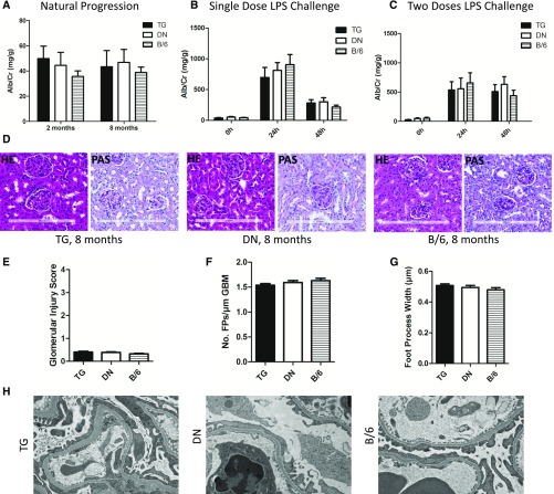
To characterize the effects of overexpressed TRPC5 on kidney filtration, we evaluated albuminuria in TG, DN, and B/6 mice (Figure 2A). At 2 months old, all mice had little albuminuria without a significant difference among the groups. By month 8, there was no change in albuminuria, which remained low in all groups. Histologic analysis by hematoxylin and eosin (HE) and periodic acid–Schiff (PAS) stain did not show any evident morphologic changes at the level of glomerulus in all mice (Figure 2, D and E). These data suggested that overexpression of TRPC5 does not lead to natural progression in kidney filtration barrier injury.
Figure 2.
Natural progression and LPS challenge caused similar level of kidney filtration injury in TG, DN, and B/6 mice. (A) No noticeable change in albuminuria over time was found for TG, DN, and B/6 mice. (B) Single dose LPS challenge induced substantial increase in albuminuria at 24 hours followed by a decrease at 48 hours. There is no difference between each of TG, DN, and B/6 mice at each time point. (C) Two doses of LPS challenge caused a similar increase in albuminuria for all three groups. (D) Representative images of hematoxylin and eosin and periodic acid–Schiff staining of kidneys from 8-month-old mice (original magnification, ×20). (E) Glomerular injury scoring on the basis of percentage of sclerosis showed no noticeable damage. (F) Analysis of TEM images exhibited equal degree of FP effacement after two doses of LPS challenge. (G) Average FP widths calculated for TG, DN, and B/6 were all similar. (H) Representative TEM images of kidneys after two doses of LPS challenge (15,000×). Analysis of TEM images exhibited equal degree of FP effacement. n≥6 per group; error bars are ±SEM. Alb/Cr, albumin/creatinine ratio; HE, hematoxylin and eosin; PAS, periodic acid–Schiff.
The LPS model was used to examine the kidney filtration barrier defects and albuminuria in our transgenic animals.6 In the first experiment, a high dose of LPS (10 mg/kg) was injected intraperitoneally (i.p.) and proteinuria was evaluated at 24 and 48 hours. As reported before,6,7 there was a significant increase in albuminuria at 24 hours, which substantially decreased at 48 hours in all three groups (Figure 2B). LPS induced a reversible change in podocyte cytoskeleton. However, the TG group responded similarly to LPS challenge as the DN or the B/6 group did. In the second experiment, two lower doses of LPS (2×5 mg/kg) were injected 24 hours apart and proteinuria was measured 24 hours after each injection. Similarly, all three groups of animals exhibited increased albuminuria after LPS challenge. But we did not observe any differences in proteinuria among all groups (Figure 2C). Kidney samples from the second experiment were used for transmission electron microscopy (TEM) to analyze podocyte foot process (FP) effacement. By counting the number of FPs and slits over a measured length of glomerular basement membrane (GBM), we detected a similar degree of effacement and FP width among all groups (Figure 2, F–H). This result was in accordance with the proteinuria findings and indicated that overexpression of TRPC5 would not result in higher susceptibility or aggravated kidney injury after LPS administration.
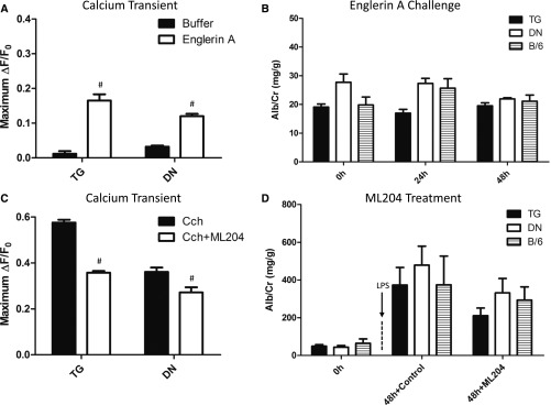
Injection of drugs either activating or inhibiting TRPC5 was implemented to further assess the relationship between TRPC5 and proteinuric disease. Englerin A, a reported potent and selective TRPC4/TRPC5 activator,8 was utilized to stimulate the TRPC5 channels in TG, DN, and B/6 animals. Five micromolar of Englerin A evoked a modest rise in intracellular Ca2+ in both TG and DN podocytes in vitro (Figure 3A). On the basis of the previous in vivo studies,9,10 we chose to inject two doses of Englerin A (2×3 mg/kg; i.p.) 24 hours apart and measure proteinuria 24 hours after each injection. As displayed in Figure 3B, activating TRPC5 by Englerin A did not cause any noticeable increase in proteinuria in all groups. Next, we tested the TRPC4/TRPC5 inhibitor ML20411 in the “two-doses” LPS model. A 20-minute preincubation of primary podocytes with 20 µM ML204 blocked Cch-induced Ca2+ transients significantly in both TG and DN groups (Figure 3C). Physiologic doses of ML204 (3×2 mg/kg) were injected (i.p.) 6 hours before the first and second LPS treatments and 6 hours after the second LPS treatment. There was no statistical distinction of albuminuria in ML204-treated groups compared with vehicle-treated groups (Figure 3D), indicating that antagonizing TRPC5 by ML204 did not rescue glomerular injury from LPS challenge.
Figure 3.
Englerin A and ML204 treatments have no effect on proteinuria in TG, DN, and B/6 mice. (A) Englerin A evoked a significant increase in Ca2+ transients in primary podocytes from TG and DN. (B) TRPC5 agonist Englerin A injection resulted in no change in albuminuria in TG, DN, and B/6 mice. (C) Average peak amplitude of Cch- and Cch+ML204-induced Ca2+ transients in TG and DN primary podocytes exhibited a markedly decreased Ca2+ transient in both groups. (D) TRPC5 antagonist ML204 injection had no significant effect on albuminuria levels between each of TG, DN, and B/6 mice. N mice ≥6 per group; N cells ≥100 per group; #P<0.05 compared with baseline or Cch; error bars are ±SEM. Alb/Cr, albumin/creatinine ratio; ΔF/F0, change in florescence normalized by the baseline fluorescence.
Ca2+ homeostasis is indispensable for orchestrating multiple cellular functions because of its role as a spatial and temporal second messenger in various cell types.12 The TRP superfamily participates in Ca2+ homeostasis and is involved in critical physiologic and pathologic cellular events.13 There is convincing evidence that dysregulation of one particular TRPC member, TRPC6, leads to FSGS by either gain- or loss-of-function mutation.3,4,14,15 Other TRPC channels are relatively less studied in the context of kidney diseases. It was reported that TRPC5 and TRPC6 had antagonistic effects on angiotensin II (AngII) receptor overexpressing podocytes upon AngII treatment.1 However, AngII-evoked TRPC5 activation cannot be seen in primary podocytes from rats or mice.16,17 Additionally, one study showed that plasma and sera from patients with recurrent FSGS produced robust effects on podocyte TRPC6 channels but had minimal effects on podocyte TRPC5 channels.18 The first direct evidence linking TRPC5 to kidney injury came recently, when a study reported that inhibition or loss of TRPC5 protected podocytes from cytoskeletal remodeling upon injury and decreased proteinuria in mice.5 Of note, unlike TRPC6, there is no evidence thus far indicating that the pathologic effect of TRPC5 is associated with patients who suffer from kidney disease. To better understand the potential role of TRPC5 in kidney injury, it is necessary to investigate TRPC5 function using a gain-of-function model of TRPC5.
Recently, we developed these novel transgenic mouse models by overexpressing either wild-type or pore mutant TRPC5 (Supplemental Figure 3). The nonfunctional, dominant negative TRPC5 was constructed with a mutation in the pore region by replacing the conserved LFW motif, which is essential for the channel function, with three alanines.19,20 This nonfunctional mutation was confirmed in our in vivo Ca2+ imaging data, where the pore mutant TRPC5 overexpression DN mice had 40% lower Cch-induced Ca2+ transient than that in wild-type TRPC5 overexpression TG mice (Figure 1F). This characteristic made our TG and DN mice an ideal pair of controls. Considering a comparable amount of TRPC6 overexpression was sufficient to produce kidney filtration injury in mice,21 this amount of TRPC5 overexpression in TG mice should have been adequate to detect a kidney phenotype.
LPS injection is a well established model to induce kidney filtration barrier defects and proteinuria independent of B or T cells.6,22 This reversible injury is associated with FP effacement and peak albuminuria 24 hours after challenge. LPS causes podocyte cytoskeletal remodeling via a Ca2+-mediated pathway.22,23 Thus, we selected this model to examine the effect of TRPC5 in vivo and found that overexpression of TRPC5 did not aggravate glomerular injury.
Next, we chose to interfere with the activity of TRPC5 by either activation or inhibition of the channel. Englerin A is a potent and selective stimulator for TRPC4/TRPC5 and is widely tested on cancer therapies.8,10 Our results showed that a tolerated dose (2×3 mg/kg) of Englerin A (i.p.) did not exhibit a change in albuminuria. Then, we tested TRPC4/TRPC5 antagonist ML204 in the LPS challenge model. It was reported that a high dose (2×20 mg/kg) of ML204 (i.p.) ameliorated LPS-induced proteinuria.5 Our physiologic dose (3×2 mg/kg) of ML20424 demonstrated a slight decrease in albuminuria compared with vehicle-treated animals. Of note, the original report on ML204 indicated that it also modestly inhibited TRPC6 channels.11 In a recent publication, it was stated that ML204 was unlikely to be a specific TRPC5 channel inhibitor because it produced an acute and potent Ca2+ release from intracellular stores.17 TRPC family members are known to be store-operated,25 which could all be affected by ML204. Therefore, inhibition of TRPC5 by ML204 did not salvage the glomerular injury and the effect of ML204 was probably not due to antagonizing TRPC5 alone.
Overall, this study reveals that overexpression or activation of TRPC5 is not the culprit for de novo or aggravating proteinuric kidney disease. Future research will be required to clarify a potential role for TRPC5 in renal pathophysiology.
Concise Methods
Mice
All animal experiments and protocols were approved by the Rush University Institutional Animal Care and Use Committee. B/6 mice were obtained from Jackson Laboratory (Bar Harbor, ME). TRPC5-overexpressing mice were generated by the Transgenic Animal Model Core at the University of Michigan. cDNA of wild-type or pore-mutated TRPC5 were constructed19,20 and cloned into the pCAGEN vector (Addgene, Cambridge, MA).26 The transgene was under the control of the CAG promoter/enhancer.27 Purified DNA was microinjected into fertilized eggs.28 The original founder mice were bred with B/6 to develop offspring. Each generation was genotyped before 6 weeks of age. All groups of experimental mice were age, weight, and sex matched.
qPCR
Total RNA was extracted from kidney with RNeasy Midi kit (QIAGEN, Germantown, MD). cDNA was synthesized using iScript cDNA Synthesis kit (Bio-Rad, Hercules, CA). qPCR was performed using SsoAdvanced SYBR Green Supermix (Bio-Rad) in a CFX Connect real-time system (Bio-Rad). The relative gene fold expression changes were determined using the comparative CT method by calculating 2–ΔΔCT for each gene.29
Western Blotting
The brain tissue was lysed in Laemmli buffer with homogenization and sonication (QSonica, Newtown, CT). Fifty-microgram samples were loaded in gels (Thermo Scientific). Target proteins were visualized and quantitated with Odyssey CLx infrared imaging system (LI-COR Biosciences). TRPC5 primary antibodies were from GenScript (customized; Piscataway, NJ) and NeuroMab (Davis, CA).
Immunofluorescence
Frozen kidney tissues were fixed with ice-cold acetone for 10 minutes. After 30 minutes of blocking (2.5% donkey normal serum and 2.5% FBS in PBS), samples were incubated with synaptopodin (1:100; Santa Cruz, Dallas, TX) and TRPC5 (1:100; GenScript) primary antibodies for 2 hours each, followed by Alexa Fluor secondary antibodies (1:1500; Molecular Probes, Eugene, OR) for 1 hour. Images were obtained and analyzed using an LSM 700 confocal microscope (Zeiss, Thornwood, NY).
In Vitro Ca2+ Imaging
Glomeruli were isolated by cell strainer and cultured in media at 37°C for 5 days. The primary podocytes were transferred to collagen-coated coverslips to grow for 3 more days. Before imaging, podocytes were loaded with Fluo-4AM (1.8 μM; Thermo Fisher Scientific, Waltham, MA) for 30 minutes in serum-free medium. A subset of coverslips were preincubated with ML204 (20 µM; Sigma-Aldrich) for 20 minutes. A balanced salt solution (140 mM NaCl, 10 mM Hepes, 2 mM CaCl2, 1 mM MgCl2, 10 mM Glucose, 5 mM KCl, pH 7.4) was used for cell rinsing and perfusing. The response of primary podocytes to 2.5 minutes of perfusion of Cch (100 µM; Abcam) or Englerin A (5 µM; PanReac AppliChem) was recorded through intracellular Ca2+ imaging. Three independent experiments from three TG and DN mice were performed. ImageJ (National Institutes of Health, Bethesda, MD) was used to determine the change in fluorescence intensity with time. Cells with spontaneous responses to perfusion buffer were excluded.
Urine Sampling and Analysis
Mouse urine samples were collected at times described in the manuscript. Urinary albumin and creatinine were measured by mouse albumin ELISA (Bethyl Laboratories, Montgomery, TX) and creatinine assay kits (Cayman Chemical, Ann Arbor, MI).
LPS Challenge
In the “single dose LPS” experiment, 10 mg/kg LPS (Sigma-Aldrich, St. Louis, MO) was injected (i.p.) to 14–16-week-old females weighing 20–25 g. Urine samples were collected 24 and 48 hours after LPS injection. In the second experiment, two doses of 5 mg/kg LPS were injected (i.p.) 24 hours apart. Urine samples were collected 24 hours after each injection. Kidneys were harvested for TEM at the end of the experiment.
Englerin A Injection
Two doses of 3 mg/kg Englerin A were injected (i.p.) to 14–16-week-old female mice (20–25 g) 24 hours apart. Urine samples were collected 24 hours after each injection.
ML204 Injection
Two-doses of LPS were injected to female mice weighing 20–25 g, as described above. Then, 2 mg/kg ML204 were injected (i.p.) 6 hours before the first and second LPS treatments and 6 hours after the second LPS treatment (three injections total). Urine samples were collected 48 hours after the first LPS injection.
TEM Analysis
Kidneys were cut into 2–3 mm3 pieces and fixed in Trumps Fixative (Electron Microscopy Sciences, Hatfield, PA). They were embedded in LX112 epoxy resin and polymerized at 60°C for 2–3 days. Ultrathin sections (70 nm) from kidney cortex were obtained using an EM UCT Ultramicrotome (Leica, Buffalo Grove, IL) and stained with 5% uranyl acetate and 0.1% lead citrate. Specimens were examined using a JEM-1220 transmission electron microscope (JEOL, Peabody, MA). Digital images were acquired using an Erlangshen ES1000W model 785 CCD camera (Gatan, Pleasanton, CA) and Digital Micrograph. The length of the GBM was measured by ImageJ and FPs and slits were counted. FP width was calculated as WFP=π∑GBM length/4∑slits.30
Statistical Analyses
Statistical analysis was performed using Prism 5.0 software (GraphPad, La Jolla, CA). Significance of the difference between two groups was assessed using the unpaired two-tailed t test. For multiple group comparisons, data were evaluated by one-way ANOVA using Tukey multiple comparison test or Bonferroni test. Data are shown as mean±SEM.
Disclosures
J.R. is cofounder and shareholder of TRISAQ, a biotechnology company in which he has financial interests and that develops kidney-protective drugs.
Supplementary Material
Acknowledgments
We acknowledge Galina Gavrilina, Wanda Filipiak, and Thom Saunders for preparation of transgenic mice and the Transgenic Animal Model Core of the University of Michigan’s Biomedical Research Core Facilities.
Core support was provided by the National Cancer Institute of the National Institutes of Health (NIH) under Award Number P30CA046592; the University of Michigan Gut Peptide Research Center, NIH grant number DK34933; and the University of Michigan George M. O’Brien Renal Core Center, NIH grant number P30DK08194. This work was supported by funds from the Department of Medicine, Rush University Medical Center. R.E.M. was supported by the NIH/National Institute of Arthritis and Musculoskeletal and Skin Diseases (K01AR070328).
Footnotes
Published online ahead of print. Publication date available at www.jasn.org.
This article contains supplemental material online at http://jasn.asnjournals.org/lookup/suppl/doi:10.1681/ASN.2017060682/-/DCSupplemental.
References
- 1.Tian D, Jacobo SM, Billing D, Rozkalne A, Gage SD, Anagnostou T, Pavenstädt H, Hsu HH, Schlondorff J, Ramos A, Greka A: Antagonistic regulation of actin dynamics and cell motility by TRPC5 and TRPC6 channels. Sci Signal 3: ra77, 2010 [DOI] [PMC free article] [PubMed] [Google Scholar]
- 2.Winn MP, Conlon PJ, Lynn KL, Farrington MK, Creazzo T, Hawkins AF, Daskalakis N, Kwan SY, Ebersviller S, Burchette JL, Pericak-Vance MA, Howell DN, Vance JM, Rosenberg PB: A mutation in the TRPC6 cation channel causes familial focal segmental glomerulosclerosis. Science 308: 1801–1804, 2005 [DOI] [PubMed] [Google Scholar]
- 3.Möller CC, Wei C, Altintas MM, Li J, Greka A, Ohse T, Pippin JW, Rastaldi MP, Wawersik S, Schiavi S, Henger A, Kretzler M, Shankland SJ, Reiser J: Induction of TRPC6 channel in acquired forms of proteinuric kidney disease. J Am Soc Nephrol 18: 29–36, 2007 [DOI] [PubMed] [Google Scholar]
- 4.Eckel J, Lavin PJ, Finch EA, Mukerji N, Burch J, Gbadegesin R, Wu G, Bowling B, Byrd A, Hall G, Sparks M, Zhang ZS, Homstad A, Barisoni L, Birbaumer L, Rosenberg P, Winn MP: TRPC6 enhances angiotensin II-induced albuminuria. J Am Soc Nephrol 22: 526–535, 2011 [DOI] [PMC free article] [PubMed] [Google Scholar]
- 5.Schaldecker T, Kim S, Tarabanis C, Tian D, Hakroush S, Castonguay P, Ahn W, Wallentin H, Heid H, Hopkins CR, Lindsley CW, Riccio A, Buvall L, Weins A, Greka A: Inhibition of the TRPC5 ion channel protects the kidney filter. J Clin Invest 123: 5298–5309, 2013 [DOI] [PMC free article] [PubMed] [Google Scholar]
- 6.Reiser J, von Gersdorff G, Loos M, Oh J, Asanuma K, Giardino L, Rastaldi MP, Calvaresi N, Watanabe H, Schwarz K, Faul C, Kretzler M, Davidson A, Sugimoto H, Kalluri R, Sharpe AH, Kreidberg JA, Mundel P: Induction of B7-1 in podocytes is associated with nephrotic syndrome. J Clin Invest 113: 1390–1397, 2004 [DOI] [PMC free article] [PubMed] [Google Scholar]
- 7.Mundel P, Reiser J: Proteinuria: An enzymatic disease of the podocyte? Kidney Int 77: 571–580, 2010 [DOI] [PMC free article] [PubMed] [Google Scholar]
- 8.Akbulut Y, Gaunt HJ, Muraki K, Ludlow MJ, Amer MS, Bruns A, Vasudev NS, Radtke L, Willot M, Hahn S, Seitz T, Ziegler S, Christmann M, Beech DJ, Waldmann H: (-)-Englerin A is a potent and selective activator of TRPC4 and TRPC5 calcium channels. Angew Chem Int Ed Engl 54: 3787–3791, 2015 [DOI] [PMC free article] [PubMed] [Google Scholar]
- 9.Carson C, Raman P, Tullai J, Xu L, Henault M, Thomas E, Yeola S, Lao J, McPate M, Verkuyl JM, Marsh G, Sarber J, Amaral A, Bailey S, Lubicka D, Pham H, Miranda N, Ding J, Tang HM, Ju H, Tranter P, Ji N, Krastel P, Jain RK, Schumacher AM, Loureiro JJ, George E, Berellini G, Ross NT, Bushell SM, Erdemli G, Solomon JM: Englerin A agonizes the TRPC4/C5 cation channels to inhibit tumor cell line proliferation. PLoS One 10: e0127498, 2015 [DOI] [PMC free article] [PubMed] [Google Scholar]
- 10.Sourbier C, Scroggins BT, Ratnayake R, Prince TL, Lee S, Lee MJ, Nagy PL, Lee YH, Trepel JB, Beutler JA, Linehan WM, Neckers L: Englerin A stimulates PKCθ to inhibit insulin signaling and to simultaneously activate HSF1: Pharmacologically induced synthetic lethality. Cancer Cell 23: 228–237, 2013 [DOI] [PMC free article] [PubMed] [Google Scholar]
- 11.Miller M, Shi J, Zhu Y, Kustov M, Tian JB, Stevens A, Wu M, Xu J, Long S, Yang P, Zholos AV, Salovich JM, Weaver CD, Hopkins CR, Lindsley CW, McManus O, Li M, Zhu MX: Identification of ML204, a novel potent antagonist that selectively modulates native TRPC4/C5 ion channels. J Biol Chem 286: 33436–33446, 2011 [DOI] [PMC free article] [PubMed] [Google Scholar]
- 12.Berridge MJ, Lipp P, Bootman MD: The versatility and universality of calcium signalling. Nat Rev Mol Cell Biol 1: 11–21, 2000 [DOI] [PubMed] [Google Scholar]
- 13.Montell C: The TRP superfamily of cation channels. Sci STKE 2005: re3, 2005 [DOI] [PubMed] [Google Scholar]
- 14.Riehle M, Büscher AK, Gohlke BO, Kaßmann M, Kolatsi-Joannou M, Bräsen JH, Nagel M, Becker JU, Winyard P, Hoyer PF, Preissner R, Krautwurst D, Gollasch M, Weber S, Harteneck C: TRPC6 G757D loss-of-function mutation associates with FSGS. J Am Soc Nephrol 27: 2771–2783, 2016 [DOI] [PMC free article] [PubMed] [Google Scholar]
- 15.Reiser J, Polu KR, Möller CC, Kenlan P, Altintas MM, Wei C, Faul C, Herbert S, Villegas I, Avila-Casado C, McGee M, Sugimoto H, Brown D, Kalluri R, Mundel P, Smith PL, Clapham DE, Pollak MR: TRPC6 is a glomerular slit diaphragm-associated channel required for normal renal function. Nat Genet 37: 739–744, 2005 [DOI] [PMC free article] [PubMed] [Google Scholar]
- 16.Kim EY, Anderson M, Dryer SE: Sustained activation of N-methyl-D-aspartate receptors in podoctyes leads to oxidative stress, mobilization of transient receptor potential canonical 6 channels, nuclear factor of activated T cells activation, and apoptotic cell death. Mol Pharmacol 82: 728–737, 2012 [DOI] [PMC free article] [PubMed] [Google Scholar]
- 17.Ilatovskaya DV, Palygin O, Levchenko V, Endres BT, Staruschenko A: The role of angiotensin II in glomerular volume dynamics and podocyte calcium handling. Sci Rep 7: 299, 2017 [DOI] [PMC free article] [PubMed] [Google Scholar]
- 18.Kim EY, Roshanravan H, Dryer SE: Changes in podocyte TRPC channels evoked by plasma and sera from patients with recurrent FSGS and by putative glomerular permeability factors. Biochim Biophys Acta 1863: 2342–2354, 2017 [DOI] [PMC free article] [PubMed] [Google Scholar]
- 19.Hofmann T, Schaefer M, Schultz G, Gudermann T: Subunit composition of mammalian transient receptor potential channels in living cells. Proc Natl Acad Sci U S A 99: 7461–7466, 2002 [DOI] [PMC free article] [PubMed] [Google Scholar]
- 20.Strübing C, Krapivinsky G, Krapivinsky L, Clapham DE: Formation of novel TRPC channels by complex subunit interactions in embryonic brain. J Biol Chem 278: 39014–39019, 2003 [DOI] [PubMed] [Google Scholar]
- 21.Krall P, Canales CP, Kairath P, Carmona-Mora P, Molina J, Carpio JD, Ruiz P, Mezzano SA, Li J, Wei C, Reiser J, Young JI, Walz K: Podocyte-specific overexpression of wild type or mutant trpc6 in mice is sufficient to cause glomerular disease. PLoS One 5: e12859, 2010 [DOI] [PMC free article] [PubMed] [Google Scholar]
- 22.Faul C, Donnelly M, Merscher-Gomez S, Chang YH, Franz S, Delfgaauw J, Chang JM, Choi HY, Campbell KN, Kim K, Reiser J, Mundel P: The actin cytoskeleton of kidney podocytes is a direct target of the antiproteinuric effect of cyclosporine A. Nat Med 14: 931–938, 2008 [DOI] [PMC free article] [PubMed] [Google Scholar]
- 23.Greka A, Mundel P: Calcium regulates podocyte actin dynamics. Semin Nephrol 32: 319–326, 2012 [DOI] [PMC free article] [PubMed] [Google Scholar]
- 24.Alawi KM, Russell FA, Aubdool AA, Srivastava S, Riffo-Vasquez Y, Baldissera L Jr, Thakore P, Saleque N, Fernandes ES, Walsh DA, Brain SD: Transient receptor potential canonical 5 (TRPC5) protects against pain and vascular inflammation in arthritis and joint inflammation. Ann Rheum Dis 76: 252–260, 2017 [DOI] [PMC free article] [PubMed] [Google Scholar]
- 25.Salido GM, Sage SO, Rosado JA: TRPC channels and store-operated Ca(2+) entry. Biochim Biophys Acta 1793: 223–230, 2009 [DOI] [PubMed] [Google Scholar]
- 26.Matsuda T, Cepko CL: Controlled expression of transgenes introduced by in vivo electroporation. Proc Natl Acad Sci U S A 104: 1027–1032, 2007 [DOI] [PMC free article] [PubMed] [Google Scholar]
- 27.Niwa H, Yamamura K, Miyazaki J: Efficient selection for high-expression transfectants with a novel eukaryotic vector. Gene 108: 193–199, 1991 [DOI] [PubMed] [Google Scholar]
- 28.Pease S, Saunders TL, International Society for Transgenic Technologies : Advanced protocols for animal transgenesis: An ISTT manual, Heidelberg, New York, Springer, 2011 [Google Scholar]
- 29.Livak KJ, Schmittgen TD: Analysis of relative gene expression data using real-time quantitative PCR and the 2(-Delta C(T)) Method. Methods 25: 402–408, 2001 [DOI] [PubMed] [Google Scholar]
- 30.Koop K, Eikmans M, Baelde HJ, Kawachi H, De Heer E, Paul LC, Bruijn JA: Expression of podocyte-associated molecules in acquired human kidney diseases. J Am Soc Nephrol 14: 2063–2071, 2003 [DOI] [PubMed] [Google Scholar]
Associated Data
This section collects any data citations, data availability statements, or supplementary materials included in this article.


