
Figure 1. Constitutively active PKC phosphorylates NP leading to impaired influenza virus polymerase activity.
(A) Expression of constitutively active PKC impairs influenza virus polymerase activity. Polymerase activity assays were performed in 293T cells in the presence or absence of the catalytic domains from classical, novel or atypical PKC isoforms. Data were averaged and normalized to the empty vector control. NP and PKC were detected by western blotting whole cell lysate. A hyper-phosphorylated form of NP was detected in some conditions. (n=3 ± standard deviation, *p<0.05 one-way ANOVA when compared to the empty vector control). (B) Polymerase activity assays were performed in the presence of PKC catalytic domains, catalytically inactive mutants, or empty vector controls. Polymerase activity and protein expression were analyzed as in (A).

Figure 1—figure supplement 1. Polymerase activity assays.
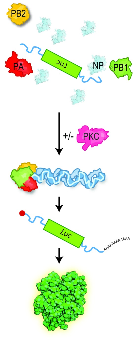
Cells are transfected with plasmids expressing a viral genome segment encoding luciferase, NP, and polymerase proteins PB1, PB2, and PA in the presence or absence of PKC. Luciferase activity output is measured as a proxy for RNP assembly and polymerase activity.
Figure 1—figure supplement 2. PKCs hyper-phosphorylate NP and only catalytically active enzymes inhibit viral polymerase activity.

(A) Whole cell lysates co-expressing NP with the indicated PKC catalytic fragments were treated with calf intestine alkaline phosphatase (CIP) or left untreated. Reaction products were analyzed by western blot with anti-NP antibody. (B) Quantification of NP phosphorylation in three separate replicates of the assay in Figure 1A (mean ±standard deviation). (C) Polymerase activity assays were performed in 293T cells co-expressing either PKC catalytic fragments (CAT) or mutants lacking catalytic activity (MUT) (n=3 ± standard deviation).