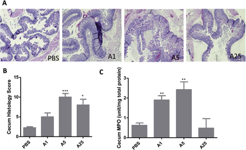
Fig. 2. Tissue damage by TcdA.
A) H&E stained ceca. Tissues were collected from mice exposed to TcdA after 24 h and fixed in 4% formalin for H&E staining. B) Average histopathological scores of 4–5 mice with each TcdA treatment; C) MPO activity assay. A small portion of cecum from each mouse was collected and lysed for MPO assay. A1: TcdA 1 µg; A5: TcdA 5 µg; A25: TcdA 25 µg *: p < 0.05, vs PBS; **: p < 0.01, vs PBS; ***: p < 0.001, vs PBS.

