Abstract
Background:
Yoga is the most popular form of alternative medicine for the management of diabetes mellitus type 2. The electro-photonic imaging (EPI) is another contribution from alternative medicine in health monitoring.
Aim:
To evaluate diabetes from EPI perspective.
Objectives:
(1) Compare various EPI parameters in normal, prediabetic and diabetic patients. (2) Find difference in controlled and uncontrolled diabetes. (3) Study the effect of 7 days diabetes-specific yoga program.
Materials and Methods:
For the first objective, there were 102 patients (normal 29, prediabetic 13, diabetic 60). In the second study, there were 60 patients (controlled diabetes 27, uncontrolled diabetes 33). The third study comprised 37 patients. EPI parameters were related to general health as well to specific organs.
Results:
In the first study, significant difference was observed between (1) Diabetics and normal: average intensity 5.978, form coefficient 3.590, immune organs 0.281 all P < 0.001; (2) Diabetics and prediabetics: average intensity 6.676, form coefficient 4.158, immune organs 5.890 P < 0.032; (3) Normal and prediabetes: immune organs (−6.171 P = 000). In the second study, remarkable difference was in the immune organs (0.201, P = 0.031). In the pre- and post-study, the mean difference was: area 630.37, form coefficient 1.78, entropy 0.03, liver 0.24, pancreas 0.17, coronary vessels 0.11, and left kidney 29, with all P < 0.02.
Conclusion:
There is a significant difference in EPI parameters between normal, prediabetics and diabetics, the prominent being average intensity, form coefficient, and immune organs. Between controlled and uncontrolled diabetes, immune organs show significant change. Intervention of yoga results in change in most parameters.
Keywords: Diabetes, electro-photonic imaging, parameter
Introduction
Diabetes poses a great threat to the world. With the changes in lifestyle in the developing and the developed countries, people are prone to this disorder. Coupled with genetic dispositions of Asians, people in those countries are predisposed to this disorder.[1] India has a major problem; the disease is spreading faster than anticipated earlier. It is shifting from older people to young adults.[2,3] The magnitude is so severe that by 2025–2030 India is expected to be the diabetic capital of the world.[4,5] The knowledge regarding this problem is known from earlier times and is mentioned in the ancient literature on Indian medical system like Ayurveda.[6] Modern medicine facilitated diagnosis and management of disease through advancement in pharmacology and research but the cure still remains a distant dream.[7] The disease is lethal in action and spreads to almost all vital organs of the body. Early detection and management would greatly help in arresting the spread of disease. The serious repercussions of the disease could thus be avoided or deferred.[8] Stress is found to be one of the main causes of diabetes. This is in line with the philosophy of Yoga and Ayurveda. As per both, the source of all the diseases is mind. Disturbances in the mind cause systemic problems and ultimately settle at the organ level. The cause and effect is known by the terms “Aadhi” and “Vyadhi,” respectively.[9,10] The modern medical system till recently was focused on the physical organs and systems for disease management. It is realized lately, that for all gross manifestations, there is a subtle undercurrent. The medicine now focuses on mind and the body rather than body only. Yoga is one blessing to the mankind which helps to calm down mind and rejuvenate the body. It is a recognized spiritual philosophy with immense health benefits and very effective in treating stress.[11,12] Thus, a holistic medicine is evolving, where wisdom of traditional practices and the modern medicine is offered simultaneously for the welfare of mankind.[13] India is taking a big lead in this direction.[14] Modern system is well supported by organized research and other systems are also trying to develop on these lines. One such system on which research has been going on for decades is electro-photonic imaging (EPI) based on Kirlian photography. This equipment is based on applied physics and tries to investigate subtle bio-energy changes in the body. The principle of working of EPI instrument is very simple. Tip of the ten fingers is placed on dielectric glass one by one, a high-voltage short duration pulse of 10 kV and frequency 1024 Hz is applied and electrons are extracted from the finger. Due to the presence of high electric field, the electrons collide with air molecules in the surroundings and photons are released around the finger. A camera fitted in the EPI captures this image which is analyzed with the help of software.[15] The captured image parameters depend on the state of health of an individual. The ten fingers represent various organs and systems as per the Chinese system of acupuncture.[16,17,18] So, EPI in the real sense is a fusion of modern physics and ancient philosophy. The software that is used for the analysis of image gives various energy diagrams and parameters. These parameters are indicative of the general state of health and the state of various organs and systems. In Russia, EPI is used in medical sciences, biometrics, sports, forensic, human behavior, etc. Healers use it for observing changes after the administration of an intervention.[19] At the moment it is a good tool in the hands of healers and those practicing alternative medicine. They can find the impact of intervention by comparing pre- and post-conditions.[20]
Aim and objectives
The aim is to study diabetes mellitus (DM) type 2 with the help of EPI parameters. The objectives are to find the changes in EPI parameters in normal, prediabetic, and diabetic patients, compare controlled and uncontrolled diabetes, and evaluate the effect of 7-day practice of yoga camp in connection with the stop diabetes movement (SDM) campaign of Swami Vivekananda Yoga Anusandhana Samsthana (S-VYASA) Yoga University, Bengaluru, India.
Materials and Methods
The study was conducted on the participants of the yoga camps held in connection with SDM campaign and Arogyadham (a residential health center) of S-VYASA Yoga University. After the scrutiny of 250 participants, following number of patients were selected for the different studies. (a) One hundred and two patients (mean age 51 ± 11) for the first objective. Out of these, 52 were males (mean age 54 ± 11) and rest females (mean age 47 ± 10). The total patients comprised 29 normal (mean age 44 ± 11), 13 prediabetic (mean age 51.2 ± 12.3) and 60 diabetic (mean age 54 ± 9.6). (b) Sixty patients (mean age 53.8 ± 9.62) for the second objective. Out of these, 35 were males (mean age 56.83 ± 8.72) and 25 females (mean age 49.56 ± 9.38). The total patients were divided into controlled diabetes n = 27 (mean age 56.04 ± 9.28) and uncontrolled diabetes n = 33 (mean age 51.97 ± 9.65). (c) Thirty-seven patients (mean age 54.46 ± 7.21) comprising 24 males (mean age 57.46 ± 7.35) and 13 females (mean age 54.62 ± 6.83). The dropouts from the initial scrutiny were on account of (1) incomplete images (2) withdrawal from the camp (3) either of the images was not available in case of pre- and post-study. EPI images were taken on the first day of the camp and in case of pre- and post-intervention study; the images were taken on the conclusion of the camp also. The categorization was based on fasting blood sugar (FBS). As per the criteria, <100 mg/dl was normal, between 100 and 125 mg/dl prediabetic and >126 mg/dl diabetic. This is as per the American Diabetes Association score.[21] Controlled and uncontrolled diabetics were classified on the basis of above scale from the participants who were confirmed diabetics from the medical history and reports. Participants who had FBS >126 mg/dl for more than 3 months, in spite of being on anti diabetes medicine, were classified as uncontrolled diabetics.[22,23] They were on medication for the management of their diabetes. The intervention was administered by yoga trainers and therapists. The EPI parameters selected for analysis were area, intensity, form coefficient, entropy, and fractality which pertain to general health.[15] Further there were parameters which were organ specific such as liver, immune organs, pancreas, coronary vessels, cerebral vessels, left kidney and right kidney. The organ-specific parameters were selected on the basis of modern medical literature.[24,25,26,27] The average of the values of the left- and right-hand fingers was considered for the EPI parameters. The EPI parameters were obtained through the software EPI diagram, EPI screening, and EPI scientific laboratory. Participants in the age range of 18–75 years, male or female and those willing to volunteer for study were selected. As diabetes is spreading to younger people and the study was broad-based covering normal, prediabetics, and diabetics, we decided for a broader range of age. All participants had to give blood for FBS test on the inaugural day of camp or have a recent blood report. Consent was taken from willing participants, and only 5 ml of blood was collected from each participant for the testing purpose. This study was cleared by the Institutional Ethics Committee at S-VYASA Yoga University, Bengaluru, India; vide RES/1EC-SVYASA/66/2015.
Exclusion criteria
Patients with comorbidities such as hypertension, dyslipidemia, and fatty liver disease and those taking any medicine in the case of normal and prediabetic participants; diabetic patients taking medicines apart from diabetes; patients suffering from any infectious or contagious disease; physically handicapped and those having missing fingers were excluded from the study. Females having menstruation or pregnancy on the day of measurement were also excluded from the study.
Sampling time
The data were taken in the morning hours with a gap of at least 3 h from the last meal. The data in the camps were mostly taken on the inaugural day of the camp. Data at Aroghyadham (residential hospital) were taken in the morning as well in the evening but ensuring gap of 3 h from the last meal. The requirement from participants was to follow yogic way of life in the matter of exercise, mental relaxation, and diet. This was monitored through regular feedbacks in the camps and records in Aroghyadham (for residential participants). EPI was calibrated each time the place of taking measurement changed or as required. Informed consent was taken from all the participants before conducting the study. The study was approved by the University's Ethics Committee.
Instrument
GDV Camera Pro with analog video camera, model number: FTDI.13.6001.110310 (Kirlionics Technologies International company, Saint-Petersburg, Russia) was used for the assessment purpose. Along with the EPI software, it provided various features such as EPI screening, EPI scientific laboratory, and EPI diagram. These are different software programs for analysis and data extraction. EPI screening allows evaluating particular sectors of different fingers related to body systems as well as to different organs. EPI scientific laboratory gives the data for each finger and the average of parameters related to general health.
Parameters analyzed
From EPI software the following parameters were analyzed: Total area is an absolute value and is measured as the number of pixels in the image having brightness above a preset threshold. Area of glow is in proportion to quantity of electrons; average intensity is evaluation of light intensity averaged over the area of image; form coefficient and fractality are measures of irregularity in the image's external contour; entropy reflects the level of nonuniformity of image, in other words, the level of stability of the energy field. It is a measure of energy disturbance in the body. From EPI screening/EPI diagram, integral area of liver, pancreas, immune organs, coronary vessels, cerebral vessels, left kidney and right kidney were analyzed. Integral area is relative value and shows the extent to which the EPI gram deviates from an ideal model. For evaluation of the functional state of particular systems and organs, these parameters are calculated for the whole EPI gram or for the sectors of particular zones. It is indicative of general health.
Data analysis
Data analysis was carried out with the help of Microsoft Office Excel 2007.lnk and R-studio version 3.2.0 along with R Cmdr version 2.1-7. Statistical tests: Independent sample t-test and paired t-test were used to compare the means.
Intervention
The intervention was yoga program based on SDM module of S-VYASA. It comprises asanas, pranayama, meditation, practices on stress management, lectures on the disease and diet regulations and the modification in lifestyle. The yoga sessions was held from 5:00 h to 7:00 h every day for 7 days under the guidance of experienced yoga trainers and therapists.
Results
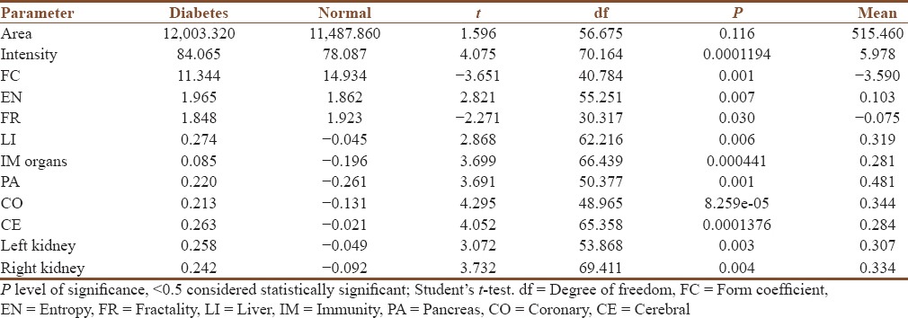
In the first study, we observed significant difference in means of average intensity (diabetes and normal = 5.978 [P = 0.0001], diabetes and prediabetes = 6.676 [P = 0.0169]); form coefficient (diabetes and normal = −3.590 [P = 0.0007], diabetes and prediabetes = −4.158 [P = 0.0315]); immune organs (diabetes and normal = 0.281 [P = 0.0004], diabetes and prediabetes = −5.890 [P = 0.0001], normal and prediabetes = −6.171 [P = 0.0001]) [Tables 1-3]. Besides, there were small differences (but very significant) observed in many other parameters as seen in Table 4. The second study observation was pertaining to immune organs (difference in means 0.201, P = 0.0319) [Table 5]. In the third, i.e. pre- and post-study, the noticeable changes were in area (mean difference 630.465, P = 0.0004); form coefficient (mean difference − 1.783, P = 0.0001); entropy (mean difference − 0.029, P = 0.0012); liver (mean difference 0.247, P = 0.0001); pancreas (mean difference 0.176, P = 0.0250); coronary vessels (mean difference 0.142, P = 0.0001); cerebral vessels (mean difference 0.192, P = 0.0002); left kidney (mean difference 0.157, P = 0.0042); right kidney (mean difference 0.248, P = 0.0001) [Table 6].
Table 1.
Independent sample t-test; normal-prediabetes
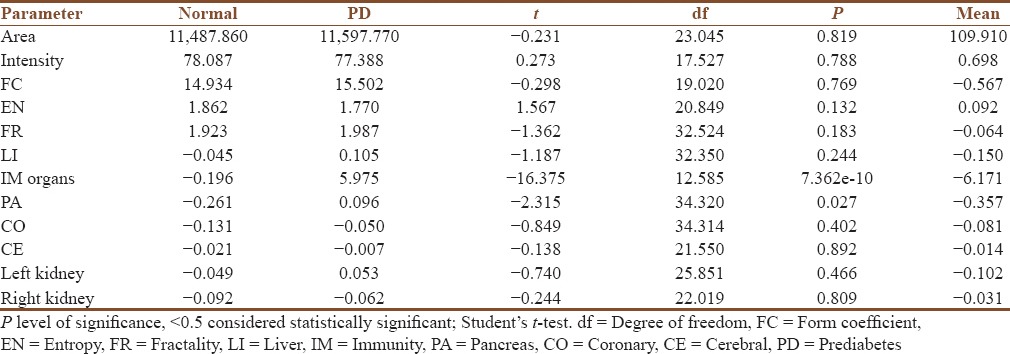
Table 3.
Independent sample t-test; diabetes and prediabetes
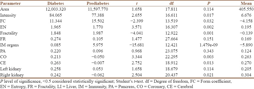
Table 4.
Combined highly significant results of independent sample t-test
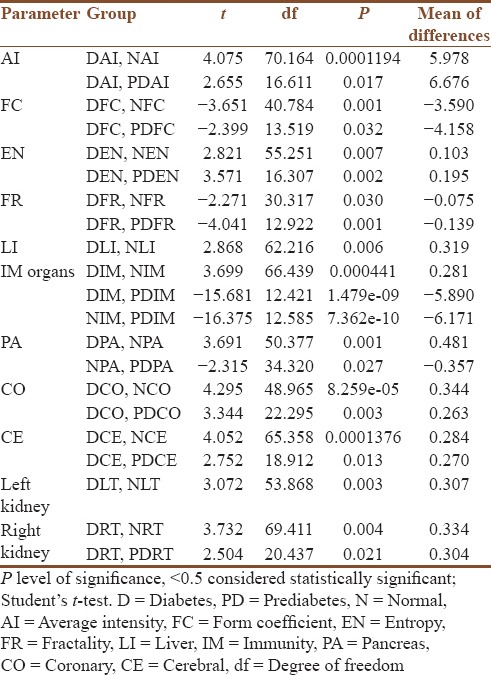
Table 5.
Independent sample t-test controlled-uncontrolled diabetes
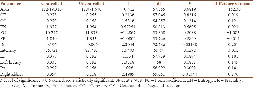
Table 6.
Pre-and post-results by paired t-test
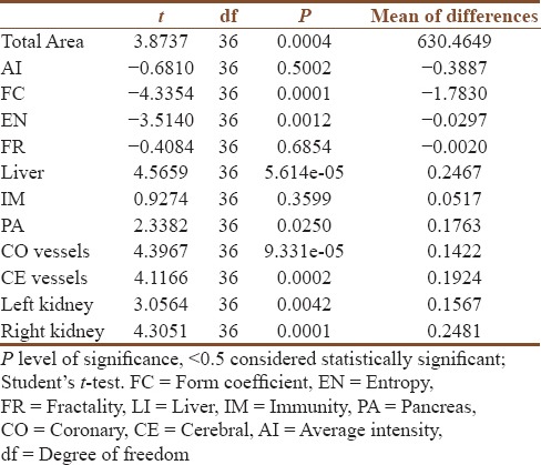
Table 2.
Independent sample t-test; diabetes-normal
Discussions
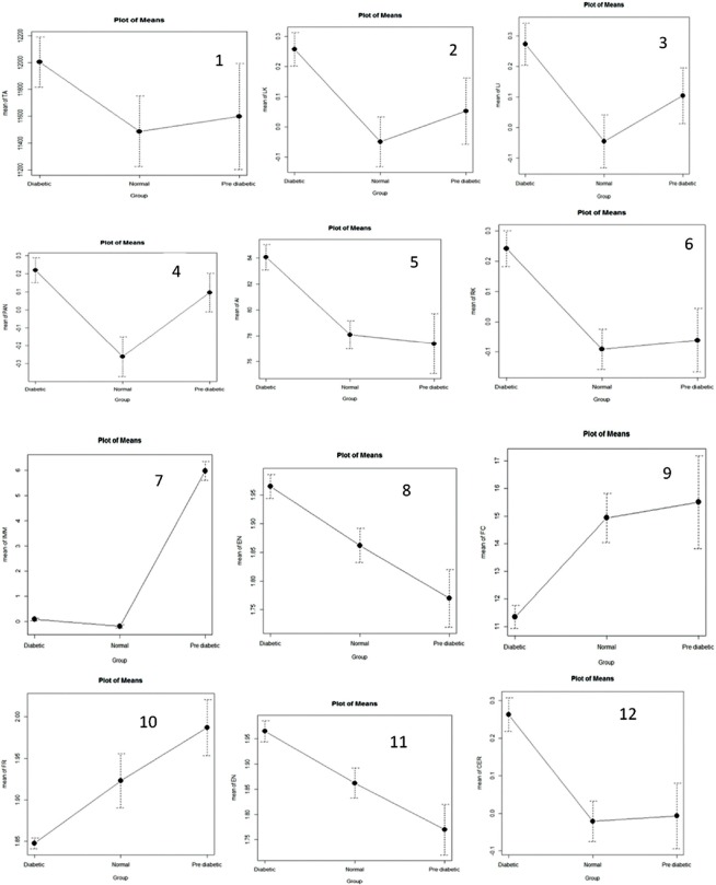
The purpose of this study was to evaluate whether the parameters of EPI can be used for diagnostic aspects of DM type 2. In the first study three stages of diabetes were considered, namely, normal, prediabetes and diabetes. Independent t-test between diabetes and normal, diabetes and prediabetes, normal and prediabetes showed significant results. There is a remarkable difference in the parameters between diabetes and normal and the difference is highly significant. Average intensity, entropy, liver, pancreas, immune organs coronary vessels, cerebral vessels, left kidney, right kidney all are more imbalanced in diabetic state than in the normal. This is on expected lines as higher values are indicative of disorder. Fractality and form coefficient which are measures of irregularity in the external contour have negative values as the images are more regular in normal condition than the diabetic and have a higher value in normal condition. This is in line with the theory of EPI that the average intensity and entropy are high with aging and progression of disease, form coefficient and fractality are lesser. Similarly, there is highly significant difference in the selected parameters in diabetic and prediabetic. However, the differences in the integral area of liver, pancreas, and left kidney are not significant. This could be due to the fact that these organs are affected at the prediabetic stage itself as they would be at the diabetic stage. Another important aspect is with respect to energy in immune organs. Results indicate that immune organs get more compromised at the prediabetic stage which paves the way for disease to progress and become devastating, the body becomes vulnerable to multiple problems.[28,29,30] The first study has revealed noticeable differences in the three stages, i.e., normal, prediabetes, and diabetes. In the second study we considered controlled diabetes and uncontrolled diabetes. The controlled diabetes as per the American Diabetes Association was considered as FBS <126 mg/dl and above this as uncontrolled. There was small noticeable but significant difference in the immune organs. It is a negative difference in line with the earlier argument that immune organs are compromised much earlier than the extreme manifestation of the disease in this case of DM type 2. In rest of the parameters there is no significant change. It perhaps shows the state of these organs in the diabetics whether the diabetes is controlled or uncontrolled. In the pre- and post-study, we observed significant differences in many parameters by the paired t-test. These are total area, form coefficient, entropy, coronary vessels, cerebral vessels, pancreas, left kidney and right kidney. The intervention of yoga makes the change. As mentioned earlier, area increases with the aging and the disturbed state of health. Reduction in the area indicates improvement. Entropy is indicative of stability of energy field and negative sign shows that a correction has happened in poststate. Results show that stability is more and hence the disturbance is less in the poststate.[15] Similarly, there is an increase in form coefficient and hence the improvement in irregularity of the image. Liver, pancreas coronary vessels, cerebral vessels, and kidneys are the organs that are affected by diabetes. We observe a reduction in the integral area of these parameters taking them towards the normal values after yoga intervention. Thus, 7-day practice of yoga related to diabetes brings general feeling of well-being and some changes at the organ level. The broad clue taken from all these studies is that EPI does indicate changes at the general level as well organ/system level in the different conditions of diabetes. There is direct evidence of progressive change in most of the selected parameters from normal to prediabetes to diabetes as seen in Table 7 and Figure 1. These findings if properly applied by the practitioners can help them to know regarding onset of the disease and advise therapy and monitor the effect of therapy.
Table 7.
One-way ANOVA
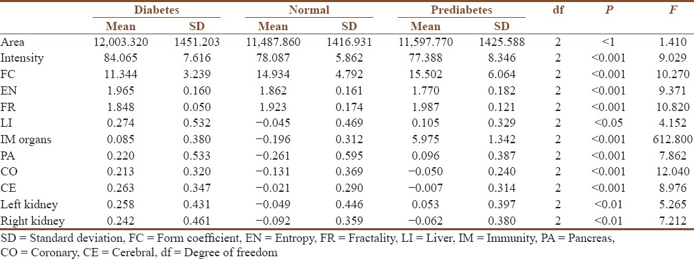
Figure 1.
1-12: Graphical representation of various parameters in normal, pre-diabetic and diabetic state
Limitations of the study
The changes in different states/conditions need to be corroborated with the modern medicine diagnostics. At the moment there is no technology in the modern science that can notice changes at the subtle level as the EPI.
Strength of the study
This is the exclusive study on various aspects of diabetes. The difference in three states, i.e., normal, prediabetic, and diabetic is demonstrated through EPI. Effect of yoga in diabetics and the changes in controlled and uncontrolled diabetes has been presented. This study shows the changes and effects in the right direction which strengthens the confidence in EPI technology.
Further research
Further research should be carried out on large sample sizes with funding from national/international medical institutes. This should be in conjunction with the practitioners/scientists of modern medicine and instrumentation. An important area is to find whether small significant changes in the general/organ-specific parameters produce noticeable changes at the physical level and correlate how much change is the desired change for the desired effect.
Conclusion
This study was meant to look at diabetes from the perspective of EPI. From the three situations considered, we infer from the first study that values of intensity, form coefficient, and immune organs can broadly classify a person into normal, prediabetic, and diabetic. From the second study, we can differentiate between controlled and uncontrolled diabetes through the EPI parameters of Immune organs. In the uncontrolled diabetes immune organs get compromised. A 7-day yoga camp on diabetes control and management produces changes which can be seen through EPI in a large number of parameters for both general and organ specific.
Financial support and sponsorship
Nil.
Conflicts of interest
There are no conflicts of interest.
Acknowledgments
We would like to thank Dr. Guru Deo for data acquisition and review. Dr. Kuldeep K Kushwaha was extremely helpful during collection of data. The support from SDM team and Aroghyadham of S-VYASA was highly valuable.
References
- 1.Zimmet P, Dowse G, Finch C, Serjeantson S, King H. The epidemiology and natural history of NIDDM – Lessons from the South Pacific. Diabetes Metab Rev. 1990;6:91–124. doi: 10.1002/dmr.5610060203. [DOI] [PubMed] [Google Scholar]
- 2.Zimmet PZ. The pathogenesis and prevention of diabetes in adults. Genes, autoimmunity, and demography. Diabetes Care. 1995;18:1050–64. doi: 10.2337/diacare.18.7.1050. [DOI] [PubMed] [Google Scholar]
- 3.Mohan V, Sandeep S, Deepa R, Shah B, Varghese C. Epidemiology of type 2 diabetes: Indian scenario. Indian J Med Res. 2007;125:217–30. [PubMed] [Google Scholar]
- 4.Misra P, Upadhyay RP, Misra A, Anand K. A review of the epidemiology of diabetes in rural India. Diabetes Res Clin Pract. 2011;92:303–11. doi: 10.1016/j.diabres.2011.02.032. [DOI] [PubMed] [Google Scholar]
- 5.Ramachandran A, Jali MV, Mohan V, Snehalatha C, Viswanathan M. High prevalence of diabetes in an urban population in South India. BMJ. 1988;297:587–90. doi: 10.1136/bmj.297.6648.587. [DOI] [PMC free article] [PubMed] [Google Scholar]
- 6.Manyam BV. Diabetes mellitus, Ayurveda, and Yoga. J Altern Complement Med. 2004;10:223–5. doi: 10.1089/107555304323062185. [DOI] [PubMed] [Google Scholar]
- 7.Nathan DM, Buse JB, Davidson MB, Ferrannini E, Holman RR, Sherwin R, et al. Medical management of hyperglycaemia in type 2 diabetes mellitus: A consensus algorithm for the initiation and adjustment of therapy: A consensus statement from the American Diabetes Association and the European Association for the study of diabetes. Diabetologia. 2009;52:17–30. doi: 10.1007/s00125-008-1157-y. [DOI] [PubMed] [Google Scholar]
- 8.Fowler MJ. Microvascular and macrovascular complications of diabetes. Clin Diabetes. 2008;26:77–82. [Google Scholar]
- 9.Surivit RS, Schneider MS, Feinglos MN. Stress and diabetes mellitus. Diabetes Care. 1992;15:1413–22. doi: 10.2337/diacare.15.10.1413. [DOI] [PubMed] [Google Scholar]
- 10.Thakar VJ. Evolution of diseases i.e. Samprapti vignana. Anc Sci Life. 1981;1:13–9. [PMC free article] [PubMed] [Google Scholar]
- 11.Nyenwe EA, Jerkins TW, Umpierrez GE, Kitabchi AE. Management of type 2 diabetes: Evolving strategies for the treatment of patients with type 2 diabetes. Metabolism. 2011;60:1–23. doi: 10.1016/j.metabol.2010.09.010. [DOI] [PMC free article] [PubMed] [Google Scholar]
- 12.Kosuri M, Sridhar GR. Yoga practice in diabetes improves physical and psychological outcomes. Metab Syndr Relat Disord. 2009;7:515–7. doi: 10.1089/met.2009.0011. [DOI] [PubMed] [Google Scholar]
- 13.Barrett B, Marchand L, Scheder J, Plane MB, Maberry R, Appelbaum D, et al. Themes of holism, empowerment, access, and legitimacy define complementary, alternative, and integrative medicine in relation to conventional biomedicine. J Altern Complement Med. 2003;9:937–47. doi: 10.1089/107555303771952271. [DOI] [PubMed] [Google Scholar]
- 14.Sharma R, Gupta N, Bijlani RL. Effect of yoga based lifestyle intervention on subjective well-being. Indian J Physiol Pharmacol. 2008;52:123–31. [PubMed] [Google Scholar]
- 15.Alexandrova RA, Fedoseev BG, Korotkov KG, Philippova NA, Zayzev S, Magidov M, et al. Analysis of the bio electro grams of bronchial asthma patients. Proceedings of conference “Measuring the human energy field: State of the science. National Institute of Health, Baltimore, MD. 2003:70–81. [Google Scholar]
- 16.Yung KT. A birdcage model for the Chinese meridian system: Part I.A channel as a transmission line. Am J Chin Med. 2004;32:815–28. doi: 10.1142/S0192415X04002417. [DOI] [PubMed] [Google Scholar]
- 17.Shu JJ, Sun Y. Developing classification indices for Chinese pulse diagnosis. Complement Ther Med. 2007;15:190–8. doi: 10.1016/j.ctim.2006.06.004. [DOI] [PubMed] [Google Scholar]
- 18.Chernyak GV, Sessler DI. Perioperative acupuncture and related techniques. Anesthesiology. 2005;102:1031–49. doi: 10.1097/00000542-200505000-00024. [DOI] [PMC free article] [PubMed] [Google Scholar]
- 19.Korotkov KG, Matravers P, Orlov DV, Williams BO. Application of electrophoton capture (EPC) analysis based on gas discharge visualization (GDV) technique in medicine: A systematic review. J Altern Complement Med. 2010;16:13–25. doi: 10.1089/acm.2008.0285. [DOI] [PubMed] [Google Scholar]
- 20.Korotkov K, Shelkov O, Shevtsov A, Mohov D, Paoletti S, Mirosnichenko D, et al. Stress reduction with osteopathy assessed with GDV electrophotonic imaging: Effects of osteopathy treatment. J Altern Complement Med. 2012;18:251–7. doi: 10.1089/acm.2010.0853. [DOI] [PubMed] [Google Scholar]
- 21.American Diabetes Association. Diagnosis and classification of diabetes mellitus. Diabetes Care. 2010;33(Suppl 1):S62–9. doi: 10.2337/dc10-S062. [DOI] [PMC free article] [PubMed] [Google Scholar]
- 22.Davidson MB, Schriger DL, Peters AL, Lorber B. Relationship between fasting plasma glucose and glycosylated hemoglobin: Potential for false-positive diagnoses of type 2 diabetes using new diagnostic criteria. JAMA. 1999;281:1203–10. doi: 10.1001/jama.281.13.1203. [DOI] [PubMed] [Google Scholar]
- 23.Hong ES, Khang AR, Yoon JW, Kang SM, Choi SH, Park KS, et al. Comparison between sitagliptin as add on therapy and insulin dose increase therapy in uncontrolled Korean type 2 diabetes – CSI study. J Pharmacol Ther. 2012;14:795–802. doi: 10.1111/j.1463-1326.2012.01600.x. [DOI] [PubMed] [Google Scholar]
- 24.Kreier F, Kap YS, Mettenleiter TC, van Heijningen C, van der Vliet J, Kalsbeek A, et al. Tracing from fat tissue, liver, and pancreas: A neuroanatomical framework for the role of the brain in type 2 diabetes. Endocrinology. 2006;147:1140–7. doi: 10.1210/en.2005-0667. [DOI] [PubMed] [Google Scholar]
- 25.Shimabukuro M, Saito T, Higa T, Nakamura K, Masuzaki H, Sata M Fukuoka Diabetologists Group. Risk stratification of coronary artery disease in asymptomatic diabetic subjects using multidetector computed tomography. Circ J. 2015;79:2422–9. doi: 10.1253/circj.CJ-15-0325. [DOI] [PubMed] [Google Scholar]
- 26.Shian Y, Lin J, Zeng M, XU P, Lin H, Yan H. Association of aortic compliance and bronchial endothelial function with cerebral small vessels disease in type 2 diabetes mellitus patients: Assessment with high resolution MRI. Biomed Res Int 2016. 2016:1609317. doi: 10.1155/2016/1609317. [DOI] [PMC free article] [PubMed] [Google Scholar]
- 27.Pecoits-Filho R, Abensur H, Betônico CC, Machado AD, Parente EB, Queiroz M, et al. Interactions between kidney disease and diabetes: Dangerous liaisons. Diabetol Metab Syndr. 2016;8:50. doi: 10.1186/s13098-016-0159-z. [DOI] [PMC free article] [PubMed] [Google Scholar]
- 28.Hirokawa K, Utsuyama M, Hayashi Y, Kitagawa M, Makinodan T, Fulop T. Slower immune system aging in women versus men in the Japanese population. Immun Ageing. 2013;10:19. doi: 10.1186/1742-4933-10-19. [DOI] [PMC free article] [PubMed] [Google Scholar]
- 29.Caruso C, Accardi G, Virruso C, Candore G. Sex, gender and immunosenescence: A key to understand the different lifespan between men and women? Immun Ageing. 2013;10:20. doi: 10.1186/1742-4933-10-20. [DOI] [PMC free article] [PubMed] [Google Scholar]
- 30.Klin SL, Flangan KL. Sex difference in immune responses. Nat Rev Immunol. 2016;16:626–38. doi: 10.1038/nri.2016.90. [DOI] [PubMed] [Google Scholar]


