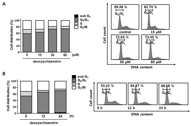
Figure 2.
Effect of deoxyschizandrin on cell cycle in A2780 cells: (A) Cell cycle analysis was performed using propidium iodide (PI) staining assay. A2780 cells were treated with the indicated concentration of deoxyschizandrin (15, 30, and 60 µM) for 48 h and then stained with propidium iodide (PI). The cell cycle distribution profiles of the cells were determined by flow cytometry (FACS); (B) Distribution of cell number in cell cycle was measured at 12 and 24 h after deoxyschizandrin (30 µM) treatment.

