Abstract
Vitamin D deficiency is common in pregnant women and may contribute to adverse events in pregnancy such as preeclampsia (PET). To date, studies of vitamin D and PET have focused primarily on serum concentrations vitamin D, 25-hydroxyvitamin D3 (25(OH)D3) later in pregnancy. The aim here was to determine whether a more comprehensive analysis of vitamin D metabolites earlier in pregnancy could provide predictors of PET. Using samples from the SCOPE pregnancy cohort, multiple vitamin D metabolites were quantified by liquid chromatography–tandem mass spectrometry in paired serum and urine prior to the onset of PET symptoms. Samples from 50 women at pregnancy week 15 were analysed, with 25 (50%) developing PET by the end of the pregnancy and 25 continuing with uncomplicated pregnancy. Paired serum and urine from non-pregnant women (n = 9) of reproductive age were also used as a control. Serum concentrations of 25(OH)D3, 25(OH)D2, 1,25(OH)2D3, 24,25(OH)2D3 and 3-epi-25(OH)D3 were measured and showed no significant difference between women with uncomplicated pregnancies and those developing PET. As previously reported, serum 1,25(OH)2D3 was higher in all pregnant women (in the second trimester), but serum 25(OH)D2 was also higher compared to non-pregnant women. In urine, 25(OH)D3 and 24,25(OH)2D3 were quantifiable, with both metabolites demonstrating significantly lower (P < 0.05) concentrations of both of these metabolites in those destined to develop PET. These data indicate that analysis of urinary metabolites provides an additional insight into vitamin D and the kidney, with lower urinary 25(OH)D3 and 24,25(OH)2D3 excretion being an early indicator of a predisposition towards developing PET.
Keywords: pregnancy, preeclampsia, vitamin D, biomarker, serum and urine
Introduction
Pre-eclampsia (PET) is a pregnancy-specific, multisystem syndrome with endothelial dysfunction and presenting with hypertension and pathologic renal protein excretion. Complicating up to 8% of pregnancies (1, 2, 3), PET is associated with significant maternal and perinatal mortality and morbidity (4, 5). Despite recognition of PET risk factors, such as raised body mass index, ethnicity, advanced maternal age, pre-existing hypertension and/or chronic renal disease, PET often complicates otherwise uncomplicated nulliparous pregnancies with severe maternal–fetal consequences (5, 6).
The key predisposing event in PET is uteroplacental maldevelopment with abnormal decidual maternal spiral artery remodelling by invading fetal extravillous trophoblast cells in the late first and second trimester. This often precedes the onset of clinical disease, which later ensues secondary to tissue hypoxia, oxidative stress and release of anti-angiogenic and pro-inflammatory factors into the maternal circulation, which result in generalised systemic endothelial dysfunction (7). Consequently, there has been considerable research interest in the identification of ‘early biomarkers’ of PET, including anti-angiogenic factors, hormones, adhesion molecules, placental perfusion measures and vasodilators such as placental soluble fms-like tyrosine kinase (sFlt-1), soluble endoglin (sEng), maternal spiral artery resistance, pulsatility index and placental growth factor (PlGF) (8, 9, 10, 11). To date, systematic analysis of the efficacy of these biomarkers has been relatively inconsistent with high intra-study heterogeneity (10, 12).
The kidney is a key organ affected by PET. In response to normal pregnancy, significant renal adaptations ensue, including marked glomerular hyperfiltration and increased effective renal plasma flow. In PET, these functional changes differ with a significantly lower glomerular filtration rate and progressive glomerular injury (13, 14, 15), as evidenced by the development of significant proteinuria. Potential pre-clinical markers of renal injury, including urinary podocytes, podocyte-specific proteinuria nephrin and urinary albumin are now also being sought (16, 17). The kidneys are also the major site for vitamin D metabolism, expressing the 25-hydroxyvitamin D3 1α-hydroxylase (1α-hydroxylase) enzyme that synthesises 1,25-dihydroxyvitamin D3 (1,25(OH)2D3) from precursor 25-hydroxyvitamin D3 (25(OH)D3), as well as 25-hydroxyvitamin D-24-hydroxylase (24-hydroxylase) and the vitamin D receptor (VDR) (18, 19). In uncomplicated pregnancy, significant changes in vitamin D physiology arise, with a surge in maternal serum 1,25(OH)2D3 from the first trimester (20). During pregnancy 1,25(OH)2D3 is also synthesised by the human placenta and decidua via localised expression of 1α-hydroxylase, which is greatest in the first and second trimester. Coincident expression of placental VDR, 24-hydroxylase, vitamin D-binding protein and vitamin D-25-hydroxylase, suggest an autocrine/paracrine role for 1,25(OH)2D3 in the placenta (21, 22, 23, 24, 25). The role of vitamin D inactivation and renal clearance in pregnancy are to be defined.
Vitamin D deficiency, defined as a serum 25(OH)D3 <50 nM, is highly prevalent in pregnant women (26, 27), and recent studies have reported association between maternal serum 25(OH)D3 levels and PET (28, 29, 30, 31, 32). A recent systematic review and meta-analysis, which included 11 observational studies, found a significant inverse relationship between maternal 25(OH)D3 and PET risk in 5 of these studies. Meta-analyses similarly suggested an inverse relationship between maternal 25(OH)D3 and PET risk, but could not prove causality (33, 34). This heterogeneity reflects our limited understanding of the effects of vitamin D during pregnancy at the cellular level and suggests that measurement of serum 25(OH)D3 alone may be too simplistic. Other vitamin D metabolites such as 1,25(OH)2D3 (35), 3-epi-25(OH)D3 (36) and 24-hydroxylated vitamin D metabolites (24,25(OH)2D3) have received recent attention, reflecting in part the significant developments in high-sensitivity analytical methods for quantification of these metabolites (21, 37). Preliminary studies by our group have shown that pregnant women with PET demonstrate aberrant vitamin D metabolism in the 3rd trimester (21). What remains unclear is whether dysregulation precedes PET, or is a consequence of the onset of this disorder. The principal objective of this study was to determine whether comprehensive analysis of vitamin D metabolites earlier in pregnancy could be utilised to predict PET. Clarification of this has major functional, prognostic and therapeutic implications for the management of PET.
Materials and methods
Ethical approval
Matched serum and urine samples were purchased from the SCOPE (Screening for Pregnancy Endpoints) Ireland study (Clinical Research Ethics Committee (REC) of the Cork Teaching Hospital: ECM5 (10) (05.02.08 approval)). The SCOPE study was conducted according to the Declaration of Helsinki guidelines. Appropriate Health Research Authority – West Midlands, Edgbaston REC (14/WM/1146 RG_14-194 (09.12.2016 approval)) and material transfer agreement (MTA) (15.04.2016 15-1386) approvals were acquired by the University of Birmingham (UoB) prior to shipment (June 2016). A non-pregnant female cohort was recruited at UoB (Birmingham, UK) (REC 14/WM/1146 RG_14-194 (09.12.2016 approval)). Written informed consent was obtained from all participants recruited.
Sample collection
Overall, 1768 participants who attended for antenatal care at Cork University Maternity Hospital, Cork, Ireland (528N) were recruited to SCOPE (Screening for Pregnancy Endpoints) Ireland study (www.anzctr.org.au; ACTRN12607000551493) early in the 2nd trimester (March 2008–January 2011). These were collected as part of a large international pregnancy cohort study with the primary aim of developing screening tests for PET, small-for gestational age (SGA) and spontaneous preterm birth. Six research centres participated in Auckland, Adelaide, London, Leeds, Manchester and Cork. The main inclusion criteria were a low-risk pregnancy, a singleton pregnancy <15 weeks of gestation and no previous pregnancy >20-week gestation. Specific exclusion criteria were applied: a predetermined high risk of PET, an SGA baby or spontaneous preterm birth due to an underlying medical condition (including chronic hypertension requiring antihypertensive drugs, diabetes, renal disease, systemic lupus erythematosus, antiphospholipid syndrome, sickle cell disease, HIV, previous cervical knife cone biopsy, ≥3 terminations of pregnancy, ≥3 miscarriages or current ruptured membranes), known major fetal anomaly or abnormal karyotype or an intervention that could modify pregnancy outcome (such as aspirin use or cervical cerclage) (38). The estimated date of delivery was calculated from a certain last menstrual period (LMP) date. Of the 1768 participants, 68 (3.8%) developed PET.
For the current study, paired serum (750 µL) and urine (900 µL) was obtained at 15-week gestation from a subset of 50 women recruited to the SCOPE study. Of these, 25 prospectively developed PET, and 25 were normotensive uncomplicated pregnancies, matched for maternal age, ethnicity and body mass index (BMI). Further detailed demographic details were obtained, including supplement and dietary intake of vitamin D, participant co-morbidities, disease course and maternofetal outcome. PET was defined as a systolic blood pressure (BP) ≥140 mmHg or diastolic ≥90 mmHg on ≥2 occasions 4 h apart after 20-week gestation. Onset was either before the onset of labour or postpartum, with either proteinuria (24-h urinary protein ≥300 mg or a spot urine protein: creatinine ratio ≥30 mg/mmol creatinine or urine dipstick protein ≥2) or any multisystem complication of PET also present (38). Samples were anonymised by the Cork SCOPE study group, with University of Birmingham researchers blinded to the clinical outcome until after completion of the serum/urine vitamin D metabolite analysis. A healthy non-pregnant female ‘control’ group (NP; n = 9) was also recruited at the University of Birmingham to provide whole blood and urine for comparative vitamin D metabolite analysis.
Serum vitamin D metabolite quantification
Using LC MS–MS technology, comprehensive analysis of serum vitamin D metabolites (25OHD3, 25OHD2, 3-epi-25OHD3, 1α,25(OH)2D3 and 24,25(OH)2D3) was performed using previously reported methods (21, 39). In brief, samples were prepared for analysis by protein precipitation and supported liquid–liquid extraction (SLE). Analysis of serum was performed on a Waters ACQUITY ultra-performance liquid chromatography (UPLC) coupled to a Waters Xevo TQ-S mass spectrometer. Analysis was carried out in multiple reaction monitoring (MRM) mode, the optimised MRM transitions are described previously (32). The LC–MS/MS method has been validated previously based on US Food and Drug Administration guidelines for analysis of these metabolites (32).
Urinary vitamin D metabolite quantification
Maternal urine samples were obtained at recruitment and stored at −80°C until use. Quantitative analysis of urinary de-conjugated vitamin D metabolites in spot urine samples was performed using a novel liquid chromatography tandem-mass spectrometry (LC–MS/MS) methodology. This was optimised from a method developed by Ogawa and coworkers, which quantified 25(OH)D3 and 24,25(OH)2D3 in spot urine samples obtained from healthy male participants using liquid chromatography/electrospray ionization-tandem mass spectrometry (LC/ESI-MS/MS) combined with derivatization using an ESI-enhancing reagent, 4-(4′-dimethylaminophenyl)-1,2,4-triazoline-3,5-dione (DAPTAD) (40).
Spot urine (700 µL) samples were de-conjugated with β-glucuronidase 1000 units and extracted by solid phase extraction as previously described (40), hydrolysis was carried out at 55°C. A Waters Xevo-MS coupled to an AQUITY UPLC was used for analysis following derivatisation with 4-phenyl-1,2,4-triazoline-3,5-dione (PTAD) to enable the required limit of detection for analysis of these less abundant samples in urine. A Waters C18 column (2.1 × 50 mm 1.7 µm) was used for separation of metabolites. The mass spectrometry conditions were desolvation temperature 500°C, capillary voltage 2.20 kV and source temperature 150°C. The mobile phase was methanol/water/0.1% formic acid. The initial mobile phase was 50% increasing to 98% methanol over 2.75 min and held until 3 min. The mobile phase was returned to 50% at 3.7 min and held until the end of the sample run at 5 min. The flow rate was 0.5 mL/min, and sample injection volume was 20 µL. Optimised multiple reaction monitoring (MRM) transitions were obtained by full and daughter scan runs of each compound. MRM transitions are listed in Supplementary Table 1 (see section on supplementary data given at the end of this article).
Urinary creatinine correction
Urinary creatinine correction was performed for all urine vitamin D metabolite measurements to account for inter-subject variations in urinary dilution and creatinine clearance. Creatinine was measured in 10 µL urine by an automated Jaffe reaction assay (R&D Systems, KJG005). This utilises a colorimetric Jaffe reaction between creatinine and picrate acid to permit accurate and rapid quantification (41). The reaction optical density was measured using a microplate reader at a wavelength of 490 nm. Urinary levels of vitamin D metabolites were then normalised to the sample creatinine content (ng/g of creatinine).
Statistics
Unless otherwise stated, data are shown as median values with interquartile ranges (IQR). All statistical analyses were carried out using GraphPad Prism, V7 Software Inc. Data were compared using either Mann–Whitney or Kruskal–Wallis (non-parametric) tests based upon ranks (P < 0.05). Spearman’s (non-parametric) correlation co-efficient was utilised to calculate R value and 95% confidence intervals (P < 0.05).
Results
Sample description
Overall, in the Ireland SCOPE data set, the incidence of PET was 3.8% (68/1768 pregnancies). Comparative analysis of 25 women from the 68 PET pregnancies and 25 from the 1768 healthy control pregnancies is presented in Table 1, with both maternal and fetal demographics reported. To minimise the potential seasonal effects upon vitamin D status, pregnant women were recruited across the calendar year, 21 in summer (June through October) and 29 in winter (November through May). Median gestational age (GA) at recruitment was 16 weeks (15.0–16 week) and 15 weeks (15.0–16.0 week) for the pregnant normotensive and PET groups, respectively. The time of urine specimen collection was not uniform, with median 10:00 h (range 9.00–14.00) and 12:00 h (range 9.00–15.00) collection in normotensive and PET pregnant groups, respectively. Concerning dietary intake of vitamin D, 15 (10 normotensive and 5 PET women) reported intake of the recommended daily dose of vitamin D (400 IU/day) pre-pregnancy. In the first trimester (≤12 week), 12 participants (9 normotensive and 3 PET) took 400 IU vitamin D daily, of which 9 (7 normotensive and 2 PET) had continued from pre-conception.
Table 1.
Summary of donor demographic analysis.
| Control (n = 25) | PET (n = 25) | |
|---|---|---|
| Maternal age, years (range) | 30.5 (24.0–38.0) | 31 (22.0–36.0) |
| Body mass index, median (25th–75th IQR), unit | 26.2 (22.9–29.2) | 25.5 (22.9–29.7) |
| Ethnicity white Caucasian, frequency (%) | 25 (100) | 25 (100) |
| Mean arterial blood pressure, median (25th–75th IQR), unit | 92.7 (89.3–96.7) | 117.3**** (113.8–124.8) |
| Vitamin D supplementation (400 IU daily); pre-pregnancy total (%), 1st trimester total (%) | 10 (40.0)9 (36.0) | 5 (20.0)3 (12.0) |
| Season at recruitment (15 weeks): summer, total (%); winter, total (%) | 10 (40.0)15 (60.0) | 11 (44.0)14 (56.0) |
| Positive smoking status at 15 week, total (%) | 2 (8.0) | 4 (16.0%) |
| Gestation at PET diagnosis, mean (range) (week) | – | 37 (31–41) |
| Term PET (gestation ≥37 week), frequency (%) | – | 14 (56.0%) |
| Preterm PET (gestation <37 week), frequency (%) | – | 11 (44.0) |
| Severe preterm PET (gestation <34 week), frequency (%) | – | 1 (4.0) |
| Gestational age at delivery, mean (25th–75th IQR) (weeks) | 41.0 (40.0–41.0) | 39.0**** (37.0–40.0) |
| Fetal birthweight, median (25th–75th IQR) (g) | 3650 (3275–4040) | 3030** (2580–3535) |
| Fetal small for gestational age, frequency (%) | 0 (0) | 3 (12.0) |
| Stillbirth, frequency (%) | 0 (0) | 1 (4.0) |
Comparison of baseline maternal–fetal clinical and disease demographics in normotensive pregnant women (n = 25) and those pregnant women who prospectively developed preeclampsia (PET; n = 25). Cases were matched for age, ethnicity and body mass index (BMI). Statistically significant variations are indicated, *P < 0.05, **P < 0.01, ***P < 0.001, ****P < 0.0001.
As summarised in Table 1, of the 25 women who developed PET, the mean GA at diagnosis was 37 week (range 31–41 week). In total, 7 (28.0%) women were diagnosed with severe PET and 6 (24.0%) developed multi-system disease. At baseline, the mean arterial blood pressure (MABP) was significantly higher in the PET group comparatively (P < 0.0001). The mean GA at delivery was significantly earlier in those with PET (P < 0.0001), with the median fetal birthweight significantly lower than those in the uncomplicated cohort (P < 0.05). A healthy non-pregnant female group (n = 9) was also recruited for comparison.
Serum vitamin D metabolite analysis
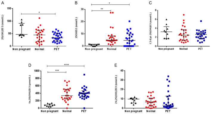
In serum, five serum vitamin D metabolites were consistently quantifiable in both the pregnant (normotensive and PET women) and non-pregnant groups: 25(OH)D3, 25(OH)D2, 1,25(OH)2D3, 24,25(OH)2D3, 3-epi-25(OH)D3, as summarised in Fig. 1 and Table 2. Considering maternal ‘vitamin D status’, the Institute of Medicine (IOM) definition of vitamin D ‘deficiency’ is 25(OH)D <20 ng/mL (50 nM/L) and ‘insufficiency’ as 25(OH)D >20 ng/mL but <30 ng/mL (75 nM/L) (42). In the normotensive pregnancy group, 14 (56.0%) women were defined as vitamin D deficient and 8 (32.0%) insufficient. In the PET group, 18 (72.0%) were vitamin D deficient and 6 (24.0%) insufficient. Although median 25(OH)D3 concentrations were lower in the PET group (median 33.1, IQR 20.5–50.8 nmol/L) compared to the control pregnancy group (44.7, 19.1–63.5 nmol/L), this was not significant (P = 0.240). Conversely, in the non-pregnant group, all women were sufficient (46.8; 42.8–91.0 nmol/L), with 25(OH)D3 levels significantly higher than values for PET (P = 0.04).
Figure 1.
Serum vitamin D metabolites in non-pregnant women and pregnant women at 15-week gestation. Serum concentrations of (A) 25-hydroxyvitamin D3 (25(OH)D3) nmol/L; (B) 25-hydroxyvitamin D2 (25(OH)D2) nmol/L; (C) 3-epi-25(OH)D3 nmol/L; (D) 1,25-dihydroxyvitamin D3 (1,25(OH)2D3) pmol/L; (E) 24,25-dihydroxyvitamin D3 (24,25(OH)2D3) nmol/L. Sample groups were non-pregnant (black; n = 9), normotensive pregnancies (red; n = 25) and those who later developed pre-eclampsia (blue; n = 25). Median with interquartile range are shown. Statistically significant variations are indicated, *P < 0.05, **P < 0.01, ***P < 0.001, ****P < 0.0001.
Table 2.
Summary of serum vitamin D metabolites in pregnant women at 15-week gestation and non-pregnant controls.
| Non-pregnant (n = 9) (median; IQR) | Control (n = 25) (median; IQR) | PET (n = 25) (median; IQR) | |
|---|---|---|---|
| 25(OH)D3 (nmol/L) | 46.8 (42.8–91) | 44.7 (19.1–63.5) | 33.1 (20.5–50.8) |
| 25(OH)D2 (nmol/L) | 1.17 (0–1.6) | 4.8 (4.2–8.3) | 4.7 (0–10.0) |
| 1,25(OH)2D3 (pmol/L) | 85.6 (47.3–117.4) | 336.3 (245.5–508.4) | 388.8 (304.2–468.4) |
| C3-Epi-25(OH)D3 (nmol/L) | 3.2 (1.7–4.4) | 2.5 (1.3–3.7) | 2.6 (1.7–3.1) |
| 24,25(OH)2D3 (nmol/L) | 9.7 (5.5–10.7) | 6.5 (2.07–10.7) | 3.2 (1.37–12.9) |
Comparison of serum concentrations of 25-hydroxyvitamin D3 (25(OH)D3) nmol/L, 25-hydroxyvitamin D2 (25(OH)D3) nmol/L, 1,25-dihydroxyvitamin D3 (1,25(OH)2D3) pmol/L, 3-epi-25(OH)D3 nmol/L, 24,25-dihydroxyvitamin D3 (24,25(OH)2D3) nmol/L in non-pregnant (n = 9), healthy normotensive pregnancies (n = 25) and those who later developed pre-eclampsia (PET; n = 25), with median values with interquartile range interval shown.
Consistent with previous studies (38), a significant seasonal difference in serum 25(OH)D3 was observed, with higher concentrations in summer (median 50.8; IQR 39.3–59.7 nmol/L) than winter (21.3; 14.1–41.7 nmol/L) (P = 0.0004). Sub-group analysis revealed that in the PET group, women pregnant during winter (24.8; 15.4–37.0 nmol/L) had significantly lower 25(OH)D3 levels than those pregnant in summer (50.7; 33.1–57.3 nmol/L) (P = 0.002). In the normotensive group, 25(OH)D3 levels were again lower in winter (20.8; 9.9–63.2 nmol/L) than summer (55.2; 42.0–64.6 nmol/L), almost reaching significance (P = 0.05).
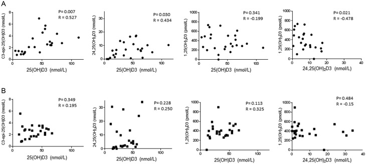
Serum concentrations of 25(OH)D2 were similar in the normotensive (4.8; 4.2–8.3 nmol/L) and PET (4.7; 0–10.0 nmol/L) groups (P = 0.352), and were, as anticipated, much lower than circulating 25(OH)D3 levels. No significant correlation between serum 25(OH)D2 and 25(OH)D3 in either the normotensive (r = −0.15, P = 0.48) or PET (r = 0.00, P > 0.10) groups was measured. 25(OH)D2 levels were significantly lower in the non-pregnant group comparative to both the PET (P = 0.02) and normotensive women (P = 0.001). There was no significant difference in serum concentrations of 3-epi-25(OH)D3 between the normotensive pregnant (2.5; 1.3–3.7 nmol/L), PET (2.6; 1.7–3.1 nmol/L) and non-pregnant (3.2; 1.7–4.4 nmol/L) cohorts. There was a significant positive correlation between 25(OH)D3 and 3-epi-25(OH)D3 in the normotensive pregnancy group (r = 0.645, P = 0.0005), but this was not evident in women who developed PET (r = 0.195, P = 0.349) (Fig. 2).
Figure 2.
Effect of maternal vitamin D status (serum 25-hydroyxvitamin D3) upon other serum vitamin D metabolites at 15-week gestation; comparative analysis in healthy pregnant controls and prospective PET cases. Serum concentrations of 25-hydroxyvitamin D3 (25(OH)D3) were correlated with C3-epi-25(OH)D3 (nmol/L), 1,25-dihydroxyvitamin D3 (1,25(OH)2D3)(pmol/L) and 24,25-dihydroxyvitamin D3 (24,25(OH)2D3) (nmol/L) in healthy pregnant controls (A) and prospective pre-eclampsia (PET) cases (B). Statistically significant correlations are indicated as P values, with Pearson R values shown.
Figure 3.
Relationship between excreted urinary vitamin D metabolites. Urine concentrations 25(OH)D3 were correlated with 24,25(OH)2D3 (nmol/L) in non-pregnant (A), healthy pregnant controls (B) and prospective pre-eclampsia (PET) cases (C). All nmol/L. Statistically significant correlations are indicated as P values, with Pearson R values shown.
Distinct from previous publications from the SCOPE study (43), 1,25(OH)2D3 and 24,25(OH)D2D3 concentrations were both quantifiable in serum samples. No significant difference in 1,25(OH)2D3 concentrations was measured in the PET (388.8; 304.2–468.4 pmol/L) comparative to normotensive pregnant group (336.3; 245.5–508.4 pmol/L). Consistent with previous reports (21), 1,25(OH)2D3 levels were significantly higher in both the PET (P < 0.0001) and normotensive (P = 0.0005) women compared to the non-pregnant group (85.6; 47.3–117.4 pmol/L).
No significant difference in serum 24,25(OH)2D3 was observed across the 3 groups. Similar to 1,25(OH)2D3, no difference in 24,25(OH)2D3 circulating concentrations in the PET (3.2; 1.4–12.9) and normotensive (6.5; 2.1–10.7) groups was measured. However, as summarised in Fig. 2, serum 25(OH)D3 levels significantly correlated with 24,25(OH)2D3 (r = 0.43, P = 0.03) in the normotensive women, whilst in those who developed PET no similar correlation (r = 0.25, P = 0.23) was observed. The significant negative relationship between 24,25(OH)2D3 and 1,25(OH)2D3 measured in the normotensive group (r = −0.48, P = 0.02) was lost in those who developed PET (r = −0.15, P = 0.484). No correlation between serum 25(OH)D3 and 1,25(OH)2D3 was observed for either group.
Urinary vitamin D analysis
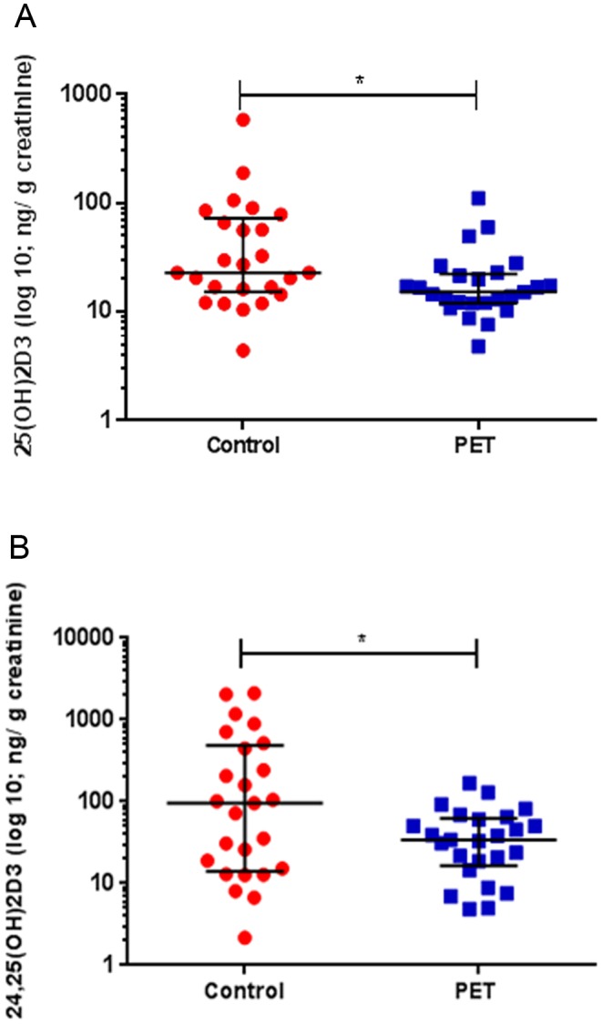
Urinary 25(OH)D3 and 24,25(OH)2D3 were consistently quantifiable in both pregnant and non-pregnant groups. A significant positive correlation between urinary 24,25(OH)2D3 and 25(OH)D3 concentrations was measured across the non-pregnant (r = 0.90, P = 0.002), normotensive (r = 0.64, P = 0.0006) and PET groups (r = 0.65, P = 0.0005) (Fig. 3). As summarised in Fig. 4, urinary 25(OH)D3 concentrations were significantly lower in the PET group (15.2; 12.0–22.2 ng/g creatinine) compared to normotensive pregnant women (22.9; 15.3–72.1 ng/g creatinine) (P = 0.018). Concentrations of urinary 24,25(OH)2D3 were similarly significantly reduced in those women who developed PET (34.1; 16.6–62.8 ng/g creatinine) (P = 0.018) (Fig. 4 and Table 3). Measurement of the metabolites 1,25(OH)2D3 and 23,25(OH)2D3 was incorporated into the method, but these analytes could not be quantified as concentrations were below the lower limit of detection.
Figure 4.

Urine vitamin D metabolite analysis in pregnant women at 15-week gestation. Urinary concentrations of (A) 25-hydroxyvitamin D3 (25(OH)D3) nmol/L and (B) 24,25-dihydroxyvitamin D3 (24,25(OH)2D3) nmol/L normalised for urinary creatinine (ng/g creatinine) for matched normotensive pregnancies (red) and those who later developed pre-eclampsia (PET; blue). Median with interquartile range values is shown. Statistically significant variations are indicated, *P < 0.05.
Table 3.
Urinary concentrations of 25(OH)D3 nmol/L and 24,25(OH)2D3 nmol/L.
| Non-pregnant | Control | PET | |
|---|---|---|---|
| 25(OH)D3 | 55.8 (14.3–84.7) | 22.9 (14.8–63.5) | 14.8 (11.9–22.5) |
| 24,25(OH)2D3 | 55.4 (22.4–118.8) | 84.1 (13.5–395.9) | 35.6 (15.5–63.7) |
Urinary vitamin D metabolite concentrations were normalised for urinary creatinine (ng/g creatinine) in non-pregnant controls (n = 9), normotensive pregnancies (n = 25) and those who later developed pre-eclampsia (PET; n = 25). Mean values with interquartile range are shown.
Discussion
Low maternal serum vitamin D concentrations in early pregnancy have been associated with an increased risk of PET (28), but the mechanisms underlying this remain unclear. Previously, we have demonstrated that in pregnant women with PET, multiple vitamin D metabolic pathways are dysregulated comparative to healthy normal pregnancy in the 3rd trimester and that this is evident for both serum and placental tissue vitamin D metabolites (21). Whilst placental vitamin D analysis offers the novel opportunity to delineate metabolism directly at the maternal–fetal interface, the clinical applications of this are limited due to the inaccessibility of this tissue throughout normal pregnancy. Given the prominent alterations in circulating vitamin D physiology across pregnancy, additional methods to ascertain vitamin D metabolism across gestation may be informative.
To advance these observations, we performed a detailed comparative analysis of serum vitamin D metabolites in a cohort of nulliparous ‘low-risk’ pregnancies at 15-week gestation of which half prospectively developed PET. To further enhance this approach, a novel urinary vitamin D metabolite quantification method was incorporated. Given the anticipated low concentrations of steroid metabolites present in urine, the analytical method employed utilised a derivatization procedure using PTAD to enhance both the sensitivity and separation of individual metabolites.
Consistent with our own data set in the West Midlands (21), vitamin D deficiency was highly prevalent in the SCOPE cohort from Ireland, particularly in winter months, with 64% (n = 32) of pregnant women having 25(OH)D3 levels <50 nmol/L at 15-week gestation. We anticipate that this would be higher still if the cohort included pregnant women with darker skin pigmentation as demonstrated in large epidemiological studies (44). Despite current clinical recommendations for pregnant women to take daily vitamin D supplementation (45, 46), in this cohort only 20% of women reported taking preconception vitamin D supplementation and by the 1st trimester adherence to supplementation advice dropped to 18% of women.
In previous published work, the SCOPE whole dataset (n = 1768) which similarly utilised LC MS–MS to measure circulating 25(OH)D3, 3-epi-25(OH)D3 and 25(OH)D2 concluded that in women with 25(OH)D3 levels >75 nM a protective effect (adjusted OR: 0.64; 95% CI: 0.43, 0.96) upon PET plus SGA outcome is evident following adjustment for potential confounding factors, including sociodemographic status, season, ethnicity, smoking status, exercise frequency and BMI (43). In the smaller subset of the SCOPE cohort assessed here only 3 (12.0%) of the pregnant cohort, none of which were in the PET group (maximum 25(OH)D3 = 60.9 nmol/L) had 25(OH)D3 levels >75 nmol/L. However, circulating 25(OH)D3 concentrations in those who developed PET were only statistically lower than the non-pregnant group. This may simply reflect the smaller cohort size resulting in a type 1 error, but may also be due to the heterogeneity of the PET cohort with respect to both timing of disease onset and progression. Our findings are however consistent with Powe and coworkers (47), who similarly concluded there was no significant difference in 25(OH)D3 levels in pregnant women who subsequently developed PET compared to those who remained normotensive (27.4 ± 1.9 vs 28.8 ± 0.80; P = 0.435). In this particular study, DBP and free 25(OH)D levels were also assessed, with no difference between the PET and normotensive group data (47). As 25(OH)D3 reflects total body stores, the levels of this metabolite may be preserved during the early stages of PET. It appears that 25(OH)D3 alone is unlikely to be an informative vitamin D metabolite within the context of ascertaining potential risk of PET.
Although we did not observe any statistically significant differences in concentrations of serum vitamin D metabolites between the two pregnancy groups, the associations between these metabolites varied significantly. In women who developed PET, there was no positive correlation between serum 25(OH)D3 and the vitamin D catabolites 3-epi-25(OH)D3 and 24,25(OH)2D3. Albeit not clearly understood, alternative metabolism of vitamin D via epimerisation to 3-epi-25(OH)D3 results in the formation of 3-epi-1,25(OH)2D3, which binds to VDR to activate target gene transcription. Importantly, 3-epi-1,25(OH)2D3 appears a less potent VDR agonist than 1,25(OH)2D3, which may have physiological consequences (48). In normal pregnancy, 3-epi-25(OH)2D3 appears directly linked to 25(OH)D3 concentrations (43); however, in the SCOPE cohort this relationship was lost only in those women who developed PET.
A trend towards elevated serum 1,25(OH)2D3 was also observed in the PET samples. This may reflect a compensatory response to lower 25(OH)D3, but may also contribute to depletion of 25(OH)D3. Healthy pregnancy is characterised by a drive towards 1,25(OH)2D3 production (21), and there is also evidence of increased upregulation of CYP27B1 (1α-hydroxylase) and CYP24A1 (24α-hydroxylase) in the placenta of women with PET (4). Increased metabolism of 25(OH)D3 to 1,25(OH)2D3 may also be secondary to decreased total serum calcium concentrations, which arise during normal human pregnancy (49). Within the context of PET, serum calcium concentrations are usually significantly reduced (50). A prospective study measuring the urine calcium/creatinine (Ca:Cr) clearance ratio also found women with PET excrete significantly less calcium (n = 60) compared to normotensive controls (51). In the current study, this may account for enhanced renal 25(OH)D3 re-uptake, limiting metabolite excretion.
Intriguingly, serum levels of 25(OH)D2 were significantly higher in both pregnancy groups relative to non-pregnant controls. The explanation for this is unclear as vitamin D2 is principally obtained from plants and mushrooms. One possibility is that enhanced circulating 25(OH)D2 is due to dietary modifications undertaken by women when they are pregnant. Furthermore, when considering 25(OH)D3 and 25(OH)D2 together, no significant difference in total vitamin D status was measured. Therefore, despite significantly lower 25(OH)D3 concentrations in those pregnant women who developed PET compared to the non-pregnant group, overall vitamin D status was not altered. This is consistent with previous data from the Osteoporotic Fractures in Men Study, which found higher 25(OH)D2 levels not to correlate with higher total 25(OH)D, and that increased 25(OH)D2 concentrations were associated with lower 25(OH)D3 levels (P < 0.01) (52). Recent supplementation data similarly indicated that 25(OH)D2 supplementation decreased serum 25(OH)D3 levels (53). In serum, 25(OH)D2 binds to the serum DBP carrier with lower affinity, and this has been postulated as an explanation for the increased serum clearance of 25(OH)D2 relative to 25(OH)D3 (54). However, in the current study, it was noticeable that 25(OH)D2 was not quantifiable in urine samples, suggesting that renal handling of 25(OH)D2 bound to DBP is efficient enough to limit urinary excretion of 25(OH)D2. Re-absorption of 25(OH)D2 from glomerular filtrates into proximal tubules may lead to increased synthesis of 24,25(OH)2D2 and 1,25(OH)2D2, but as neither of these metabolites was measured in the current study, this remains to be confirmed.
To our knowledge, this is the first report of vitamin D metabolite quantification in the urine of pregnant women. The method utilised was adapted from that reported by Ogawa and coworkers who quantified 25(OH)D3 and 24,25(OH)2D3 in spot urine samples (1 mL) from healthy male subjects (n = 20) pre and post vitamin D supplementation (40). This method used the derivatization agent PTAD to quantify 25OHD3 and 24,25(OH)2D3 in a 700 µL spot urine. This optimised method presents a reference range for urinary vitamin D during pregnancy, along with comparison to circulating serum levels. To our knowledge, this is the first time circulating and urinary vitamin D has been compared for clinical analysis, describing changes in both circulating and excreted levels of vitamin D on pregnancy outcome. This method combined with serum analysis will provide a comprehensive assessment in vitamin D metabolism in clinical conditions related to vitamin D deficiency. This method was capable of quantifying urinary 25OHD3 and 24,25(OH)2D3; however, under the current method conditions, levels of 1α,25(OH)2D3 and 23,25(OH)2D3 could not be measured following derivatization, as these concentration were below the limits of detection. To assess the ability to quantify these analytes in urine, method development will be required on a later generation mass spectrometer that will enable reduced detection limits.
In pregnancy, urinary 24,25(OH)2D3 concentrations were approximately 3-fold higher than 25(OH)D3, representing the predominant excreted vitamin D metabolite. This was not evident in the non-pregnant group, for which urinary 25(OH)D3 and 24,25(OH)2D3 concentrations were comparable. Furthermore, in the healthy non-pregnant females, median urinary 25(OH)D3 concentrations were at least 2-fold higher than both pregnant groups, in particular, those who developed PET. Together, these findings are consistent with an increased role for 25(OH)D3 in pregnancy, with significantly enhanced classical and non-classical placental 1,25(OH)2D3 production and turnover from the 1st trimester (21, 22). Reduced 25(OH)D3 excretion may also reflect increased neonatal vitamin D metabolism, as 25(OH)D3 readily diffuses across the placenta principally to permit later fetal bone development and growth (55). In rat models, VDR expression is demonstrated from a very early stage in fetal development (56).
Urinary 25(OH)D3 and 24,25(OH)2D3 were significantly correlated in both the normotensive and PET groups. This is consistent with serum 25(OH)D3 and 24,25(OH)2D3 in normal pregnancy, as evidenced here and our previous study (21). However, in normal pregnancy, serum concentrations of 25(OH)D3 and 24,25(OH)2D3 did not correlate with their respective urinary concentrations. Given the number of known extra-renal sites of vitamin D storage and metabolism, including the placenta, this may be anticipated. Furthermore, the kidney has the capacity to actively reabsorb vitamin D metabolites in a DBP-megalin-dependent manner (57), and this may contribute to the lack of correlation between serum and urinary vitamin D metabolites.
In the current study, urinary vitamin D metabolite analysis suggests that dysregulation of vitamin D metabolism occurs at an early stage in women who go on to develop PET. Within the context of PET, both urinary 25(OH)D3 and 24,25(OH)2D3 concentrations were significantly decreased compared to those who were normotensive throughout pregnancy. As discussed, alongside this increased serum concentrations of 1,25(OH)2D3 were measured in those with PET. This increased vitamin D metabolism to 1,25(OH)2D3 may arise secondary to decreased serum calcium concentrations. Within the context of PET, serum calcium concentrations are significantly reduced. A prospective study measuring the urine calcium/creatinine (Ca:Cr) clearance ratio also found women with PET excrete significantly less calcium (n = 60) compared to normotensive controls (51). This may account for enhanced renal 25(OH)D3 re-uptake, limiting metabolite excretion.
Urinary metabolite concentrations are susceptible to variation by factors including hydration status and overall renal function. Twenty-four-hour urine sample collections are considered the gold-standard measurement, but are cumbersome and reliant upon strict compliance. Spot samples and first morning void are widely deemed acceptable for analyte measures, provided the effect of sample dilution is quantified and appropriately adjusted (58, 59). At present, no consensus upon which is most appropriate adjustment technique; however, creatinine remains commonly applied. This simply calculates the ratio to creatinine concentrations and does not account for temporal variations in creatinine excretion rates (59). The potential value of a urinary marker of vitamin D status has wider clinical implications outside of pregnancy, and certainly, this method will be highly transferrable.
Moving forward, serial serum and urinary analyses at a set time-point would provide a more detailed insight into the pathogenesis of vitamin D dysregulation. It would have been interesting to include in this cohort more early-onset PET cases (<34 week) (n = 1), which are typically more severe (60) and women of greater ethnic diversity (61). Subgroup analysis of those women who developed PET pre-term (≤37 week) (n = 9) did not reveal any significant differences with regards to the 5 major serum vitamin D metabolites measured (data not shown), but numbers were too small to draw any robust conclusions from this. The current study was based simply on the availability of sample material. In this setting, urinary 25(OH)D3 measurements with n = 25 per sample group, and a difference of means of 13.86 ng/g creatinine resulted in statistical power of 0.588 at α = 0.05. To reach power of >0.8 would have required n = 41 per sample group. Nevertheless, our preliminary data indicate dysregulation of vitamin D metabolism may precede clinical onset of PET. This was evident from spot urinary analyses, which offer novel insights into the underlying pathogenesis of vitamin D dysregulation in PET. From the data presented, we demonstrate that routine measurement of serum 25(OH)D3 alone provides only a limited perspective on the requirement for vitamin D in pregnancy. Detailed analysis of vitamin D metabolism, including renal catabolism and excretion is required.
Supplementary Material
Declaration of interest
The authors declare that there is no conflict of interest that could be perceived as prejudicing the impartiality of the research reported.
Funding
This study was supported by funding from NIH (AR063910, M H), Wellbeing of Women (RTF401, J A T) and a Royal Society Wolfson Merit Award (WM130118, M H).
Acknowledgements
The authors would like thank Prof. Louise Kenny (University College Cork) for facilitating access to the SCOPE samples. They would also like to acknowledge help from Prof. Cedric Shackleton in establishing the urinary vitamin D metabolite analyses. The authors declare that there is no conflict of interest that could be perceived as prejudicing the impartiality of the research reported. Vitabiotics Pregnacare generously supported this work through the charity Wellbeing of Women, which independently selected the research through its AMRC accredited peer review process led by its Research Advisory Committee. Dr Tamblyn and her work were not impacted or influenced in any way by its sources of funding.
References
- 1.Tranquilli AL, Brown MA, Zeeman GG, Dekker G, Sibai BM. The definition of severe and early-onset preeclampsia statements from the International Society for the Study of Hypertension in Pregnancy (ISSHP). Pregnancy Hypertension 2013. 3 44–47. ( 10.1016/j.preghy.2012.11.001) [DOI] [PubMed] [Google Scholar]
- 2.Khan KS, Wojdyla D, Say L, Gülmezoglu AM, Van Look PFA. WHO analysis of causes of maternal death: a systematic review. Lancet 2006. 367 1066–1074. ( 10.1016/S0140-6736(06)68397-9) [DOI] [PubMed] [Google Scholar]
- 3.Steegers EA, von Dadelszen P, Duvekot JJ, Pijnenborg R. Pre-eclampsia. Lancet 2010. 376 631–644. ( 10.1016/S0140-6736(10)60279-6) [DOI] [PubMed] [Google Scholar]
- 4.Mol BW, Roberts CT, Thangaratinam S, Magee LA, de Groot CJ, Hofmeyr GJ. Pre-eclampsia. Lancet 2016. 387 999–1011. ( 10.1016/S0140-6736(15)00070-7) [DOI] [PubMed] [Google Scholar]
- 5.Say L, Chou D, Gemmill A, Tunçalp Ö, Moller A-B, Daniels J, Gülmezoglu AM, Temmerman M, Alkema L. Global causes of maternal death: a WHO systematic analysis. Lancet Global Health 2014. 2 e323–e333. ( 10.1016/S2214-109X(14)70227-X) [DOI] [PubMed] [Google Scholar]
- 6.Carr A, Kershaw T, Brown H, Allen T, Small M. Hypertensive disease in pregnancy: an examination of ethnic differences and the Hispanic paradox. Journal of Neonatal-Perinatal Medicine 2013. 6 11–15. ( 10.3233/NPM-1356111) [DOI] [PubMed] [Google Scholar]
- 7.Fisher SJ. Why is placentation abnormal in preeclampsia? American Journal of Obstetrics and Gynecology 2015. 213 S115–S122. ( 10.1016/j.ajog.2015.08.042) [DOI] [PMC free article] [PubMed] [Google Scholar]
- 8.Rădulescu C, Bacârea A, Huţanu A, Gabor R, Dobreanu M. Placental growth factor, soluble fms-like tyrosine kinase 1, soluble endoglin, IL-6, and IL-16 as biomarkers in preeclampsia. Mediators of Inflammation 2016. 2016 3027363 ( 10.1155/2016/3027363) [DOI] [PMC free article] [PubMed] [Google Scholar]
- 9.Ghosh SK, Raheja S, Tuli A, Raghunandan C, Agarwal S. Combination of uterine artery Doppler velocimetry and maternal serum placental growth factor estimation in predicting occurrence of pre-eclampsia in early second trimester pregnancy: a prospective cohort study. European Journal of Obstetrics, Gynecology, and Reproductive Biology 2012. 161 144–151. ( 10.1016/j.ejogrb.2011.12.031) [DOI] [PubMed] [Google Scholar]
- 10.Wu P, van den Berg C, Alfirevic Z, O’Brien S, Röthlisberger M, Baker PN, Kenny LC, Kublickiene K, Duvekot JJ. Early pregnancy biomarkers in pre-eclampsia: a systematic review and meta-analysis. International Journal of Molecular Sciences 2015. 16 23035–23056. ( 10.3390/ijms160923035) [DOI] [PMC free article] [PubMed] [Google Scholar]
- 11.Ghosh SK, Raheja S, Tuli A, Raghunandan C, Agarwal S. Serum placental growth factor as a predictor of early onset preeclampsia in overweight/obese pregnant women. Journal of the American Society of Hypertension 2013. 7 137–148. ( 10.1016/j.jash.2012.12.006) [DOI] [PubMed] [Google Scholar]
- 12.Zhu XL, Wang J, Jiang RZ, Teng YC. Pulsatility index in combination with biomarkers or mean arterial pressure for the prediction of pre-eclampsia: Systematic literature review and meta-analysis. Annals of Medicine 2015. 47 414–422. ( 10.3109/07853890.2015.1059483) [DOI] [PubMed] [Google Scholar]
- 13.Lafayette RA, Druzin M, Sibley R, Derby G, Malik T, Huie P, Polhemus C, Deen WM, Myers BD. Nature of glomerular dysfunction in pre-eclampsia. Kidney International 1998. 54 1240–1249. ( 10.1046/j.1523-1755.1998.00097.x) [DOI] [PubMed] [Google Scholar]
- 14.Hussein W, Lafayette RA. Renal function in normal and disordered pregnancy. Current Opinion in Nephrology and Hypertension 2014. 23 46–53. ( 10.1097/01.mnh.0000436545.94132.52) [DOI] [PMC free article] [PubMed] [Google Scholar]
- 15.Ayansina D, Black C, Hall SJ, Marks A, Millar C, Prescott GJ, Wilde K, Bhattacharya S. Long term effects of gestational hypertension and pre-eclampsia on kidney function: record linkage study. Pregnancy Hypertension 2016. 6 344–349. ( 10.1016/j.preghy.2016.08.231) [DOI] [PMC free article] [PubMed] [Google Scholar]
- 16.Garovic VD. The role of the podocyte in preeclampsia. Clinical Journal of the American Society of Nephrology 2014. 9 1337–1340. ( 10.2215/CJN.05940614) [DOI] [PMC free article] [PubMed] [Google Scholar]
- 17.Müller-Deile J, Schiffer M. Preeclampsia from a renal point of view: insides into disease models, biomarkers and therapy. World Journal of Nephrology 2014. 3 169–181. ( 10.5527/wjn.v3.i4.169) [DOI] [PMC free article] [PubMed] [Google Scholar]
- 18.Kumar R, Tebben PJ, Thompson JR. Vitamin D and the kidney. Archives of Biochemistry and Biophysics 2012. 523 77–86. ( 10.1016/j.abb.2012.03.003) [DOI] [PMC free article] [PubMed] [Google Scholar]
- 19.Kumar R, Schaefer J, Grande JP, Roche PC. Immunolocalization of calcitriol receptor, 24-hydroxylase cytochrome P-450, and calbindin D28k in human kidney. American Journal of Physiology 1994. 266 F477–F485. ( 10.1152/ajprenal.1994.266.3.F477) [DOI] [PubMed] [Google Scholar]
- 20.Turner M, Barre PE, Benjamin A, Goltzman D, Gascon-Barre M. Does the maternal kidney contribute to the increased circulating 1,25-dihydroxyvitamin D concentrations during pregnancy? Mineral and Electrolyte Metabolism 1988. 14 246–252. [PubMed] [Google Scholar]
- 21.Tamblyn JA, Susarla R, Jenkinson C, Jeffery LE, Ohizua O, Chun RF, Chan SY, Kilby MD, Hewison M. Dysregulation of maternal and placental vitamin D metabolism in preeclampsia. Placenta 2017. 50 70–77. ( 10.1016/j.placenta.2016.12.019) [DOI] [PMC free article] [PubMed] [Google Scholar]
- 22.Evans KN, Bulmer JN, Kilby MD, Hewison M. Vitamin D and placental-decidual function. Journal of the Society for Gynecologic Investigation 2004. 11 263–271. ( 10.1016/j.jsgi.2004.02.002) [DOI] [PubMed] [Google Scholar]
- 23.Zehnder D, Evans KN, Kilby MD, Bulmer JN, Innes BA, Stewart PM, Hewison M. The ontogeny of 25-hydroxyvitamin D(3) 1alpha-hydroxylase expression in human placenta and decidua. American Journal of Pathology 2002. 161 105–114. ( 10.1016/S0002-9440(10)64162-4) [DOI] [PMC free article] [PubMed] [Google Scholar]
- 24.Ma R, Gu Y, Zhao S, Sun J, Groome LJ, Wang Y. Expressions of vitamin D metabolic components VDBP, CYP2R1, CYP27B1, CYP24A1, and VDR in placentas from normal and preeclamptic pregnancies. American Journal of Physiology: Endocrinology and Metabolism 2012. 303 E928–E935. ( 10.1152/ajpendo.00279.2012) [DOI] [PMC free article] [PubMed] [Google Scholar]
- 25.Tamblyn J, Hewison M, Wagner C, Bulmer J, Kilby M. Immunological role of vitamin D at the maternal–fetal interface. Journal of Endocrinology 2015. 224 R107–R121. ( 10.1530/JOE-14-0642) [DOI] [PubMed] [Google Scholar]
- 26.Saraf R, Morton SM, Camargo CA, Jr, Grant CC. Global summary of maternal and newborn vitamin D status – a systematic review. Maternal and Child Nutrition 2016. 12 647–668. ( 10.1111/mcn.12210) [DOI] [PMC free article] [PubMed] [Google Scholar]
- 27.Hollis BW, Wagner CL. Vitamin D deficiency during pregnancy: an ongoing epidemic. American Journal of Clinical Nutrition 2006. 84 273. [DOI] [PubMed] [Google Scholar]
- 28.Bodnar LM, Catov JM, Simhan HN, Holick MF, Powers RW, Roberts JM. Maternal vitamin D deficiency increases the risk of preeclampsia. Journal of Clinical Endocrinology and Metabolism. 2007. 92 3517–3522. ( 10.1210/jc.2007-0718) [DOI] [PMC free article] [PubMed] [Google Scholar]
- 29.Djekic-Ivankovic M, Weiler H, Jones G, Kaufmann M, Kaludjerovic J, Aleksic-Velickovic V, Mandić LM, Glibetic M. Vitamin D status in mothers with pre-eclampsia and their infants: a case-control study from Serbia, a country without a vitamin D fortification policy. Public Health Nutrition 2016. 20 1825–1835. ( 10.1017/S1368980016000409) [DOI] [PMC free article] [PubMed] [Google Scholar]
- 30.Burris HH, Rifas-Shiman SL, Kleinman K, Litonjua AA, Huh SY, Rich-Edwards JW, Camargo CA, Jr, Gillman MW. Vitamin D deficiency in pregnancy and gestational diabetes mellitus. American Journal of Obstetrics and Gynecology 2012. 207 182e1–182e8. ( 10.1016/j.ajog.2012.05.022) [DOI] [PMC free article] [PubMed] [Google Scholar]
- 31.Bodnar LM, Catov JM, Zmuda JM, Cooper ME, Parrott MS, Roberts JM, Marazita ML, Simhan HN. Maternal serum 25-hydroxyvitamin D concentrations are associated with small-for-gestational age births in white women. Journal of Nutrition 2010. 140 999–1006. ( 10.3945/jn.109.119636) [DOI] [PMC free article] [PubMed] [Google Scholar]
- 32.Wagner CL, Baggerly C, McDonnell S, Baggerly KA, French CB, Baggerly L, Hamilton SA, Hollis BW. Post-hoc analysis of vitamin D status and reduced risk of preterm birth in two vitamin D pregnancy cohorts compared with South Carolina March of Dimes 2009–2011 rates. Journal of Steroid Biochemistry and Molecular Biology 2016. 155 245–251. ( 10.1016/j.jsbmb.2015.10.022) [DOI] [PMC free article] [PubMed] [Google Scholar]
- 33.Hypponen E, Cavadino A, Williams D, Fraser A, Vereczkey A, Fraser WD, Bánhidy F, Lawlor D, Czeizel AE. Vitamin D and pre-eclampsia: original data, systematic review and meta-analysis. Annals of Nutrition and Metabolism 2013. 63 331–340. ( 10.1159/000358338) [DOI] [PubMed] [Google Scholar]
- 34.Harvey NC, Holroyd C, Ntani G, Javaid K, Cooper P, Moon R, Cole Z, Tinati T, Godfrey K, Dennison E, et al Vitamin D supplementation in pregnancy: a systematic review. Health Technology Assessment 2014. 18 1–190. ( 10.3310/hta18450) [DOI] [PMC free article] [PubMed] [Google Scholar]
- 35.Thota C, Menon R, Fortunato SJ, Brou L, Lee JE, Al-Hendy A. 1,25-Dihydroxyvitamin D deficiency is associated with preterm birth in African American and Caucasian women. Reproductive Sciences 2014. 21 244–250. ( 10.1177/1933719113493513) [DOI] [PMC free article] [PubMed] [Google Scholar]
- 36.Bennett SE, Casey C, McPeake J, McCance DR, Manderson JG, McGinty A. 3-Epi-25 hydroxyvitamin D in pregnancy. Pregnancy Hypertension 2014. 4 236 ( 10.1016/j.preghy.2014.03.021) [DOI] [PubMed] [Google Scholar]
- 37.Shah AD, Hsiao EC, O’Donnell B, Salmeen K, Nussbaum R, Krebs M, Baumgartner-Parzer S, Kaufmann M, Jones G, Bikle DD, et al Maternal hypercalcemia due to failure of 1,25-dihydroxyvitamin-D3 catabolism in a patient with CYP24A1 mutations. Journal of Clinical Endocrinology and Metabolism 2015. 100 2832–2836. ( 10.1210/jc.2015-1973) [DOI] [PMC free article] [PubMed] [Google Scholar]
- 38.Kenny LC, Black MA, Poston L, Taylor R, Myers JE, Baker PN, McCowan LM, Simpson NA, Dekker GA, Roberts CT, et al Early pregnancy prediction of preeclampsia in nulliparous women, combining clinical risk and biomarkers: the Screening for Pregnancy Endpoints (SCOPE) international cohort study. Hypertension 2014. 64 644–652. ( 10.1161/HYPERTENSIONAHA.114.03578) [DOI] [PubMed] [Google Scholar]
- 39.Jenkinson C, Taylor AE, Hassan-Smith ZK, Adams JS, Stewart PM, Hewison M, Keevil BG. High throughput LC–MS/MS method for the simultaneous analysis of multiple vitamin D analytes in serum. Journal of Chromatography B 2016. 1014 56–63. ( 10.1016/j.jchromb.2016.01.049) [DOI] [PMC free article] [PubMed] [Google Scholar]
- 40.Ogawa S, Ooki S, Shinoda K, Higashi T. Analysis of urinary vitamin D(3) metabolites by liquid chromatography/tandem mass spectrometry with ESI-enhancing and stable isotope-coded derivatization. Analytical and Bioanalytical Chemistry 2014. 406 6647–6654. ( 10.1007/s00216-014-8095-y) [DOI] [PubMed] [Google Scholar]
- 41.Peake M, Whiting M. Measurement of serum creatinine – current status and future goals. Clinical Biochemist Reviews 2006. 27 173–184. [PMC free article] [PubMed] [Google Scholar]
- 42.Holick MF, Binkley NC, Bischoff-Ferrari HA, Gordon CM, Hanley DA, Heaney RP, Heaney RP, Murad MH, Weaver CM. & Endocrine Society. Evaluation, treatment, and prevention of vitamin D deficiency: an Endocrine Society Clinical Practice Guideline. Journal of Clinical Endocrinology and Metabolism 2011. 96 1911–1930. ( 10.1210/jc.2011-0385) [DOI] [PubMed] [Google Scholar]
- 43.Kiely ME, Zhang JY, Kinsella M, Khashan AS, Kenny LC. Vitamin D status is associated with uteroplacental dysfunction indicated by pre-eclampsia and small-for-gestational-age birth in a large prospective pregnancy cohort in Ireland with low vitamin D status. American Journal of Clinical Nutrition 2016. 104 354–361. ( 10.3945/ajcn.116.130419) [DOI] [PubMed] [Google Scholar]
- 44.Bodnar LM, Simhan HN, Powers RW, Frank MP, Cooperstein E, Roberts JM. High prevalence of vitamin D insufficiency in black and white pregnant women residing in the northern United States and their neonates. Journal of Nutrition 2007. 137 447–452. [DOI] [PMC free article] [PubMed] [Google Scholar]
- 45.National Institute for Health and Care Excellence. Antenatal care for uncomplicated preganancies. NICE clinical guideline 62. London, UK: NICE, 2008. (available at: https://www.nice.org.uk/guidance/cg62) [Google Scholar]
- 46.O’Reilly-Marshall A. Antenatal Care. Nutrition in pregnancy. Dublin, Ireland: Irish College of General Practitioners, 2010. (available at: https://www.icgp.ie/assets/21/7FB21F16-19B9-E185-83915661D103354F_document/DL23-28.pdf) [Google Scholar]
- 47.Powe CE, Seely EW, Rana S, Bhan I, Ecker J, Karumanchi SA, Thadhani R. First trimester vitamin D, vitamin D binding protein, and subsequent preeclampsia. Hypertension 2010. 56 758–763. ( 10.1161/HYPERTENSIONAHA.110.158238) [DOI] [PMC free article] [PubMed] [Google Scholar]
- 48.Messerlian S, Gao X, St-Arnaud R. The 3-epi- and 24-oxo-derivatives of 1alpha,25 dihydroxyvitamin D(3) stimulate transcription through the vitamin D receptor. Journal of Steroid Biochemistry and Molecular Biology 2000. 72 29–34. ( 10.1016/S0960-0760(99)00148-X) [DOI] [PubMed] [Google Scholar]
- 49.Soma-Pillay P, Catherine N-P, Tolppanen H, Mebazaa A, Tolppanen H, Mebazaa A. Physiological changes in pregnancy. Cardiovascular Journal of Africa 2016. 27 89–94. ( 10.5830/CVJA-2016-021) [DOI] [PMC free article] [PubMed] [Google Scholar]
- 50.Kanagal DV, Rajesh A, Rao K, Devi UH, Shetty H, Kumari S, Shetty PK. Levels of serum calcium and magnesium in pre-eclamptic and normal pregnancy: a study from coastal India. Journal of Clinical and Diagnostic Research 2014. 8 Obib1–Obib4. ( 10.7860/JCDR/2014/8872.4537) [DOI] [PMC free article] [PubMed] [Google Scholar]
- 51.Ozcan T, Kaleli B, Ozeren M, Turan C, Zorlu G. Urinary calcium to creatinine ratio for predicting preeclampsia. American Journal of Perinatology 1995. 12 349–351. ( 10.1055/s-2007-994494) [DOI] [PubMed] [Google Scholar]
- 52.Swanson CM, Nielson CM, Shrestha S, Lee CG, Barrett-Connor E, Jans I, Cauley JA, Boonen S, Bouillon R, Vanderschueren D, et al Higher 25(OH)D(2) is associated with lower 25(OH)D(3) and 1,25(OH)(2)D(3). Journal of Clinical Endocrinology and Metabolism 2014. 99 2736–2744. ( 10.1210/jc.2014-1069) [DOI] [PMC free article] [PubMed] [Google Scholar]
- 53.Demetriou ET, Travison TG, Holick MF. Treatment with 50,000 IU vitamin D(2) every other week and effect on serum 25-hydroxyvitamin D(2), 25-hydroxyvitamin D(3), and total 25-hydroxyvitamin D in a clinical setting. Endocrine Practice 2012. 18 399–402. ( 10.4158/EP11268.OR) [DOI] [PubMed] [Google Scholar]
- 54.Jones KS, Assar S, Harnpanich D, Bouillon R, Lambrechts D, Prentice A, Schoenmakers I. 25(OH)D(2) half-life is shorter than 25(OH)D(3) half-life and is influenced by dbp concentration and genotype. Journal of Clinical Endocrinology and Metabolism 2014. 99 3373–3381. ( 10.1210/jc.2014-1714) [DOI] [PMC free article] [PubMed] [Google Scholar]
- 55.Nicolaidou P, Hatzistamatiou Z, Papadopoulou A, Kaleyias J, Floropoulou E, Lagona E, Tsagris V, Costalos C, Antsaklis A. Low vitamin D status in mother-newborn pairs in Greece. Calcified Tissue International 2006. 78 337–342. ( 10.1007/s00223-006-0007-5) [DOI] [PubMed] [Google Scholar]
- 56.Salle BL, Delvin EE, Lapillonne A, Bishop NJ, Glorieux FH. Perinatal metabolism of vitamin D. American Journal of Clinical Nutrition 2000. 71 1317s–1324s. [DOI] [PubMed] [Google Scholar]
- 57.Nykjaer A, Dragun D, Walther D, Vorum H, Jacobsen C, Herz J, Melsen F, Christensen EI, Willnow TE. An endocytic pathway essential for renal uptake and activation of the steroid 25-(OH) vitamin D3. Cell 1999. 96 507–515. ( 10.1016/S0092-8674(00)80655-8) [DOI] [PubMed] [Google Scholar]
- 58.Rivera-Núñez Z, Meliker JR, Linder AM, Nriagu JO. Reliability of spot urine samples in assessing arsenic exposure. International Journal of Hygiene and Environmental Health 2010. 213 259–264. ( 10.1016/j.ijheh.2010.03.003) [DOI] [PMC free article] [PubMed] [Google Scholar]
- 59.Hidalgo AA, Deeb KK, Pike JW, Johnson CS, Trump DL. Dexamethasone enhances 1alpha,25-dihydroxyvitamin D3 effects by increasing vitamin D receptor transcription. Journal of Biological Chemistry 2011. 286 36228–36237. ( 10.1074/jbc.M111.244061) [DOI] [PMC free article] [PubMed] [Google Scholar]
- 60.Lisonkova S, Joseph KS. Incidence of preeclampsia: risk factors and outcomes associated with early- versus late-onset disease. American Journal of Obstetrics and Gynecology 2013. 209 544.e1–544.e12. ( 10.1016/j.ajog.2013.08.019) [DOI] [PubMed] [Google Scholar]
- 61.Goodwin AA, Mercer BM. Does maternal race or ethnicity affect the expression of severe preeclampsia? American Journal of Obstetrics and Gynecology 2005. 193 973–978. ( 10.1016/j.ajog.2005.05.047) [DOI] [PubMed] [Google Scholar]
Associated Data
This section collects any data citations, data availability statements, or supplementary materials included in this article.


 This work is licensed under a

