Abstract
Pachyonychia congenita (PC) is a cutaneous disorder primarily characterized by nail dystrophy and painful palmoplantar keratoderma. PC is caused by mutations in KRT6A, KRT6B, KRT6C, KRT16, and KRT17, a set of keratin genes expressed in the nail bed, palmoplantar epidermis, oral mucosal epithelium, hair follicle and sweat gland. RNA-seq analysis revealed that all PC-associated keratins (except for Krt6c that does exist in the mouse genome) are expressed in the mouse enamel organ. We further demonstrated that these keratins are produced by ameloblasts and are incorporated into mature human enamel. Using genetic and intraoral examination data from 573 adults and 449 children, we identified several missense polymorphisms in KRT6A, KRT6B and KRT6C that lead to a higher risk for dental caries. Structural analysis of teeth from a PC patient carrying a p.Asn171Lys substitution in keratin-6a (K6a) revealed disruption of enamel rod sheaths resulting in altered rod shape and distribution. Finally, this PC-associated substitution as well as more frequent caries-associated SNPs, found in two of the KRT6 genes, that result in p.Ser143Asn substitution (rs28538343 in KRT6B and rs151117600 in KRT6C), alter the assembly of K6 filaments in ameloblast-like cells. These results identify a new set of keratins involved in tooth enamel formation, distinguish novel susceptibility loci for tooth decay and reveal additional clinical features of pachyonychia congenita.
Author summary
Tooth decay, more commonly known as dental cavities, is the most common chronic disease worldwide, both in children and in adults. It consists in the destruction of tooth enamel, the outer layer of the teeth, by acid-producing bacteria. Enamel is the hardest tissue in the body, comprised of 96% minerals. However, it contains a small fraction of proteins that is important for its resistance to mechanical stress and decay. Here we show that this protein fraction contains a set of structural proteins (K6a, K6b, K6c, K16 and K17) that belong to the keratin family and are present specifically in the skin of the palms and soles, as well as in nails. We further show that common genetic mutations that affect the composition of these proteins lead to an increased number of cavities. Rare mutations in these keratins lead to a human disease called pachyonychia congenita (PC) and characterized by severe nail malformations and lesions in the skin of the palms and soles. Analysis of wisdom teeth from one of these patients showed that their enamel exhibited structural defects. These results demonstrate that these keratins are important components of tooth enamel and that common genetic variants in the genes that encode them influence tooth decay risk in the general population.
Introduction
Tooth enamel is made of 96% hydroxyapatite minerals, which makes it the hardest tissue in the human body. Enamel is also the first compartment of the tooth to be attacked by dental caries, a chronic disease that affects 42% of children and 92% of adults, with various degrees of severity (number of teeth and tooth surfaces affected) in the general population. Dental caries is initiated at the surface of the tooth by bacteria metabolizing food residues and releasing acids that dissolve enamel minerals [1]. Even though dental caries is influenced by environmental and behavioral factors, there is clear evidence that susceptibility to caries is also driven by host genetic factors [1–3], and genome-wide association studies (GWASs) have revealed genetic variants associated with increased susceptibility to tooth decay [4–8]. These genetic factors may influence the quality of dental tissues and ability to resist carious attacks, may impact other aspects of the oral environment such as the quality of the saliva, enamel pellicle and oral microbiome, and may differ between the primary and permanent dentitions [9].
Tooth enamel is produced in two phases [10]: first, a secretion phase during which enamel matrix proteins are secreted and deposited in a highly structured manner to form enamel rods; and second, a maturation phase during which most enamel matrix proteins are degraded to make space for the full expansion of hydroxyapatite minerals. After maturation, the enamel is left with only 1% of proteins that are abundant near the dentin-enamel junction (DEJ) and expand throughout the enamel as thin layers of enamel rod sheaths located at the interrod region [11]. This organic material has been shown to play a crucial role in the biomechanical properties of enamel [12, 13] and in the resistance to caries [14, 15]. Until recently, the exact composition of the insoluble fraction in this organic material had been uncharacterized, even though there was strong evidence that the highly crosslinked proteins present in mature enamel had biochemical properties similar to those of keratins [11, 16–18]. We showed previously that the organic material in mature enamel is in part composed of epithelial hair keratins, and that missense mutations in KRT75, previously linked to common hair disorders, were associated with increased susceptibility to dental caries [19].
In the present study, we focus on the presence in enamel of another set of keratins encoded by genes mutated in pachyonychia congenita (PC), a cutaneous disorder characterized by nail dystrophy and painful palmoplantar keratoderma [20, 21]. Additional features of this disease may include oral leukokeratosis, follicular keratosis, cysts, hyperhidrosis, and natal teeth. Some of these phenotypic traits are consistent with the expression pattern of the keratins involved. Herein we present novel findings that relate this set of keratins to the development of tooth enamel and to the susceptibility to tooth decay.
Results
Pachyonychia congenita-associated keratins are produced by ameloblasts and incorporated into mature tooth enamel
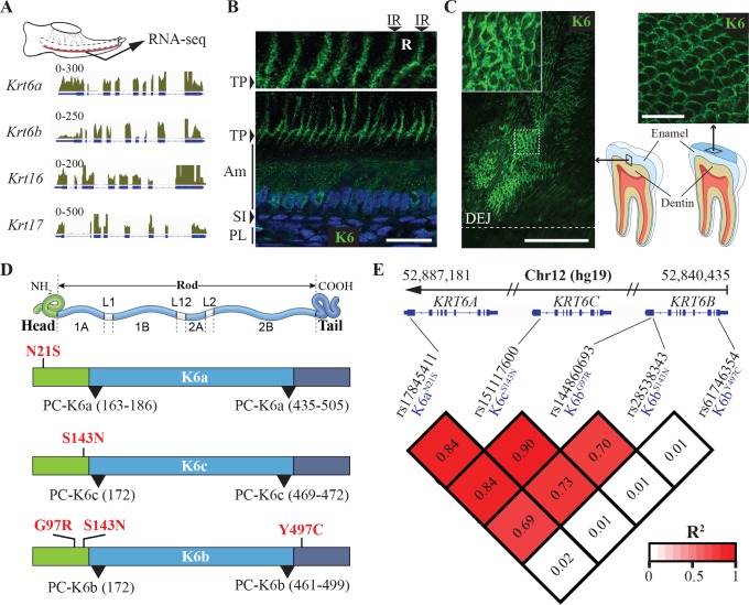
In a previous study, we determined through RNA-seq analysis that subsets of epithelial keratins were expressed in the enamel organ in mouse [19]. Of particular interest was the expression of Krt6a, Krt6b, Krt16 and Krt17 (Fig 1A), a set of keratin genes encoding keratin-6a (K6a), keratin-6b (K6b), keratin-16 (K16) and keratin-17 (K17), respectively, and in which mutations in humans lead to pachyonychia congenita (PC-K6a, OMIM #615726; PC-K6b, OMIM #615728; PC-K16, OMIM #167200; PC-K17, OMIM #167210), characterized by nail dystrophy and painful palmoplantar keratoderma [20, 21]. In humans, the KRT6 family includes a third member (KRT6C, encoding K6c), mutations in which have been associated with a milder form of PC with no/minor nail defects (PC-K6c) that was initially reported as palmoplantar keratoderma, non-epidermolytic, focal or diffuse (PPKNEFD, OMIM #615735).
Fig 1. K6 proteins in enamel and genetic association between missense SNPs in KRT6 genes and tooth decay experience.
(A) RNA-seq data from mouse enamel organ showing expression of Krt6a, Krt6b, Krt16 and Krt17 in the tissue. The schematics at the top shows a mouse mandible at postnatal day 10 from which the enamel organ (red) was micro-dissected for RNA extraction. Tracks represent the alignment of the RNA-seq reads to the mouse genome for the Krt6a, Krt6b, Krt16 and Krt17 loci. (B) Immunohistochemical analysis of K6 distribution in rat enamel organ at postnatal day 10. Anti-K6 antibody recognizes all members of the K6 family (K6a and K6b in rodents). Nuclei are stained with DAPI (blue). Inset at the top shows enlarged view of Tomes’ processes. PL, papillary layer; SI, stratum intermedium; Am, ameloblasts; TP, Tomes’ processes; R, rod; IR, interrod. Scale bar: 50 μm. (C) Immunohistochemical detection of K6 on polished sections of human third molars. Antibody raised in guinea pig against the N-terminal of K6 (recognizes K6a, K6b and K6c in humans) was used (green). As indicated by the schematics on the bottom right corner, the image in the left panel was acquired from mesial-distal sections of human third molars and shows the area of enamel adjacent to dentin, whereas the image in the right panel was acquired from transverse section of the crown near the cusps. DEJ, dentin-enamel junction. Scale bars: 100 μm for left panel, 20 μm for right panel. (D) Schematics showing the structure of keratin proteins with a central rod domain flanked by a head domain and a tail domain. Shown in red are the position of the five missense SNPs in KRT6A, KRT6B and KRT6C that were found to have significant genetic association with tooth decay risk as measured by three indices of dental caries experience. All SNPs associated with high caries experience in adults resulted in substitutions in the head domain of K6 proteins, while the unique SNP that showed association with tooth decay risk in children only results in a substitution in the tail domain of K6b (Y497C). Areas where mutations leading to PC have so far been identified are highlighted by black arrowheads and are exclusively at the beginning and at the end of the rod domain. (E) Linkage disequilibrium data measured by R2 between the five missense polymorphisms highlighted in D.
Immunohistochemical analysis revealed that K6 (using an antibody recognizing all K6 proteins) and K17 are produced by rodent ameloblasts but exhibit very distinct distributions (Fig 1B and S1A Fig). K6 distribution is relatively diffuse in secretory ameloblasts (Fig 1B) while K17 forms characteristic keratin filaments that run throughout the ameloblasts and underlying tissues (stratum intermedium and papillary layer) (S1A Fig). At the apex of the ameloblasts and outside the Tomes’ processes, highly specialized structures where the deposition of enamel is coordinated, K6 was detected primarily at the interrod region (Fig 1B, magnification top panel). In the same area, K17 staining resulted in parallel transverse bands within the rods in addition to a longitudinal interrod distribution similar to that obtained with K6 staining (S1A Fig, magnification top panel). These distributions indicate that K6 and K17 are both incorporated into the enamel matrix but with distinct patterns. To determine whether these keratins were part of the organic material present in mature human enamel, we performed immunohistochemical staining on polished sections of human third molars (Fig 1C and S1B Fig). Consistent with its distribution near the apex of rodent ameloblasts, K6 was detected primarily where the enamel rod sheaths are located, at the periphery of the enamel rods (Fig 1C). More intense staining was detected at regular intervals near the DEJ, along structures that are likely to correspond to enamel tufts, areas of higher accumulation of organic material (Fig 1C, left panel). In addition to intense staining near the DEJ, K6 was detected throughout the thickness of enamel and restricted to the periphery of the rods (Fig 1C, right panel). K16 and K17 were detected near the DEJ where they were not restricted to the interrod regions but also present in the core of the rods (S1B Fig), a pattern consistent with the distribution of K17 near the apex of rodent ameloblasts. The restricted pattern of K6 distribution at the enamel rod sheaths was confirmed with two different antibodies, a polyclonal antibody raised in guinea-pig against the C-terminus of the protein and a monoclonal antibody raised in mouse against the N-terminus (S2 Fig). These results indicate that PC-associated keratins are part of the organic material present in mature enamel but exhibit distinct distributions.
Several missense SNPs in KRT6 genes are associated with susceptibility to dental caries in a dentition-dependent manner (primary vs. permanent)
In order to determine if the presence of K6a, K6b, K6c, K16 and K17 in mature human enamel had an impact on the susceptibility to tooth decay, we tested the association between SNPs in the genes encoding these keratins and three measures of dental caries experience assessed in the primary dentition of 449 children (mixed European descent, 6–12 years) and permanent dentition of 573 adults (mixed European descent, 25–50 years). We focused our attention to common missense SNPs that occur at a sufficient frequency (minor allele frequency > 1%) allowing for statistical testing in our unselected population-based cohort. Three missense SNPs in KRT6A, eight in KRT6C, and seven in KRT6B responded to these criteria (Table 1). Across all 18 missense SNPs, seven SNPs showed nominal evidence of association (p < 0.05) with at least one measure of dental caries experience in either adults or children, and the following five SNPs exhibited associations with all three measures of dental caries experience (Table 1):
Table 1. Genetic association of missense SNPs in KRT6A, KRT6B and KRT6C with dental caries in permanent dentition of adults (N = 573, 25–50 years) and primary dentition of children (N = 449, 6–12 years).
| Permanent | Primary | ||||||||
|---|---|---|---|---|---|---|---|---|---|
| Gene | SNP | Phenotype | β estimate | SE | p-value | Phenotype | β estimate | SE | p-value |
| KRT6A | rs17845411 (K6aN21S) |
DS | 1.527 | 2.454 | 0.0144 | ds | -0.126 | -0.563 | 0.5734 |
| DMFS | 3.502 | 2.505 | 0.0125 | dfs | -0.404 | -0.690 | 0.4904 | ||
| PF-DMFS | 0.818 | 1.983 | 0.0478 | pf-dfs | 0.013 | 0.049 | 0.9606 | ||
| rs62617089 (K6aR443W) |
DS | -2.968 | -1.423 | 0.1552 | ds | 0.3068 | 0.451 | 0.6522 | |
| DMFS | -7.3 | -1.557 | 0.1199 | dfs | -0.2014 | -0.1131 | 0.9100 | ||
| PF-DMFS | -1.337 | -0.9675 | 0.3337 | pf-dfs | -0.5381 | -0.6993 | 0.4847 | ||
| rs62617088 (K6aV532I/F) |
DS | -3.962 | -0.7643 | 0.4450 | ds | -1.15 | -0.6674 | 0.5048 | |
| DMFS | 24.88 | 2.142 | 0.0326 | dfs | 4.396 | 0.9748 | 0.3301 | ||
| PF-DMFS | 4.09 | 1.193 | 0.2334 | pf-dfs | 4.702 | 2.030 | 0.0430 | ||
|
KRT6C |
rs373213028 (K6cY62M/I) |
DS | -0.3984 | -0.7322 | 0.4643 | ds | -0.2299 | -1.178 | 0.2392 |
| DMFS | -0.7495 | -0.6127 | 0.5403 | dfs | -0.09136 | -0.1786 | 0.8583 | ||
| PF-DMFS | -0.04437 | -0.1233 | 0.9019 | pf-dfs | -0.1664 | -0.7417 | 0.4587 | ||
| rs411107 (K6cG88R) |
DS | -0.3576 | -0.6481 | 0.5172 | ds | -0.1539 | -0.7534 | 0.4516 | |
| DMFS | -0.6542 | -0.5273 | 0.5982 | dfs | 0.3151 | 0.589 | 0.5561 | ||
| PF-DMFS | -0.07503 | -0.2056 | 0.8372 | pf-dfs | 0.02438 | 0.1035 | 0.9176 | ||
| rs200653200 (K6cG97R) |
DS | 2.174 | 2.361 | 0.0186 | ds | 0.093 | 0.2671 | 0.7895 | |
| DMFS | 2.029 | 0.976 | 0.3294 | dfs | -0.564 | -0.618 | 0.5370 | ||
| PF-DMFS | 0.844 | 1.380 | 0.1680 | pf-dfs | -0.119 | -0.299 | 0.7651 | ||
| rs394598 (K6cG111D) |
DS | -0.07541 | -0.1363 | 0.8916 | ds | -0.1852 | -0.9114 | 0.3625 | |
| DMFS | -0.3352 | -0.2695 | 0.7876 | dfs | -0.2154 | -0.4046 | 0.6859 | ||
| PF-DMFS | 0.07602 | 0.2077 | 0.8355 | pf-dfs | -0.2583 | -1.11 | 0.2678 | ||
| rs151117600 (K6cS143N) |
DS | 1.552 | 2.479 | 0.0185 | ds | -0.091 | -0.403 | 0.6874 | |
| DMFS | 3.756 | 2.670 | 0.0078 | dfs | -0.449 | -0.763 | 0.4461 | ||
| PF-DMFS | 0.855 | 2.060 | 0.0399 | pf-dfs | -0.028 | -0.109 | 0.9133 | ||
| rs11608915 (K6cR182Q) |
DS | -0.7027 | -1.313 | 0.1897 | ds | 0.05696 | 0.31 | 0.7567 | |
| DMFS | -1.844 | -1.534 | 0.1257 | dfs | -0.05705 | -0.1186 | 0.9057 | ||
| PF-DMFS | -0.1604 | -0.4524 | 0.6512 | pf-dfs | -0.2227 | -1.068 | 0.2862 | ||
| rs412533 (K6cV481I) |
DS | -0.4038 | -0.7407 | 0.4592 | ds | -0.2487 | -1.281 | 0.2006 | |
| DMFS | -8959 | -0.7309 | 0.4651 | dfs | -0.08172 | -0.1606 | 0.8725 | ||
| PF-DMFS | -0.09512 | -0.2637 | 0.7921 | pf-dfs | -0.1556 | -0.697 | 0.4862 | ||
| Rs71453291 (K6cG538A) |
DS | -2.262 | -0.9653 | 0.3348 | ds | -1.169 | -1.397 | 0.163 | |
| DMFS | -6.548 | -1.243 | 0.2143 | dfs | -3.483 | -1.591 | 0.1123 | ||
| PF-DMFS | -0.5726 | -0.3688 | 0.7124 | pf-dfs | -1.396 | -1.514 | 0.1308 | ||
|
KRT6B |
rs428894 (K6bN21S) |
DS | -0.7057 | -1.32 | 0.1874 | ds | 0.2388 | 1.291 | 0.1972 |
| DMFS | -1.626 | -1.353 | 0.1766 | dfs | 0.2836 | 0.5851 | 0.5588 | ||
| PF-DMFS | -0.5035 | -1.426 | 0.1543 | pf-dfs | 0.1425 | 0.671 | 0.5026 | ||
| rs144860693 (K6bG97R) |
DS | 1.626 | 2.611 | 0.0093 | ds | -0.102 | -0.452 | 0.6512 | |
| DMFS | 3.434 | 2.452 | 0.0145 | dfs | -0.471 | -0.800 | 0.4242 | ||
| PF-DMFS | 0.809 | 1.958 | 0.0507 | pf-dfs | -0.041 | -0.161 | 0.8720 | ||
| rs61745883 (K6bG111D) |
DS | 0.7205 | 1.037 | 0.3000 | ds | -0.3064 | -1.123 | 0.2619 | |
| DMFS | -0.946 | -0.6055 | 0.5451 | dfs | 0.2102 | 0.294 | 0.7689 | ||
| PF-DMFS | -0.084 | -0.1826 | 0.8552 | pf-dfs | 0.07402 | 0.2365 | 0.8182 | ||
| rs28538343 (K6bS143N) |
DS | 2.404 | 3.574 | 0.0004 | ds | -0.106 | -0.451 | 0.6525 | |
| DMFS | 4.276 | 2.816 | 0.0050 | dfs | -0.387 | -0.631 | 0.5286 | ||
| PF-DMFS | 0.960 | 2.141 | 0.0327 | pf-dfs | -0.101 | -0.378 | 0.7058 | ||
| rs652423 (K6bN227S/I) |
DS | -0.05969 | -0.1027 | 0.9182 | ds | -0.2959 | -1.464 | 0.1438 | |
| DMFS | -0.1918 | -0.1469 | 0.8833 | dfs | -0.0427 | -0.08051 | 0.9359 | ||
| PF-DMFS | 0.01969 | 0.05124 | 0.9591 | pf-dfs | -0.1598 | -0.6853 | 0.4935 | ||
| rs61746354 (K6bY497C) |
DS | -0.265 | -0.213 | 0.8315 | ds | 1.047 | 2.182 | 0.0296 | |
| DMFS | -1.116 | -0.398 | 0.6905 | dfs | 2.917 | 2.322 | 0.0206 | ||
| PF-DMFS | -1.097 | -1.343 | 0.1797 | pf-dfs | 1.742 | 3.152 | 0.0017 | ||
| rs61746355 (K6bG499S) |
DS | -3.274 | -1.395 | 0.1636 | ds | 0.2434 | 0.2896 | 0.7723 | |
| DMFS | 0.2415 | 0.04569 | 0.9636 | dfs | -0.03597 | -0.01634 | 0.987 | ||
| PF-DMFS | 1.947 | 1.253 | 0.2109 | pf-dfs | -0.1073 | -0.1114 | 0.9114 | ||
Note: All models are adjusted for age and sex.
DS and ds, Number of tooth surfaces with untreated decay; DMFS and dfs, number of decayed, missing
due to decay, and filled surfaces; PF-DMFS and pf-dfs, partial DMFS and dfs limited to molars and premolars pit and fissure surfaces.
KRT6A:c.61A>G (rs17845411) leading to p.Asn21Ser (K6aN21S).
KRT6C:c.428G>A (rs151117600) leading to p.Ser143Asn (K6cS143N).
KRT6B:c.289G>A (rs144860693) leading to p.Gly97Arg (K6bG97R).
KRT6B:c.428G>A (rs28538343) leading to p.Ser143Asn (K6bS143N).
KRT6B:c.1490A>G (rs61746354) leading to p.Tyr497Cys (K6bY497C).
The SNPs identified in KRT6A and KRT6C were associated with increased caries experience in adults only. Among the missense polymorphisms identified in KRT6B, rs61746354 (K6bY497C) was associated with higher caries experience in children, while rs144860693 (K6bG97R) and rs28538343 (K6bS143N) were associated with higher caries experience in adults (Table 1). These results indicate that the effect of specific polymorphisms in keratin genes may differ across dentition (primary vs. permanent).
Only one common missense SNP in KRT16, rs111383277 (KRT16:c.121C>T; K16p.Arg41Cys), was at a frequency higher than 1%, while none were found in KRT17. rs111383277 did not show significant association with dental caries experience in the cohorts tested. Due to this limited number of common SNPs in KRT16 and KRT17, we were not able to conclude on the potential implication of these two keratins in caries risk.
Caries-associated missense SNPs in KRT6 genes result in substitutions in the head and tail domains of the proteins
Keratins are structured into three major domains with a central “rod” domain, directly involved in the dimerization and further assembly of keratin filaments, flanked by a “head” domain and a “tail” domain on the N-terminal side and C-terminal side, respectively (Fig 1D). Interestingly, all the missense polymorphisms that showed significant association with higher caries experience in KRT6A, KRT6B and KRT6C result in amino acid substitutions in the head or tail domains, while all the mutations that have been associated so far with PC are located at the beginning or at the end of the rod domain (Fig 1D). The KRT6B polymorphism associated with higher caries experience in children (rs61746354, K6bY497C) is the only SNP that results in an amino acid substitution in the tail domain (Fig 1D). The missense SNPs in KRT6 genes were present at various frequencies in the cohorts studied (S1 Table). Moderate to high linkage disequilibrium (R2 between 0.69 and 0.89) was observed between rs17845411 (K6aN21S), rs151117600 (K6cS143N), rs144860693 (K6bG97R), and rs28538343 (K6bS143N) (Fig 1E).
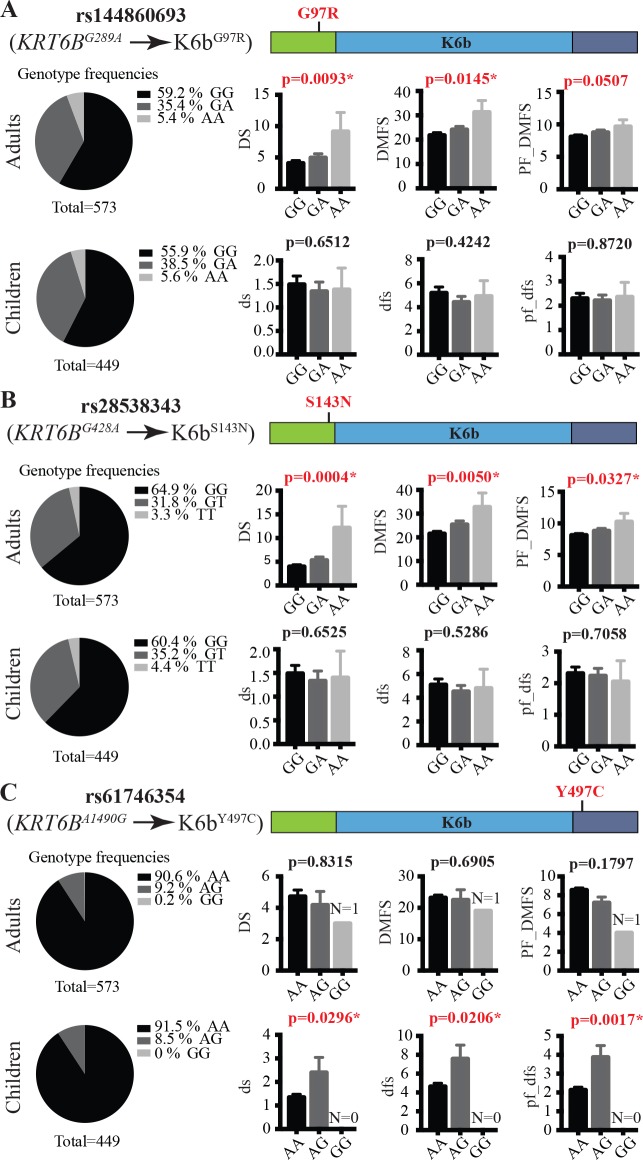
Genotype frequencies and quantifications of caries experience per genotype group for the three missense SNPs identified in KRT6B are shown in Fig 2. The frequencies of rs144860693 (K6bG97R) and rs28538343 (K6bS143N), the two variants that exhibited the most significant association with dental caries risk in adults, are high in the cohorts studied (Fig 2A and 2B; S1 Table). These two SNPs have a major impact on caries experience, with an estimated increase in the average number of carious tooth surfaces of 1.6 and 2.4 surfaces per copy of the risk allele, respectively (Table 1). These variants did not demonstrate a statistically significant effect on average caries experience in children (Fig 2A and 2B). rs61746354 (K6bY497C), the missense SNP in KRT6B that was associated with higher caries risk in children and occurs at a frequency higher that 4% in our cohorts (S1 Table), was associated with an estimated 1-surface increase in the average number of carious tooth surfaces (Table 1 and Fig 2C). Genotype frequencies and quantifications of caries experience per genotype for the other missense SNPs identified in KRT6A and KRT6C are shown in S3 Fig.
Fig 2. Genetic association between missense SNPs in KRT6B and tooth decay experience.
Genotype frequencies and quantification of caries experience per genotype for missense SNPs rs144860693 (A), rs28538343 (B) and rs61746354 (C). Pie charts on the left show the frequencies of all three genotypes for each SNP in the cohorts of 573 adults and 449 children that were evaluated for their caries experience. Bar graphs on the right represent the average and SEM (error bar) measures of three indices standardly used to assess caries experience: left, the number of tooth surfaces with untreated decay (DS and ds); center, the number of decayed, missing due to decay, and filled surfaces (DMFS and dfs); right, partial DMFS and dfs indices limited to molars and premolars pit and fissure surfaces (PF_DMFS and pf_dfs).
Statistically significant interaction effects are observed between caries-associated missense SNPs in KRT6 genes
Given that KRT6B harbors three missense SNPs showing significant association with caries experience, we wanted to further quantify the genetic relationship of missense variants in this gene on dental caries. To do so, we examined pairwise interactions between rs144860693 (K6bG97R), rs28538343 (K6bS143N) and rs61746354 (K6bY497C). Statistically significant interaction effects were observed between rs144860693 (K6bG97R) and rs28538343 (K6bS143N) on the number of surfaces with untreated decay (DS) model, between rs144860693 (K6bG97R) and rs61746354 (K6bY497C), and rs28538343 (K6bS143N) and rs61746354 (K6bY497C) on the number of decayed, missing due to decay, and filled surfaces (DMFS), adjusting for age, sex, and all the other SNPs in KRT6B (S2 Table). Even though rs61746354 (K6bY497C) was associated with higher caries risk in children only, this SNP exhibited a significant statistical interaction effect with rs144860693 (K6bG97R) and rs28538343 (K6bS143N) in adults (S2 Table). Therefore, the effect of the two SNPs that result in amino acid substitutions in the head domain of K6b on caries risk in adults may be influenced by the presence or absence of the p.Tyr497Cys substitution in the tail domain of the same keratin, a SNP that by itself is associated to higher caries risk only in children. The two SNPs resulting in p.Ser143Asn substitution in KRT6B and KRT6C (rs28538343 and rs151117600, respectively) also exhibited statistically significant interaction effect on the number of surfaces with untreated decay (DS) in adults (p-value = 0.044). When focusing on the 4 SNPs that lead to higher caries risk in adults, we found a significant cumulative effect of the number of risk alleles on caries experience (S4 Fig).
Given that KRT75 is adjacent and phylogenetically related to the KRT6 genes in the human genome, we explored potential linkage disequilibrium and interaction effects between the KRT75 SNP previously shown to increase caries experience in adults [19] and the newly identified SNPs in KRT6 genes. The previously reported SNP rs2232387 (K75A161T) was not in linkage disequilibrium with any of the KRT6 SNPs (S5 Fig) and there was no statistical interaction (all p-values >0.05) between the same SNPs.
Altogether, our data support genetic association between SNPs in KRT6A, KRT6B and KRT6C and tooth decay risk, in a way that is dentition-specific, and with statistical interaction between various loci in these three genes.
PC patient with mutation in KRT6A exhibits structural enamel defects
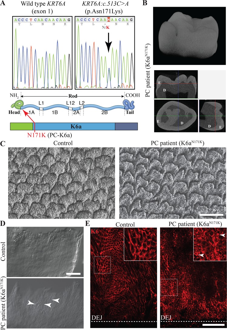
In order to assess how mutations in KRT6 genes may affect enamel structure, we analyzed third molars that were extracted from a PC patient who is heterozygous for the KRT6A:c.513C>A transversion that results in p.Asn171Lys amino acid substitution (K6aN171K) (Fig 3A). This patient is a white male who was 18 years old at the time his third molars were extracted, and is the member of a family in which the mutation in KRT6A was previously reported [22]. The patient experienced 20/20 nail dystrophy, very painful palmoplantar keratoderma, oral leukokeratosis, follicular keratosis, but did not have natal teeth. The overall shape and structure of the third molar enamel did not appear defective based on micro-computed tomography analysis (Fig 3B). However, scanning electron microscopy analysis of polished sections of the teeth (section plane transverse to the enamel rods) revealed alteration of the distribution and shape of enamel rods when compared to third molars extracted from healthy patients (Fig 3C).
Fig 3. Structural enamel defects in third molars from a patient with PC-causing mutation in KRT6A.
(A) Chromatograph on the left shows wild type sequence of KRT6A exon 1 at position c.505–519. Chromatograph on the right shows equivalent region for a PC patient with heterozygous mutation c.513C>A leading to missense mutation p.Asn171Lys (K6aN171K). Schematics in lower panel shows the position of the N171K amino acid substitution at the beginning of the rod domain in K6a. (B) Micro-computed tomography analysis of a wisdom tooth extracted from a PC patient who is heterozygous for the mutation described in A. The upper panel shows the 3D reconstruction of the tooth crown. The bottom panel shows 3-axes sections of the molar on which enamel (E) and dentin (D) can be distinguished. (C) Scanning electron microscopy analysis of polished transverse section of wisdom teeth from PC and control patients. Sections were taken in the area of the cusps and in a plane perpendicular to the orientation of the enamel rods. Scale bar: 10 μm. (D) Differential interference contrast imaging of insoluble organic material isolated from a third molar from PC and control patients. White arrowheads indicate curls formed by the rod sheaths. Scale bar: 50 μm. (E) Immunohistochemical detection of K6 (red) on polished section of human third molar extracted from PC and control patients. White arrowheads indicate aggregates. Scale bar: 50 μm. DEJ, dentin-enamel junction.
The insoluble organic material present in mature enamel can be isolated after full demineralization of a tooth in EDTA. When isolated from molars extracted from this PC patient, the insoluble material exhibited uneven alignment of the enamel rod sheaths that tended to form curls (Fig 3D). To assess the effects of the K6aN171K mutant protein on K6 distribution in enamel, we performed immunohistochemical staining using anti-K6 antibody on polished sections of the patient’s tooth. K6 staining was still found to be stronger in the tuft areas near the DEJ (Fig 3E). However, K6 distribution was no longer restricted to the interrod but could also be found as smaller rings or clumps within rods (Fig 3E). These results indicate that this PC-causing mutation in KRT6A leads to improper incorporation of the K6a protein into enamel rod sheaths, which results in altered shape and arrangement of enamel rods.
Caries-associated p.Ser143Asn and p.Tyr497Cys substitutions alter K6 protein behavior in ameloblast-like cells
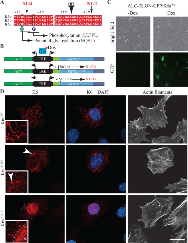
Missense mutations in keratins may affect their assembly, modify their subcellular localization and/or affect their interaction with keratin-associated proteins. Phosphorylation and glycosylation of the head and tail domain of intermediate filaments proteins have been shown to influence their interaction with other proteins and their subcellular localization [23, 24]. When comparing the position of the SNPs we determined to be associated with increased caries experience and potential sites for post-translational modifications in K6 proteins, we observed that the p.Ser143Asn substitution (rs28538343 in KRT6B and rs151117600 in KRT6C) is immediately adjacent to an LLS/TPL consensus phosphorylation site that is highly conserved in type II keratins [24, 25], and within a potential N-linked glycosylation site (Fig 4A and S6 Fig). Although it remains to be determined how K6 proteins interact with and are deposited into the enamel matrix in the context of a secretory stage ameloblast in vivo, we assessed the effect of the p.Ser143Asn substitution in K6 in a context in which the mutant protein is overexpressed in ameloblast-like cells (ALC) [26]. In this assay, we also analyzed the behavior of K6N171K mutant protein carried by the PC patient included in this study (Fig 3), a mutation located in the rod domain and known to have a severe effect on keratin filament assembly [27, 28]. Given the high degree of sequence identity between K6 proteins (S7 Fig) and the fact that the mutations of interest are located in highly conserved regions (Fig 4A), we used K6a as a model protein for this assay. We used site-directed mutagenesis to introduce the c.428G>A transition (results in p.Ser143Asn substitution) and the c.513C>A transversion (results in p.Asn171Lys substitution) into the KRT6A cDNA, and cloned the different isoforms (KRT6AWT, KRT6Ac.428G>A and KRT6Ac.513C>A) into a vector that allows for tetracycline inducible co-expression of KRT6A isoforms and GFP (Fig 4B).
Fig 4. Effects of S143N and N171K mutations on the assembly of K6a in ameloblast-like cells.
(A) Alignment of K6a, K6b and K6c protein sequences between amino acids 138 and 175 showing the position of serine 143 and asparagine 171 on both sides of the transition between the head and the rod domain (black box labeled HR). Serine 143 is right upstream of an LLS/TPL consensus phosphorylation site (blue circle labeled P) that is highly conserved in type II keratins. Serine 143 is also part of an NQSL potential N-glycosylation site (green circle labeled G). (B) Schematic representation of pBi4-GFP/K6a bidirectional constructs used for tetracycline inducible expression of GFP with KRT6AWT, KRT6AG428A (produces K6aS143N) or KRT6AC513A (produces K6aN171K mutant). When transfected into cells expressing the transactivator rTA, transgene expression through binding of rTA to the tetracycline responsive element (TRE) can be induced by addition of doxycycline (Dox) to the culture medium. (C) Detection of GFP induction in ameloblasts-like cells stably expressing rTA (ALC-TetON) transfected with pBi4-GFP/K6a constructs and grown with or without Dox. (D) Immunohistochemical analysis of K6a subcellular distribution (red) in ALC-TetON cells producing K6aWT, K6aS143N or K6aN171K. Nuclei are stained with DAPI (blue) and actin filaments are stained with phalloidin conjugated to Alexa Fluor 647 (white).
These constructs were used to transfect ALC-TetON cells in which expression of KRT6A isoforms and GFP can be induced by addition of doxycycline to the culture medium (Fig 4C). Immunohistochemical analysis using anti-K6 antibody was used to determine the distribution of K6a isoforms in ALC-TetON cells (Fig 4D). While K6aWT formed thick and relatively short bundles of keratin filaments in ALC-TetON cells, K6aS143N tended to form a web of thinner filaments together with large aggregates (Fig 4D). These large aggregates were not seen with K6a proteins harboring the p.Asn21Ser and p.Gly97Arg substitutions (S8A and S8B Fig) caused by the other SNPs that are associated with higher caries in adults (rs17845411 and rs144860693, respectively) and are in partial linkage disequilibrium with the SNPs leading to p.Ser143Asn substitution (rs28538343 in KRT6B and rs151117600 in KRT6C). These results suggest that the p.Ser143Asn substitution may contribute most significantly to the caries-prone phenotype in adults. The behavior of the K6aN171K mutant protein fused to a YFP tag has been previously studied in the context of human hepatoma PLC cells in which the mutant protein was shown to form aggregates primarily located in the cytoplasm [27, 28]. In the context of ALC-TetON cells, K6aN171K formed aggregates that showed heightened accumulation in the nucleus (Fig 4D). These results confirm a severe impairment of K6a assembly in PC patients with p.Asn171Lys substitution. The fact that the aggregates in PLC cells were mostly in the cytoplasm may reflect a cell-specific behavior of the mutant protein or may be due to the YFP tag that was fused to K6a in these experiments [27, 28].
Given that the SNP leading to p.Tyr497Cys substitution in the tail domain of K6b (rs61746354) is the only one we found associated with higher caries risk in children, we tested its effect on K6a assembly. Similarly to K6aS143N, the K6aY497C isoform tended to form large aggregates in ALC-TetON cells (S8C Fig), which suggests that this SNP may be the cause of the caries-prone phenotype in children. Even though this substitution is not found near a potential posttranslational modification site, the presence of a new cysteine in the tail domain may result in the formation of disruptive disulfide bonds. Given the interaction effects measured between the SNPs that lead to the p.Ser143Asn and p.Tyr497Cys substitutions, we generated a DNA construct for the expression of a K6a protein that harbors both substitutions (K6aS143N-Y497C). This double mutant tends to form aggregates to the same extent as the single mutant proteins (S8D Fig).
Taken together, these results confirm that the PC-associated p.Asn171Lys substitution results in profound impairment of K6a protein assembly, and reveal that the caries-associated p.Ser143Asn and p.Tyr497Cys substitutions in K6 proteins also affect the behavior of the proteins, when overexpressed in an ameloblast cell line.
Discussion
The present report highlights the contribution of specific sets of keratins to the organic fraction of mature tooth enamel and demonstrates through genetic and analytical studies their crucial function in the formation of enamel and its resistance to decay. K75, an epithelial hair keratin in which mutations have been associated with hair disorders, was the first keratin we investigated in this context [19]. K6 proteins, that are the focus of the present study, are expressed in epithelia that withstand particularly high levels of mechanical strain (palmoplantar skin, oral epithelium) as well as in the supporting layers of the hair follicle where their function is similar to that of K75 which is not expressed in palmoplantar epidermis and oral epithelium. Our findings demonstrate that, as K75, K6 proteins play a crucial role in the enamel rod sheath and that mutations in the genes encoding these keratins may impair the stability of the organic structural component of mature enamel. We propose that, with their unique biochemical properties, K75 and K6 contribute to the toughness, elasticity and resistance to degradation of the enamel rod sheaths, which contributes to establishing proper shape and arrangement of enamel rods and enhances the biomechanical properties of tooth enamel [12, 13]. Moreover, since it has long been suggested that the stability of the proteins in mature enamel influences the resistance of enamel to carious attack [14, 15], we propose that keratins contribute to the stability of enamel rod sheaths and therefore to the resistance of enamel to decay.
Since K6 proteins are also expressed in the oral epithelium, and patients with PC may exhibit oral leukokeratosis, there could be a partial involvement of the oral cavity in the increased susceptibility to caries measured in this study. However, the fact that we found SNPs that lead to a higher number of caries in children and not in adults (same oral cavity but different set of teeth) strongly suggests that defects in the dental tissue itself are the major factor leading to this effect. The structure and chemical composition of tooth enamel is known to be different between primary and permanent teeth. Primary teeth exhibit thinner and whiter enamel with a smoother surface and higher content in calcium and phosphate when compared to permanent teeth [29, 30]. Enamel from primary teeth also has a greater susceptibility to demineralization [31]. Moreover, it has been proposed that the genetic factors influencing dental caries differ between primary and permanent dentition [9]. However, there has been no study comparing the composition of the organic material present in the enamel from primary and permanent teeth. The dentition-specific effect we report here for SNPs in KRT6 genes, which we previously observed for two SNPs in KRT75, with one affecting adults and the other one affecting children [19], suggests that the combination and/or the mode of incorporation of these keratins in the enamel rod sheaths is different in primary and permanent teeth.
Even though this is the first evidence of K6 proteins being incorporated into the enamel matrix, a previous yeast-two-hybrid study determined that K6 could interact with enamel matrix proteins such as amelogenin and tuftelin [32]. It is therefore likely that K6 proteins interact with enamel matrix proteins during the process of enamel secretion. However, the mode of incorporation of keratins into the enamel matrix remains to be elucidated. The interaction of keratins with other proteins is known to involve their head and tail domains rather than the rod domain through which heterodimerization of acidic and basic keratins is established. These interactions are regulated by posttranslational modifications such as phosphorylation and glycosylation [23, 24], and mutations impairing such modifications have been linked to skin diseases [33], as well as diseases related to liver and pancreatic injury [34]. Interestingly, all the caries-associated missense SNPs we identified in KRT6 genes result in substitutions in the head and tail domains of the proteins, which suggests that they may affect their interaction with other proteins rather than their heterodimerization. We further demonstrate that the p.Ser143Asn substitution that may affect phosphorylation and/or glycosylation of the head domain of K6 proteins affects the behavior of K6A in the context of an ameloblast cell line, which suggests that this substitution found in both K6b and K6c may contribute most significantly to the caries-prone phenotype in adults. Functional studies will be required to elucidate the effects of the p.Ser143Asn substitution on the biochemical properties of K6 proteins, in particular on its ability to undergo posttranslational modifications that may affect interaction with enamel matrix proteins and incorporation into the enamel in vivo. Interestingly, in a recent clinical report, an isolated case of PC was proposed to be caused by de novo c.428G>A mutation in KRT6A that leads to the p.Ser143Asn substitution [35]. This is so far the only report of PC-causing mutation outside of the rod domain. Based on its location in the head domain and on the high frequency of the same substitution in K6b and K6c, the p.Ser143Asn substitution in K6a is unlikely to be the sole cause for the PC phenotype in this patient.
In epidermal tissues, K6 proteins (Type II, basic or neutral) form heterodimers with K16 or K17 (Type I, acidic) to assemble in larger polymeric structures. Interestingly, the subcellular distribution of K16 and K17 proteins is distinct from the distribution of K6 in the enamel organ, which suggests that they do not follow their canonical mode of assembly in this tissue. Due to the low number of frequent missense SNPs in KRT16 and KRT17 in our cohorts, the present study did not allow us to make any conclusion on the potential genetic association between variants in these two genes and dental caries experience. However, the striking difference in the way K16 and K17 proteins are incorporated into enamel suggests that their function in this tissue is distinct from that of K6 proteins. Based on the restricted localization of K16 and K17 near the DEJ and in the core of the enamel rods, these keratins may be involved in shock absorption and protection against fracture [13] rather than in the resistance to caries. Structural analysis of enamel from PC patients with mutations in KRT16 and KRT17 will help address this question.
In conclusion, we show for the first time that (i) K6 proteins are incorporated into mature tooth enamel at the rod sheaths, (ii) SNPs in KRT6 genes are associated with increased susceptibility to dental caries, (iii) a PC patient with a mutation in KRT6A exhibits defects in enamel structure, and (iv) caries-associated p.Ser143Asn substitution in K6 proteins impairs proper protein interactions.
Materials and methods
Ethics statement
We thank the Pachyonychia Congenita Project and Ms. Holly Evans for providing us with clinical information and extracted third molars from a PC patient (20040468–1057496), and the NIDCR dental clinic for providing extracted third molars from healthy patients (NCT01805869).
For the COHRA study, written informed consent was provided by all adult participants, and verbal assent with parental written consent was provided by all child participants. All procedures, forms and protocols were approved by the Institutional Review Boards of the University of Pittsburgh and West Virginia University.
Written informed consent was obtained from the pachyonychia congenita patient, as part of a research registry approved by an institutional review board that complies with all principles of the Helsinki Accord (Western IRB study no. 20040468).
All animal work was approved by the NIAMS Animal Care and Use Committee.
Human subject recruitment for the COHRA study
The Center for Oral Health Research in Appalachia (COHRA) study was initiated to investigate the community-, family-, and individual-level contributors to oral health [36]. Participants from rural counties of Pennsylvania and West Virginia were enrolled via a household-based recruitment strategy, with eligible households required to include at least one biological parent-child pair. All other members of eligible households were invited to participate without regard to biological or legal relationships, or oral health status. Written informed consent was provided by all adult participants, and verbal assent with parental written consent was provided by all child participants. All procedures, forms and protocols were approved by the Institutional Review Boards of the University of Pittsburgh and West Virginia University.
Data collection for the COHRA study
Intra-oral examinations of all participants were performed by licensed dentists and/or research dental hygienists. Each surface of each tooth (excluding third molars) was examined for evidence of decay, from which dental caries indices were generated. Three measurements of caries experience were considered: (1) the number of surfaces with untreated decay (DS/ds); (2) the traditional DMFS/dfs indices which represent the number of decayed (D/d), missing due to decay (M), and filled (F/f) tooth surfaces (S/s) in the permanent (DMFS) and primary (dfs) dentitions; and (3) the partial DMFS and dfs indices limited to the molars and premolar pit and fissure surfaces which are at high risk of decay. DNA samples were collected via blood, saliva or buccal swab. Genotyping for approximately 600,000 polymorphisms was performed by the Center for Inherited Disease Research at Johns Hopkins University using the Illumina Human610-Quadv1_B BeadChip (Illumina). Extensive data cleaning and quality assurance analyses were performed as previously reported [4]. Imputation of approximately 16 million unobserved genetic variants was performed using the 1000 Genome Project (phase 1 June 2011 release) as reference. In brief, pre-phasing of haplotypes was performed via SHAPEIT2 [37] and imputation was performed via IMPUTE2 [38].
Statistical analysis for the COHRA study
Linear regression was used to test the association of dental caries experience with genetic polymorphisms under the additive genetic model while adjusting for age and sex. Pairwise SNP-by-SNP interaction effects were tested in the same modeling framework by including main effects of each SNP and their product term for selected variants within the same gene region, along with age and sex. Analyses were performed separately for dental caries in the permanent dentition of adults (ages 25–50 years) and the primary dentition of children (ages 6–12 years). All analyses were limited to self-reported non-Hispanic whites (mixed European descent); self-reported race was consistent with genetically-determined ancestry. Analyses were performed using PLINK (v1.9) (http://www.cog-genomics.org/plink/1.9/)[39] and R (R Foundation for Statistical Computing).
Collection of third molars from patient with pachyonychia congenita
Third molars were obtained from a patient involved in the International Pachyonychia Congenita Research Registry (IPCRR), under the IRB Protocol number 20040468 and IRB Study number 1057496. Third molars from healthy patients were obtained from the NIDCR OP-1 Dental Clinic that were collected under the IRB Protocol number NCT01805869.
RNA-seq analysis
RNA-seq analysis was performed as described previously [40]. Briefly, mandibles were dissected from P10 mice, transferred to RNAlater solution (Life Technologies) and stored at 4°C. Enamel organs were dissected from mandibles and homogenized in Trizol reagent (Invitrogen) using the Precellys 24 (Bertin Technologies). Total RNA was extracted and further purified using the RNAeasy mini kit (Qiagen). RNA-seq analysis was performed using the Mondrian SP kit (Illumina) and the Illumina HiSeq 2000 sequencing system.
Immunohistochemical analysis on rat mandible section
Rat mandibles at postnatal day 10 were fixed overnight at 4°C in 4% paraformaldehyde in 1X PBS, dehydrated and embedded in paraffin and 10 μm-thick sections were prepared. Immunohistochemical analysis was performed using a blocking solution containing 5% goat serum and 7.5% BlockHen II (Aves Labs, Tigard, OR) in 1X PBS. Enzymatic antigen retrieval was performed using Ultravision Trypsin (Thermo Fisher Scientific, Waltham MA). Primary antibodies used: Guinea-pig anti-K6 (Progen Biotechnik GmbH, Germany), guinea-pig anti-K17 (Progen Biotechnik GmbH, Germany). Alexa-488 anti-guinea-pig (Thermo Fisher Scientific, Waltham MA) was used as secondary antibody. Images were acquired on a Leica LS5 confocal microscope (Leica Microsystems Inc., Buffalo Grove, IL).
Immunohistochemical analysis on polished human tooth sections
Ground, polished and etched human teeth were stained with guinea-pig anti-K6 (Progen Biotechnik GmbH, Germany), mouse anti-K6 (Abcam, Cambridge MA), guinea-pig anti-K16 (Progen Biotechnik GmbH, Germany) or guinea-pig anti-K17 (Progen Biotechnik GmbH, Germany) antibody. Alexa-488 anti-guinea-pig and Alexa-555 anti-mouse (Thermo Fisher Scientific, Waltham MA) were used as secondary antibodies.
Scanning electron microscopy
Ground, polished and etched human teeth were prepared for SEM as described previously [19]. Samples were fixed overnight at 4°C in 2% glutaraldehyde, 2% PFA in 0.1M phosphate buffer pH 7.4 and dehydrated through a series of 50%, 70%, 95% and 100% ethanol solutions. They were incubated for 10 min in hexamethyldisilazane, air-dried for 30 min, mounted on aluminum specimen mount stubs covered with conductive carbon adhesive tabs (Electron Microscopy Sciences, Hatfield, PA), sputter-coated with gold and analyzed under a Field Emission Scanning Electron Microscope S4800 (Hitashi, Toronto, Canada) at 10 kV.
Micro-CT analysis and 3-D reconstructions
Micro-CT analysis of fixed molars was performed as described previously [19] using the Skyscan 1172 desktop X-ray microfocus CT scanner and the following parameters: 0.5mm AI + 0.1mm Cu filters, 100 kV, 100 uA, 8.00 micron resolution, 0.4 degrees rotation step over 180 degrees). CTvox software (Bruker microCT) was used for 3D reconstruction.
Plasmids
The cDNAs encoding the, K6aN21S, K6aG97R, K6aS143N, K6aY497C and K6aN171K mutant proteins were generated by site-directed mutagenesis using the QuikChange Site-directed Mutagenesis Kit (Stratagene). The following primers were used: N21S-forward: GGGGTTTCAGTGCCAgCTCAGCCAGGC, N21S-reverse: GCCTGGCTGAGcTGGCACTGAAACCCC, G97R-forward: GGCTTTGGTGGCGCCaGGAGTGGATTGG, G97R-reverse: CCAATCCACTCCtGGCGCCACCAAAGCC, S143N-forward: GTCAACCAGAaTCTCCTGACTCCCCTC, S143N-reverse: GAGGGGAGTCAGGAGAtTCTGGTTGAC, Y497C-forward: CCGTCTCCAGTGGCTgTGGCGGTGCCAG, Y497C-reverse: CTGGCACCGCCAcAGCCACTGGAGACGG, N171K-forward: GATCAAGACCCTCAAaAACAAGTTTGCC, N171K-reverse: GGCAAACTTGTTtTTGAGGGTCTTGATC (lower cases indicate the position of the mutations). The pBi4-GFP vector was used to simultaneously express the reporter protein EGFP with K6aWT, K6aN21S, K6aG97R, K6aS143N, K6aY497C, K6aS143N-Y497C, or K6aN171K under control of a unique tetracycline responsive element.
Cell culture and transfections
Murine Ameloblast-like cells (ALC) were kindly provided by Dr. Sugiyama [26] and used to produce a tetracycline inducible ameloblast cell line. These cells were stably transfected with a prtTA2-M2/IRES-Neo plamid obtained after subcloning of the rtTA2-M2 cassette [41] into the pCMV-IRES-Neo (Clontech). rtTA-M2 is a mutagenized form of rtTA that shows a lower basal activity and a higher sensitivity to doxycycline (Dox) than the original rtTA [41]. The presence of the IRES cassette before the neomycin (Neo) resistance gene allowed coexpression of the rtTA-M2 transactivator and the Neo resistance gene, increasing the chance to select clones that express sufficient amounts of the transactivator in Neo-resistant cells. Clones were isolated and functionality of the tet system was screened by transient transfection with pTRE2-luc (expression of luciferase under the control of tetracycline responsive element). Cells were grown in the presence or absence of 2 ug/ml Dox and luciferase activity was estimated. One clone exhibiting a low basal activity of the transactivator in the absence of Dox and a strong induction in the presence of Dox (+Dox/-Dox ratio) was selected for subsequent experiments and named ALC-TetON. ALC-TetON cells were grown in Dulbecco’s modified Eagle’s medium (10% fetal bovine serum, 1% penicillin/streptomycin and 1 ug/ml G418). For transfections, the cells were grown to at least 70% confluence. 2 million cells were used per transfection with either the pBi4-GFP/K6aWT, pBi4-GFP/K6aN21S, pBi4-GFP/K6aG97R, pBi4-GFP/K6aS143N, pBi4-GFP/K6aY497C, pBi4-GFP/K6aS143N-Y497C, or pBi4-GFP/K6aN171K plasmid and the pCMV-K16WT plasmid (Amaxa Nucleofactor).
Immunocytochemistry
Transfected cells were seeded on glass coverslips coated with 0.1% gelatin and immediately induced with 2ug/ml doxycycline. Twenty-four hours after induction, cells were washed three times in 1X PBS and fixed with 4% paraformaldehyde in PBS for 15 min at room temperature. A 5 min incubation in 0.2% Triton in 1X PBS was used to permeabilize the cells before blocking unspecific sites using 3% BSA in PBS for 1 h. Primary antibodies diluted in blocking solution were applied for 1 h. Primary antibody used: guinea-pig anti-K6 (Progen). Secondary antibodies diluted in blocking solution were applied for 30 min. Secondary antibodies used: Alexa Fluor 555 anti-guinea pig IgG (Life Technologies). Nuclei were stained using DAPI and coverslips were mounted on glass slides using Mowiol (Calbiochem). Images were acquired using a LEICA Sp5 confocal microscope.
Supporting information
(DOCX)
(DOCX)
(A) Immunohistochemical analysis of K17 distribution in rat enamel organ at postnatal day 10. Nuclei are stained with DAPI (blue). Inset at the top shows enlarged view of Tomes' processes. PL, papillary layer; SI, stratum intermedium; Am, ameloblasts; TP, Tomes’ processes; R, rod; IR, interrod. Scale bar: 50 μm. (B) Immunohistochemical detection of K16 and K17 on polished sections of human third molars. As indicated by the schematics on the left, the images were acquired from mesial-distal sections of human third molars and show the area of enamel adjacent to dentin. DEJ, dentin-enamel junction. Scale bars: 100 μm.
(TIF)
Immunohistochemical detection of K6 on polished sections of human third molars. Two different antibodies against K6 (recognize K6a, K6b and K6c in humans) were used: a polyclonal antibody raised in guinea-pig against the C-terminus of the protein (green) and a monoclonal antibody raised in mouse against the N-terminus of the protein (red). As indicated by the schematics on the left, the images in the upper panels were acquired from mesial-distal sections of human third molars and show the area of enamel adjacent to dentin, whereas the images in the lower panels were acquired from transverse section of the crown near the cusps. DEJ, dentin-enamel junction (dashed lines). Scale bars: 100 μm for upper panels, 20 μm for lower panels.
(TIF)
Genotype frequencies and quantification of caries experience per genotype for missence SNPs rs17845411 and rs151117600 (locations shown on the left). Pie charts in the center show the frequencies of all three genotypes for each SNP in the cohorts of 573 adults and 449 children that were evaluated for their caries experience. Bar graphs on the right represent the average and SEM (error bar) measures of three incices standardly used to assess caries experience: left, the number of tooth surfaces with untreated decay (DS and ds); center, the number of decayed, missing due to decay, and filled surfaces (DMFS and dfs); right, partial DMFS and dfs indices limited to molars and premolars pit and fissure surfaces (PF_DMFS and pf_dfs).
(TIF)
Number of caries for three phenotypes measured in adults (DS, DMFS, PF_DMFS) were plotted against the number of risk alleles carried for 4 SNPs in KRT6A, KRT6B and KRT6C that are associated with higher caries risk: rs17845411 (K6aN21S; risk allele: G), rs144860693 (K6bG97R; risk allele: A), rs28538343 (K6bS143N; risk allele A), and rs151117600 (K6cS143N; risk allele: A).
(TIF)
Linkage disequilibrium data measured by R2 between rs2232387, a missense SNP in KRT75 that was previously associated with higher caries risk, and five SNPs associated with higher caries risk in KRT6A, KRT6B and KRT6C. The KRT75 gene is located right downstream of KRT6B on chromosome 12. rs2232387 is a c.481G>A transition in KRT75 that causes an alanine-to-threonine substitution at position 161 (K75A161T).
(TIF)
The presence of potential N-Glycosylation sites in K6a was determined using NetNGlyc 1.0 Server. (A) A potential glycosylation site was found for the arginine at position 141 in a consensus NQSL site that includes serine 143. (B) The potential glycosylation site at position 141 was not called when serine 143 was replaced with an arginine.
(TIF)
(TIF)
Immunohistochemical analysis of K6a subcellular distribution (red) in ALC-TetON cells producing K6aN21S (A), K6aG97R (B), K6aY497C (C) or K6aS143N-Y497C (D). Nuclei are stained with DAPI (blue), actin filaments are stained with phalloidin conjugated to Alexa Fluor 647 (grey), and transfected cells express GFP (green). Scale bar: 20 μm.
(TIF)
Acknowledgments
We thank Dr. Juliane Lessard for helpful suggestions at the initiation of this project. At the NIAMS, we thank Gustavo Gutierrez-Cruz form the High Throughput Sequencing Core facility; Hong-Wei Sun and Stephen Brooks from the Biodata Mining core facility; Evelyn Ralston and Kristina Zaal from the Light Imaging Facility. At the NIH, we thank Dr. Richard Leapman (NIBIB) for giving us access to their scanning electron microscope. We thank Dr. Toshihiro Sugiyama from the Akika University (Japan) for providing us with their mouse ameloblast-like cells (ALC). For their involvement in the implementation of the COHRA study, we thank Dr. Toshiki Soejima and Dr. Robert J. Weyant from the University of Pittsburgh, and Dr. Daniel W. McNeil and Dr. Richard Crout from West Virginia University. We would like to express our gratitude to the COHRA participants and field research teams whose contributions made this work possible.
Data Availability
All genotype and phenotype data from the COHRA study cohort are available in dbGaP (accession number phs000095.v3.p1).
Funding Statement
This work was supported by the Intramural Research Program of the National Institute of Arthritis and Musculoskeletal and Skin Diseases of the National Institutes of Health (ZIA-AR041124 and ZIA-AR041171 to MIM), and by grants from the National Institute of Dental and Craniofacial Research (R01-DE014899 and U01-DE018903 to MLM, and R03-DE024264 to JRS). The funders had no role in study design, data collection and analysis, decision to publish, or preparation of the manuscript.
References
- 1.Shaw JH (1987) Causes and control of dental caries. N Engl J Med 317: 996–1004. https://www.ncbi.nlm.nih.gov/pubmed/3309652 doi: 10.1056/NEJM198710153171605 [DOI] [PubMed] [Google Scholar]
- 2.Werneck RI, Mira MT, Trevilatto PC (2010) A critical review: an overview of genetic influence on dental caries. Oral Dis 16: 613–23. https://www.ncbi.nlm.nih.gov/pubmed/20846151 doi: 10.1111/j.1601-0825.2010.01675.x [DOI] [PubMed] [Google Scholar]
- 3.Vieira AR, Modesto A, Marazita ML (2014) Caries: review of human genetics research. Caries Res 48: 491–506. https://www.ncbi.nlm.nih.gov/pubmed/24853115 doi: 10.1159/000358333 [DOI] [PMC free article] [PubMed] [Google Scholar]
- 4.Shaffer JR, Wang X, Feingold E, Lee M, Begum F, Weeks DE, et al. (2011) Genome-wide association scan for childhood caries implicates novel genes. J Dent Res 90: 1457–62. https://www.ncbi.nlm.nih.gov/pubmed/21940522 doi: 10.1177/0022034511422910 [DOI] [PMC free article] [PubMed] [Google Scholar]
- 5.Wang X, Shaffer JR, Zeng Z, Begum F, Vieira AR, Noel J, et al. (2012) Genome-wide association scan of dental caries in the permanent dentition. BMC Oral Health 12: 57 https://www.ncbi.nlm.nih.gov/pubmed/23259602 doi: 10.1186/1472-6831-12-57 [DOI] [PMC free article] [PubMed] [Google Scholar]
- 6.Shaffer JR, Feingold E, Wang X, Lee M, Tcuenco K, Weeks DE, et al. (2013) GWAS of dental caries patterns in the permanent dentition. J Dent Res 92: 38–44. https://www.ncbi.nlm.nih.gov/pubmed/23064961 doi: 10.1177/0022034512463579 [DOI] [PMC free article] [PubMed] [Google Scholar]
- 7.Zeng Z, Shaffer JR, Wang X, Feingold E, Weeks DE, Lee M, et al. (2013) Genome-wide association studies of pit-and-fissure- and smooth-surface caries in permanent dentition. J Dent Res 92: 432–7. https://www.ncbi.nlm.nih.gov/pubmed/23470693 doi: 10.1177/0022034513481976 [DOI] [PMC free article] [PubMed] [Google Scholar]
- 8.Morrison J, Laurie CC, Marazita ML, Sanders AE, Offenbacher S, Salazar CR, et al. (2016) Genome-wide association study of dental caries in the Hispanic Communities Health Study/Study of Latinos (HCHS/SOL). Hum Mol Genet 25: 807–16. https://www.ncbi.nlm.nih.gov/pubmed/26662797 doi: 10.1093/hmg/ddv506 [DOI] [PMC free article] [PubMed] [Google Scholar]
- 9.Wang X, Shaffer JR, Weyant RJ, Cuenco KT, DeSensi RS, Crout R, et al. (2010) Genes and their effects on dental caries may differ between primary and permanent dentitions. Caries Res 44: 277–84. https://www.ncbi.nlm.nih.gov/pubmed/20516689 doi: 10.1159/000314676 [DOI] [PMC free article] [PubMed] [Google Scholar]
- 10.Simmer JP, Papagerakis P, Smith CE, Fisher DC, Rountrey AN, Zheng L, et al. (2010) Regulation of dental enamel shape and hardness. J Dent Res 89: 1024–38. http://www.ncbi.nlm.nih.gov/pubmed/20675598 doi: 10.1177/0022034510375829 [DOI] [PMC free article] [PubMed] [Google Scholar]
- 11.Robinson C, Lowe NR, Weatherell JA (1975) Amino acid composition, distribution and origin of "tuft" protein in human and bovine dental enamel. Arch Oral Biol 20: 29–42. https://www.ncbi.nlm.nih.gov/pubmed/1054568 [DOI] [PubMed] [Google Scholar]
- 12.Baldassarri M, Margolis HC, Beniash E (2008) Compositional determinants of mechanical properties of enamel. J Dent Res 87: 645–9. https://www.ncbi.nlm.nih.gov/pubmed/18573984 doi: 10.1177/154405910808700711 [DOI] [PMC free article] [PubMed] [Google Scholar]
- 13.Chai H, Lee JJ, Constantino PJ, Lucas PW, Lawn BR (2009) Remarkable resilience of teeth. Proc Natl Acad Sci U S A 106: 7289–93. https://www.ncbi.nlm.nih.gov/pubmed/19365079 doi: 10.1073/pnas.0902466106 [DOI] [PMC free article] [PubMed] [Google Scholar]
- 14.Pincus P (1948) Relation of enamel protein to dental caries. Nature 161: 1014 https://www.ncbi.nlm.nih.gov/pubmed/18865798 [DOI] [PubMed] [Google Scholar]
- 15.Little K (1962) Caries-prone and caries-resistant teeth. Nature 193: 388–9. https://www.ncbi.nlm.nih.gov/pubmed/14465897 [DOI] [PubMed] [Google Scholar]
- 16.Lesot H, Smith AJ, Matthews JB, Ruch JV (1988) An extracellular matrix protein of dentine, enamel, and bone shares common antigenic determinants with keratins. Calcif Tissue Int 42: 53–7. https://www.ncbi.nlm.nih.gov/pubmed/2452004 [DOI] [PubMed] [Google Scholar]
- 17.Robinson C, Shore RC, Kirkham J (1989) Tuft protein: its relationship with the keratins and the developing enamel matrix. Calcif Tissue Int 44: 393–8. https://www.ncbi.nlm.nih.gov/pubmed/2475232 [DOI] [PubMed] [Google Scholar]
- 18.Robinson C, Hudson J (2011) Tuft protein: protein cross-linking in enamel development. Eur J Oral Sci 119 Suppl 1: 50–4. https://www.ncbi.nlm.nih.gov/pubmed/22243226 doi: 10.1111/j.1600-0722.2011.00906.x [DOI] [PubMed] [Google Scholar]
- 19.Duverger O, Ohara T, Shaffer JR, Donahue D, Zerfas P, Dullnig A, et al. (2014) Hair keratin mutations in tooth enamel increase dental decay risk. J Clin Invest 124: 5219–24. http://www.ncbi.nlm.nih.gov/pubmed/25347471 doi: 10.1172/JCI78272 [DOI] [PMC free article] [PubMed] [Google Scholar]
- 20.McLean WH, Hansen CD, Eliason MJ, Smith FJ (2011) The phenotypic and molecular genetic features of pachyonychia congenita. J Invest Dermatol 131: 1015–7. https://www.ncbi.nlm.nih.gov/pubmed/21430705 doi: 10.1038/jid.2011.59 [DOI] [PubMed] [Google Scholar]
- 21.Wilson NJ, Leachman SA, Hansen CD, McMullan AC, Milstone LM, Schwartz ME, et al. (2011) A large mutational study in pachyonychia congenita. J Invest Dermatol 131: 1018–24. https://www.ncbi.nlm.nih.gov/pubmed/21326300 doi: 10.1038/jid.2011.20 [DOI] [PubMed] [Google Scholar]
- 22.Smith FJ, Liao H, Cassidy AJ, Stewart A, Hamill KJ, Wood P, et al. (2005) The genetic basis of pachyonychia congenita. J Investig Dermatol Symp Proc 10: 21–30. https://www.ncbi.nlm.nih.gov/pubmed/16250206 doi: 10.1111/j.1087-0024.2005.10204.x [DOI] [PubMed] [Google Scholar]
- 23.Omary MB, Ku NO, Tao GZ, Toivola DM, Liao J (2006) "Heads and tails" of intermediate filament phosphorylation: multiple sites and functional insights. Trends Biochem Sci 31: 383–94. https://www.ncbi.nlm.nih.gov/pubmed/16782342 doi: 10.1016/j.tibs.2006.05.008 [DOI] [PubMed] [Google Scholar]
- 24.Snider NT, Omary MB (2014) Post-translational modifications of intermediate filament proteins: mechanisms and functions. Nat Rev Mol Cell Biol 15: 163–77. https://www.ncbi.nlm.nih.gov/pubmed/24556839 doi: 10.1038/nrm3753 [DOI] [PMC free article] [PubMed] [Google Scholar]
- 25.Toivola DM, Zhou Q, English LS, Omary MB (2002) Type II keratins are phosphorylated on a unique motif during stress and mitosis in tissues and cultured cells. Mol Biol Cell 13: 1857–70. https://www.ncbi.nlm.nih.gov/pubmed/12058054 doi: 10.1091/mbc.01-12-0591 [DOI] [PMC free article] [PubMed] [Google Scholar]
- 26.Nakata A, Kameda T, Nagai H, Ikegami K, Duan Y, Terada K, et al. (2003) Establishment and characterization of a spontaneously immortalized mouse ameloblast-lineage cell line. Biochem Biophys Res Commun 308: 834–9. https://www.ncbi.nlm.nih.gov/pubmed/12927794 [DOI] [PubMed] [Google Scholar]
- 27.Hickerson RP, Smith FJ, McLean WH, Landthaler M, Leube RE, Kaspar RL (2006) SiRNA-mediated selective inhibition of mutant keratin mRNAs responsible for the skin disorder pachyonychia congenita. Ann N Y Acad Sci 1082: 56–61. https://www.ncbi.nlm.nih.gov/pubmed/17145926 doi: 10.1196/annals.1348.059 [DOI] [PubMed] [Google Scholar]
- 28.Hickerson RP, Smith FJ, Reeves RE, Contag CH, Leake D, Leachman SA, et al. (2008) Single-nucleotide-specific siRNA targeting in a dominant-negative skin model. J Invest Dermatol 128: 594–605. https://www.ncbi.nlm.nih.gov/pubmed/17914454 doi: 10.1038/sj.jid.5701060 [DOI] [PubMed] [Google Scholar]
- 29.De Menezes Oliveira MA, Torres CP, Gomes-Silva JM, Chinelatti MA, De Menezes FC, Palma-Dibb RG, et al. (2010) Microstructure and mineral composition of dental enamel of permanent and deciduous teeth. Microsc Res Tech 73: 572–7. https://www.ncbi.nlm.nih.gov/pubmed/19937744 doi: 10.1002/jemt.20796 [DOI] [PubMed] [Google Scholar]
- 30.Lucchese A, Storti E (2011) Morphological characteristics of primary enamel surfaces versus permanent enamel surfaces: SEM digital analysis. Eur J Paediatr Dent 12: 179–83. https://www.ncbi.nlm.nih.gov/pubmed/22077687 [PubMed] [Google Scholar]
- 31.Wang LJ, Tang R, Bonstein T, Bush P, Nancollas GH (2006) Enamel demineralization in primary and permanent teeth. J Dent Res 85: 359–63. https://www.ncbi.nlm.nih.gov/pubmed/16567559 doi: 10.1177/154405910608500415 [DOI] [PMC free article] [PubMed] [Google Scholar]
- 32.Paine CT, Paine ML, Snead ML (1998) Identification of tuftelin- and amelogenin-interacting proteins using the yeast two-hybrid system. Connect Tissue Res 38: 257–67;discussion 95–303. https://www.ncbi.nlm.nih.gov/pubmed/11063033 [DOI] [PubMed] [Google Scholar]
- 33.Knobel M, O'Toole EA, Smith FJ (2015) Keratins and skin disease. Cell Tissue Res 360: 583–9. https://www.ncbi.nlm.nih.gov/pubmed/25620412 doi: 10.1007/s00441-014-2105-4 [DOI] [PubMed] [Google Scholar]
- 34.Ku NO, Toivola DM, Strnad P, Omary MB (2010) Cytoskeletal keratin glycosylation protects epithelial tissue from injury. Nat Cell Biol 12: 876–85. https://www.ncbi.nlm.nih.gov/pubmed/20729838 doi: 10.1038/ncb2091 [DOI] [PMC free article] [PubMed] [Google Scholar]
- 35.Luo S, Luo Q, Zhang H, Wan C (2015) A novel H1 mutation in keratin 6a in an infant with pachyonychia congenita. Indian J Dermatol Venereol Leprol 81: 385–7. https://www.ncbi.nlm.nih.gov/pubmed/26087092 doi: 10.4103/0378-6323.158651 [DOI] [PubMed] [Google Scholar]
- 36.Polk DE, Weyant RJ, Crout RJ, McNeil DW, Tarter RE, Thomas JG, et al. (2008) Study protocol of the Center for Oral Health Research in Appalachia (COHRA) etiology study. BMC Oral Health 8: 18 https://www.ncbi.nlm.nih.gov/pubmed/18522740 doi: 10.1186/1472-6831-8-18 [DOI] [PMC free article] [PubMed] [Google Scholar]
- 37.Delaneau O, Marchini J, Zagury JF (2011) A linear complexity phasing method for thousands of genomes. Nat Methods 9: 179–81. https://www.ncbi.nlm.nih.gov/pubmed/22138821 doi: 10.1038/nmeth.1785 [DOI] [PubMed] [Google Scholar]
- 38.Howie B, Fuchsberger C, Stephens M, Marchini J, Abecasis GR (2012) Fast and accurate genotype imputation in genome-wide association studies through pre-phasing. Nat Genet 44: 955–9. https://www.ncbi.nlm.nih.gov/pubmed/22820512 doi: 10.1038/ng.2354 [DOI] [PMC free article] [PubMed] [Google Scholar]
- 39.Chang CC, Chow CC, Tellier LC, Vattikuti S, Purcell SM, Lee JJ (2015) Second-generation PLINK: rising to the challenge of larger and richer datasets. Gigascience 4: 7 https://www.ncbi.nlm.nih.gov/pubmed/25722852 doi: 10.1186/s13742-015-0047-8 [DOI] [PMC free article] [PubMed] [Google Scholar]
- 40.Duverger O, Ohara T, Bible PW, Zah A, Morasso MI (2017) DLX3-Dependent Regulation of Ion Transporters and Carbonic Anhydrases is Crucial for Enamel Mineralization. J Bone Miner Res 32: 641–53. https://www.ncbi.nlm.nih.gov/pubmed/27760456 doi: 10.1002/jbmr.3022 [DOI] [PMC free article] [PubMed] [Google Scholar]
- 41.Duverger O, Lee D, Hassan MQ, Chen SX, Jaisser F, Lian JB, et al. (2008) Molecular consequences of a frameshifted DLX3 mutant leading to Tricho-Dento-Osseous syndrome. J Biol Chem 283: 20198–208. http://www.ncbi.nlm.nih.gov/pubmed/18492670 doi: 10.1074/jbc.M709562200 [DOI] [PMC free article] [PubMed] [Google Scholar]
Associated Data
This section collects any data citations, data availability statements, or supplementary materials included in this article.
Supplementary Materials
(DOCX)
(DOCX)
(A) Immunohistochemical analysis of K17 distribution in rat enamel organ at postnatal day 10. Nuclei are stained with DAPI (blue). Inset at the top shows enlarged view of Tomes' processes. PL, papillary layer; SI, stratum intermedium; Am, ameloblasts; TP, Tomes’ processes; R, rod; IR, interrod. Scale bar: 50 μm. (B) Immunohistochemical detection of K16 and K17 on polished sections of human third molars. As indicated by the schematics on the left, the images were acquired from mesial-distal sections of human third molars and show the area of enamel adjacent to dentin. DEJ, dentin-enamel junction. Scale bars: 100 μm.
(TIF)
Immunohistochemical detection of K6 on polished sections of human third molars. Two different antibodies against K6 (recognize K6a, K6b and K6c in humans) were used: a polyclonal antibody raised in guinea-pig against the C-terminus of the protein (green) and a monoclonal antibody raised in mouse against the N-terminus of the protein (red). As indicated by the schematics on the left, the images in the upper panels were acquired from mesial-distal sections of human third molars and show the area of enamel adjacent to dentin, whereas the images in the lower panels were acquired from transverse section of the crown near the cusps. DEJ, dentin-enamel junction (dashed lines). Scale bars: 100 μm for upper panels, 20 μm for lower panels.
(TIF)
Genotype frequencies and quantification of caries experience per genotype for missence SNPs rs17845411 and rs151117600 (locations shown on the left). Pie charts in the center show the frequencies of all three genotypes for each SNP in the cohorts of 573 adults and 449 children that were evaluated for their caries experience. Bar graphs on the right represent the average and SEM (error bar) measures of three incices standardly used to assess caries experience: left, the number of tooth surfaces with untreated decay (DS and ds); center, the number of decayed, missing due to decay, and filled surfaces (DMFS and dfs); right, partial DMFS and dfs indices limited to molars and premolars pit and fissure surfaces (PF_DMFS and pf_dfs).
(TIF)
Number of caries for three phenotypes measured in adults (DS, DMFS, PF_DMFS) were plotted against the number of risk alleles carried for 4 SNPs in KRT6A, KRT6B and KRT6C that are associated with higher caries risk: rs17845411 (K6aN21S; risk allele: G), rs144860693 (K6bG97R; risk allele: A), rs28538343 (K6bS143N; risk allele A), and rs151117600 (K6cS143N; risk allele: A).
(TIF)
Linkage disequilibrium data measured by R2 between rs2232387, a missense SNP in KRT75 that was previously associated with higher caries risk, and five SNPs associated with higher caries risk in KRT6A, KRT6B and KRT6C. The KRT75 gene is located right downstream of KRT6B on chromosome 12. rs2232387 is a c.481G>A transition in KRT75 that causes an alanine-to-threonine substitution at position 161 (K75A161T).
(TIF)
The presence of potential N-Glycosylation sites in K6a was determined using NetNGlyc 1.0 Server. (A) A potential glycosylation site was found for the arginine at position 141 in a consensus NQSL site that includes serine 143. (B) The potential glycosylation site at position 141 was not called when serine 143 was replaced with an arginine.
(TIF)
(TIF)
Immunohistochemical analysis of K6a subcellular distribution (red) in ALC-TetON cells producing K6aN21S (A), K6aG97R (B), K6aY497C (C) or K6aS143N-Y497C (D). Nuclei are stained with DAPI (blue), actin filaments are stained with phalloidin conjugated to Alexa Fluor 647 (grey), and transfected cells express GFP (green). Scale bar: 20 μm.
(TIF)
Data Availability Statement
All genotype and phenotype data from the COHRA study cohort are available in dbGaP (accession number phs000095.v3.p1).


