Abstract
Anion channels perform a diverse range of functions and have been implicated in ATP release, volume regulation, and phosphatidylserine exposure. Platelets have been shown to express several anion channels but their function is incompletely understood. Due to a paucity of specific pharmacological blockers, we investigated the effect of extracellular chloride substitution on platelet activation using aggregometry and flow cytometry. In the absence of extracellular chloride, we observed a modest reduction of the maximum aggregation response to thrombin or collagen-related peptide. However, the rate of aggregation was substantially reduced in a manner that was dependent on the extracellular chloride concentration and aggregation in the absence of chloride was noticeably biphasic, indicative of impaired secondary signaling. This was further investigated by targeting secondary agonists with aspirin and apyrase or by blockade of the ADP receptor P2Y12. Under these conditions, the rates of aggregation were comparable to those recorded in the absence of extracellular chloride. Finally, we assessed platelet granule release by flow cytometry and report a chloride-dependent element of alpha, but not dense, granule secretion. Taken together these data support a role for anion channels in the efficient induction of platelet activation, likely via enhancement of secondary signaling pathways.
Keywords: ADP, aggregation, chloride, ion channels platelets, thrombin
Introduction
In contrast to the role of cations (i.e., Ca2+, K+, and Zn2+) [1–3], the contribution(s) of anions to platelet activation remains unclear. Anion channels perform diverse functions including regulatory volume decrease [4], phosphatidylserine exposure [5], and ATP release [6]. Early patch clamp recordings demonstrated functional expression of Ca2+-activated Cl− channels in platelets [7, 8] and a megakaryocyte-like DAMI cell line [9]. Proteomic [10] and transcriptomic [11] analyses have since identified numerous anion channels that may be expressed by platelets. Of these, functional expressions of CLIC1 (Intracellular Cl− channel-1) [12], TMEM16F [13], and pannexin-1 [14] have been confirmed. Indicative of a hemostatic role for anion channels, CLIC1- and TMEM16F-deficient mice have associated platelet-related bleeding phenotypes [12, 13]. Additionally, pannexin-1 channels have been shown to amplify platelet activation responses to threshold agonist concentrations [14]. These ATP-permeable channels are associated with inflammatory conditions and may contribute to atherosclerosis [15].
Given the lack of specific anion channel blockers, we focus on the effect of extracellular Cl− ([Cl−]o) substitution on platelet activation. Our experiments highlight a role for anion channels in modulating the rate of platelet aggregation.
Methods
Materials
Aspirin, apyrase, and thrombin were from Sigma (Poole, UK). AR-C66096 was from Tocris Bioscience (Bristol, UK). Cross-linked collagen-related peptide (CRP-XL) was prepared as described previously [16] and supplied by R. Farndale (Cambridge, UK). Unless indicated, all other reagents were from Sigma.
Washed platelet preparation
This study was approved by the local Ethics Committee at Anglia Ruskin University. Human blood was collected from healthy volunteers following informed consent in accordance with the Declaration of Helsinki. Blood was collected into 11 mM sodium citrate and washed platelets were prepared as described previously [17]. Platelets were resuspended in a nominally calcium-free buffer containing (in mM) 145 NaCl, 5 KCl, 1 MgCl2, 10 glucose, 10 HEPES, titrated to pH 7.35 with NaOH. Where indicated, [Cl−]o was substituted by equimolar gluconate.
Aggregometry
Platelet aggregation was monitored as described previously using an AggRam aggregometer (Helena Biosciences, Gateshead, UK) [17]. In the experiments of Figure 1c, aliquots of 151 and 1 mM [Cl−]o-containing platelet suspensions were mixed 5 min prior to agonist addition. Platelets were preincubated with each drug(s) for 5 min at 37°C.
Granule release
Thrombin-evoked alpha and dense granule release was assessed by flow cytometry using fluorescently conjugated CD62P and CD63 antibodies (BD Biosciences, Oxford, UK), respectively. Antibody binding was monitored for 5 min using an Accuri C6 Flow cytometer (BD Biosciences) and the percentage of positive cells was calculated within FlowJo (V10.2, Oregon, USA).
Data analysis and statistics
Maximum aggregation (%) and the initial rate of aggregation (% s−1) were calculated in Excel (Microsoft, Redmond, Washington, USA), where rate was determined as the change in aggregation (%) in the first 30 s following shape change. Data were analyzed in GraphPad Prism by two-way ANOVA or Student’s t test as indicated and are representative of a minimum of four independent experiments. ***, **, *, and ns denote P < 0.001, P < 0.01, P < 0.05, and not significant, respectively.
Results
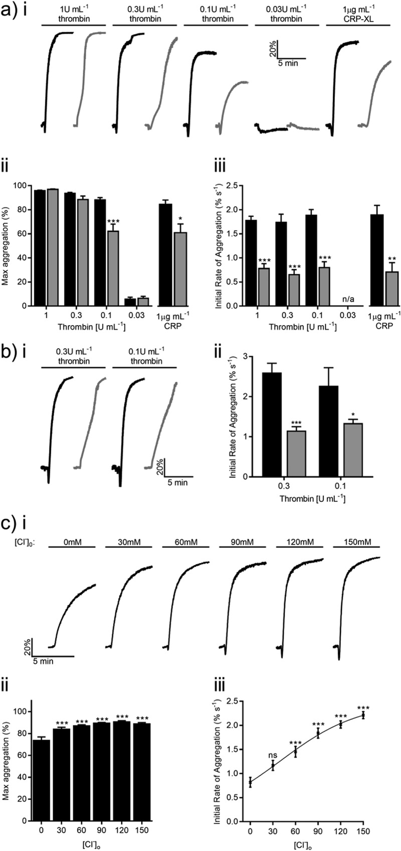
Platelet aggregation was assessed in the presence/absence of [Cl−]o in response to increasing thrombin concentrations (Figure 1a). Removal of [Cl−]o did not affect the maximum aggregation recorded across a 5-min time course in response to 0.03, 0.3, or 1 U mL−1 thrombin (Figure 1aii). A reduction from 82.3 ± 1.9% to 62.2 ± 6.0% (P < 0.001, Figure 1aii) was observed at an intermediate concentration (0.1 U mL−1). Cl− substitution affected the kinetics of platelet activation as the aggregation rate decreased by 1.1 ± 0.2%, 1.1 ± 0.2%, and 1.0 ± 0.2% s−1 in response to stimulation by 0.1, 0.3, and 1 U mL−1 thrombin, respectively (P < 0.01, Figure 1aiii). Similar effects were observed in the presence of 2 mM Ca2+ (Figure 1b), suggesting that this effect was not due to Ca2+ buffering. This observation was not exclusive to thrombin-evoked aggregation; removal of [Cl−]o reduced maximal CRP-XL-induced (1 µg mL−1) aggregation by 23.5 ± 3.9% (P < 0.01, Figure 1aii) and the rate decreased by 1.2 ± 0.1% s−1 (P < 0.001, Figure 1aiii). The sensitivity of thrombin-evoked (0.1 U mL−1) aggregation to Cl− was assessed by increasing [Cl−]o from 1 to 151 mM (30 mM increments; Figure 1c). There was no change in maximum aggregation beyond 31 mM [Cl−]o yet rates increased across the concentration range (EC50 = 46.5 ± 23.3 mM).
Figure 1.

Extracellular chloride is required for efficient platelet activation. (a) Washed platelets were stimulated by increasing concentrations of thrombin or 1 µg mL−1 CRP-XL in the presence of 151 mM (black) or 1 mM (gray) [Cl−]o. Representative aggregation traces are shown for each condition (ai). Maximum aggregation (aii) and the initial rate of aggregation (aiii) were calculated for each condition. (b) In the presence of 2 mM [Ca2+]o, [Cl−]o substitution continued to reduce the initial rate of thrombin-evoked aggregation. (c) The sensitivity of thrombin-evoked (0.1 U mL−1) aggregation to Cl− was assessed by increasing [Cl−]o from 1 to 151 mM in 30 mM increments. Representative traces (ci), maximum (cii) and initial rate of thrombin-induced (0.1 U mL−1) aggregation (ciii) for each [Cl−]o are shown. The rate of aggregation increased across the concentration range with an EC50 = 46.5 ± 23.3 mM. Data are representative of a minimum of four independent experiments. Thrombin and CRP-XL data sets were analyzed by two-way ANOVA and Student’s t test, respectively.
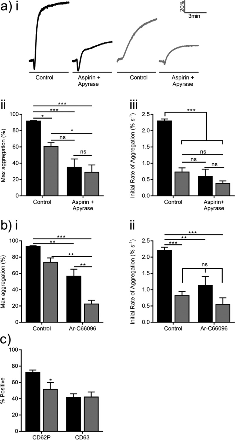
Differences in aggregation rate may be due to reduced secondary signaling in the absence of [Cl−]o. Platelets were preincubated with 100 µM aspirin and 5U mL−1 apyrase to assess contributions by thromboxane A2 and extracellular nucleotides. These compounds reduced maximum aggregation in response to 0.1 U mL−1 thrombin by 56.6 ± 10.2% (P < 0.001) and 31.6 ± 10.2% (P < 0.05, Figure 2ai, ii) in the presence of 151 and 1 mM [Cl−]o, respectively. The rate of platelet aggregation following aspirin and apyrase treatment in the presence of 151 mM (0.6 ± 0.2% s−1) or 1 mM (0.38 ± 0.08% s−1) [Cl−]o was comparable to that of 1 mM [Cl−]o control (0.7 ± 0.1% s−1; P > 0.05, Figure 2aiii).
P2Y12-mediated signaling is a major step during integrin αIIbβ3 activation [18, 19] and reduced ADP availability may account for the observed aggregation defect. 0.1 U mL−1 thrombin-evoked aggregation in the presence of a P2Y12 inhibitor (1 µM AR-C66096) decreased by 36.7 ± 8.2% and 51.2 ± 7.4% in the presence of 151 and 1 mM [Cl−]o (P < 0.01, Figure 2bi), respectively. It is noteworthy that no differences between the aggregation rate of AR-C66096-treated platelets in 151 or 1 mM [Cl−]o and the 1 mM [Cl−]o control were observed (P > 0.05, Figure 2bii), suggesting a role for P2Y12 during Cl–-dependent aggregation. Finally, we assessed thrombin-induced granule release by flow cytometry. Peak alpha granule release was reduced from 72.4 ± 2.9% to 59.9 ± 1.6% (P < 0.001, Figure 2c), while dense granule release was unaffected by Cl− substitution (P > 0.05, Figure 2c).
Conclusions
Here we demonstrate that [Cl−]o enhances the rate of platelet aggregation in a concentration-dependent manner (Figure 1). This effect was equivalent to blockade of secondary mediators and P2Y12 inhibition (Figure 2). One possible explanation of these data is that [Cl−]o is required for efficient release of ATP and/or ADP from the platelet, but we failed to observe a change in dense granule secretion (Figure 2c). Pannexin-1 has been shown to activate in response to elevation of intracellular Ca2+ ([Ca2+]i) [20], facilitating cytosolic ATP release [14]. It has been suggested that ADP release may occur via a similar mechanism [21]. Given that release of alpha granule cargo (e.g., fibrinogen, thrombin, and Zn2+) is required for aggregation and is enhanced by P2Y12 signaling [22–24], it is possible that [Cl−]o enhances platelet activation by mediating efficient alpha granule secretion.
Figure 2.

Role for secondary signaling during Cl–-dependent platelet aggregation. (a) Washed platelets were preincubated with 100 µM aspirin (cyclooxygenase inhibitor) and 5 U mL−1 apyrase (ectonucleotidase) prior to performing aggregometry in the presence of 151 mM (black) or 1 mM (gray) [Cl−]o. Representative traces (ai), maximum (aii), and initial rate (aiii) of thrombin-induced (0.1 U mL−1) aggregation are shown in the presence of vehicle control (0.1% ethanol) or aspirin plus apyrase. (b) Summary data for maximum (bi) and initial rate of thrombin-evoked (0.1 U mL−1) platelet aggregation (bii) in the presence of vehicle control (H2O) or 1 µM Ar-C66096 (P2Y12 inhibitor). (c) Alpha (i) and dense (ii) granule release before (unstimulated) and after 0.1 U mL−1 thrombin stimulation was assessed by flow cytometry using fluorescently labeled CD62P and CD63 antibodies, respectively. Data are representative of a minimum of four independent experiments and data were analyzed by two-way ANOVA.
Reduction of [Cl−]o has been shown to substantially reduce thrombin-plus-CRP-XL-mediated elevation of [Ca2+]i in a similar manner to that of Cl− channel blockers [25]. It has been suggested that Cl− currents hyperpolarize the cell, increasing driving force for Ca2+ influx [9]. However, the platelet Cl− equilibrium is ≈35 mV in the platelet [7], meaning activation of a Cl− conductance would depolarize rather than hyperpolarize platelets. Reduced Ca2+ influx may be due to reduced secondary signaling, rather than altered membrane potential.
We have focused on the contribution of [Cl−]o by way of ionic substitution experiments because of the paucity of specific pharmacological tools to study anion channels. This may also explain why anion channels have previously received much less attention than cation channels. It is worth noting that anion channels have been associated with cystic fibrosis, bleeding phenotypes, and inflammatory conditions [12, 13, 15, 26, 27] and may represent valuable therapeutic targets, as demonstrated by clinical use of CFTR modulators [28]. Further work will be required to investigate the contribution(s) by the cohort of platelet anion channels during platelet activation.
Funding Statement
This work was funded by BHF Project Grant (PG/14/47/30912) and a Wellcome Trust Vacation Scholarship (202641/Z/16/Z).
Declaration of interest
The authors report no declarations of interest.
Funding
This work was funded by BHF Project Grant (PG/14/47/30912) and a Wellcome Trust Vacation Scholarship (202641/Z/16/Z).
References
- 1. Varga-Szabo D, Braun A, Nieswandt B.. Calcium signaling in platelets. J Thromb Haemost 2009;7(7):1057–1066. [DOI] [PubMed] [Google Scholar]
- 2. McCloskey C, Jones S, Amisten S, Snowden RT, Kaczmarek LK, Erlinge D, Goodall AH, Forsythe ID, Mahaut-Smith MP.. Kv1.3 is the exclusive voltage-gated K+ channel of platelets and megakaryocytes: roles in membrane potential, Ca2+ signalling and platelet count. J Physiol 2010;588(Pt9):1399–1406. [DOI] [PMC free article] [PubMed] [Google Scholar]
- 3. Taylor KA, Pugh N. The contribution of zinc to platelet behaviour during haemostasis and thrombosis. Metallomics Integr Biometal Sci 2016;8(2):144–155. [DOI] [PubMed] [Google Scholar]
- 4. Livne A, Grinstein S, Rothstein A. Volume-regulating behavior of human platelets. J Cell Physiol 1987;131(3):354–363. [DOI] [PubMed] [Google Scholar]
- 5. Suzuki J, Umeda M, Sims PJ, Nagata S. Calcium-dependent phospholipid scrambling by TMEM16F. Nature 2010;468(7325):834–838. [DOI] [PubMed] [Google Scholar]
- 6. Bao L, Locovei S, Dahl G. Pannexin membrane channels are mechanosensitive conduits for ATP. FEBS Lett 2004;572(1–3):65–68. [DOI] [PubMed] [Google Scholar]
- 7. Mahaut-Smith MP. Chloride channels in human platelets: evidence for activation by internal calcium. J Membr Biol 1990;118(1):69–75. [DOI] [PubMed] [Google Scholar]
- 8. MacKenzie AB, Mahaut-Smith MP. Chloride channels in excised membrane patches from human platelets: effect of intracellular calcium. Biochim Biophys Acta 1996;1278(1):131–136. [DOI] [PubMed] [Google Scholar]
- 9. Sullivan R, Kunze DL, Kroll MH. Thrombin receptors activate potassium and chloride channels. Blood 1996;87(2):648–656. [PubMed] [Google Scholar]
- 10. Burkhart JM, Vaudel M, Gambaryan S, Radau S, Walter U, Martens L, Geiger J, Sickmann A, Zahedi RP. The first comprehensive and quantitative analysis of human platelet protein composition allows the comparative analysis of structural and functional pathways. Blood 2012;120(15):e73–82. [DOI] [PubMed] [Google Scholar]
- 11. Wright JR, Amisten S, Goodall AH, Mahaut-Smith MP. Transcriptomic analysis of the ion channelome of human platelets and megakaryocytic cell lines. Thromb Haemost 2016;116(2):272–284. [DOI] [PMC free article] [PubMed] [Google Scholar]
- 12. Qiu MR, Jiang L, Matthaei KI, Schoenwaelder SM, Kuffner T, Mangin P, Joseph JE, Low J, Connor D, Valenzuela SM, et al. Generation and characterization of mice with null mutation of the chloride intracellular channel 1 gene. Genesis New York, N.Y.: 2000 2010;48(2):127–136. [DOI] [PubMed] [Google Scholar]
- 13. Yang H, Kim A, David T, Palmer D, Jin T, Tien J, Huang F, Cheng T, Coughlin SR, Jan YN, et al. TMEM16F forms a Ca2+-activated cation channel required for lipid scrambling in platelets during blood coagulation. Cell 2012;151(1):111–122. [DOI] [PMC free article] [PubMed] [Google Scholar]
- 14. Taylor KA, Wright JR, Vial C, Evans RJ, Mahaut-Smith MP. Amplification of human platelet activation by surface pannexin-1 channels. Jth 2014;12(6):987–998. [DOI] [PMC free article] [PubMed] [Google Scholar]
- 15. Velasquez S, Eugenin EA. Role of Pannexin-1 hemichannels and purinergic receptors in the pathogenesis of human diseases. Front Physiol 2014;5:96. [DOI] [PMC free article] [PubMed] [Google Scholar]
- 16. Asselin J, Gibbins JM, Achison M, Lee YH, Morton LF, Farndale RW, Barnes MJ, Watson SP. A collagen-like peptide stimulates tyrosine phosphorylation of syk and phospholipase C gamma2 in platelets independent of the integrin alpha2beta1. Blood 1997;89(4):1235–1242. [PubMed] [Google Scholar]
- 17. Watson BR, White NA, Taylor KA, Howes J-M, Malcor J-DM, Bihan D, Sage SO, Farndale RW, Pugh N. Zinc is a transmembrane agonist that induces platelet activation in a tyrosine phosphorylation-dependent manner. Metallomics Integr Biometal Sci 2016;8(1):91–100. [DOI] [PubMed] [Google Scholar]
- 18. Foster CJ, Prosser DM, Agans JM, Zhai Y, Smith MD, Lachowicz JE, Zhang FL, Gustafson E, Monsma FJ, Wiekowski MT, et al. Molecular identification and characterization of the platelet ADP receptor targeted by thienopyridine antithrombotic drugs. J Clin Invest 2001;107(12):1591–1598. [DOI] [PMC free article] [PubMed] [Google Scholar]
- 19. Offermanns S. Activation of platelet function through G protein-coupled receptors. Circ Res 2006;99(12):1293–1304. [DOI] [PubMed] [Google Scholar]
- 20. Locovei S, Wang J, Dahl G. Activation of pannexin 1 channels by ATP through P2Y receptors and by cytoplasmic calcium. FEBS Lett 2006;580(1):239–244. [DOI] [PubMed] [Google Scholar]
- 21. Taylor KA, Wright JR, Mahaut-Smith MP. Regulation of pannexin-1 channel activity. Biochem Soc Trans 2015;43(3):502–507. [DOI] [PubMed] [Google Scholar]
- 22. Marx G, Korner G, Mou X, Gorodetsky R. Packaging zinc, fibrinogen, and factor XIII in platelet alpha-granules. J Cell Physiol 1993;156(3):437–442. [DOI] [PubMed] [Google Scholar]
- 23. Deppermann C, Cherpokova D, Nurden P, Schulz J-N, Thielmann I, Kraft P, Vögtle T, Kleinschnitz C, Dütting S, Krohne G, et al. Gray platelet syndrome and defective thrombo-inflammation in Nbeal2-deficient mice. J Clin Invest 2013;123(8):3331–3342. [DOI] [PMC free article] [PubMed] [Google Scholar]
- 24. Harper MT, Van Den Bosch MT, Hers I, Poole AW. Platelet dense granule secretion defects may obscure α-granule secretion mechanisms: evidence from Munc13-4-deficient platelets. Blood 2015;125(19):3034–3036. [DOI] [PMC free article] [PubMed] [Google Scholar]
- 25. Harper MT, Poole AW. Chloride channels are necessary for full platelet phosphatidylserine exposure and procoagulant activity. Cell Death Dis 2013;4:e969. [DOI] [PMC free article] [PubMed] [Google Scholar]
- 26. Castoldi E, Collins PW, Williamson PL, Bevers EM. Compound heterozygosity for 2 novel TMEM16F mutations in a patient with Scott syndrome. Blood 2011;117(16):4399–4400. [DOI] [PubMed] [Google Scholar]
- 27. Verkman AS, Galietta LJV. Chloride channels as drug targets. Nature reviews. Drug Discovery 2009;8(2):153–171. [DOI] [PMC free article] [PubMed] [Google Scholar]
- 28. Quon BS, Rowe SM. New and emerging targeted therapies for cystic fibrosis. Bmj Clinical research ed. 2016;352:i859. [DOI] [PMC free article] [PubMed] [Google Scholar]


