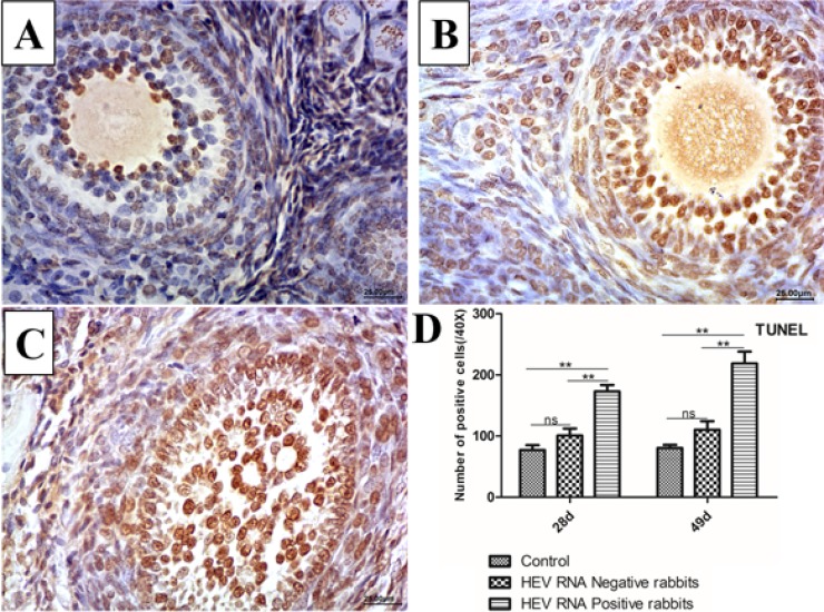
Figure 4. Representative TUNEL-stained histological sections of ovaries from control group and Swine HEV inoculated rabbits.
TUNEL-positive cells have brown nuclei. The number of TUNEL-positive cells were remarkably higher in HEV RNA positive ovaries at 28dpi (B) and 49dpi (C) than in the control (A). (magnification:40×). Quantitative analysis of TUNEL-positive cells in ovaries of rabbits (D). The data were expressed as the percentage (mean ± SD). (*P < 0.05) or (**P < 0.01), indicated statistical significance versus the control group (n = 5).

