



Abstract
Both preclinical and clinical studies demonstrate that depression is strongly associated with reduced light availability, which in turn contributes to decreased function of brain regions that control mood. Here, we review findings that support a critical pathway for the control of mood that depends upon ambient light. We put forward a novel hypothesis, functionally linking retina to locus coeruleus (LC) in depression, and discuss the role of norepinephrine in affective disease. Finally, we discuss how utilizing the chemogenetic tool Designer Receptors Exclusively Activated by Designer Drugs (DREADDs) to precisely control this retina–LC circuit may be used as a novel therapeutic to treat depression.
Keywords: antidepressants, anxiety/anxiety disorders, depression, mood disorders, pharmacotherapy, seasonal affective disorder, sleep disorders, stress, treatment
Depression is a complex mood disorder with a lifetime prevalence of 16% (Kessler et al., 2005), and a 12-month prevalence of over 9% in the United States (Demyttenaere et al., 2004). It is characterized by persistent low mood, loss of pleasure or interest, disrupted sleep and appetite, and feelings of guilt or low self-worth (Serretti, Mandelli, Lattuada, & Smeraldi, 2004). Depression is associated with impaired cognitive (Hammar & Ardal, 2009) and social function (Rubin & Burgess, 2001; Setterfield, Walsh, Frey, & McCabe, 2016), and is predicted to be the leading cause of disease burden by 2030 (Lépine & Briley, 2011), possibly due to its comorbidity with other psychiatric disorders such as anxiety (Fava et al., 2008), drug addiction (Volkow, 2004), and attention-deficit hyperactivity disorder (Waxmonsky, 2003).
Despite extensive research efforts, the precise etiology of depression remains elusive. The conceptualization of depression as a physical illness was prompted by the serendipitous discovery of the first psychopharmacological treatments in the 1950s. Since then, the formation of the monoamine (Prange, 1964; Schildkraut, 1965) and neurohormonal (Nemeroff, 1988) theories of depression have undoubtedly enhanced our understanding of the mechanisms underlying the disease, and indeed, have arguably led to relative enhancements of treatment outcomes. Yet despite these and other credible theories underlying the biology of depression (for review see Ordway, Klimek, & Mann, 2002), many important questions remain unanswered: no consistent genetic associations between loci and depression diagnosis or treatment responsivity have been identified Fried, 2015 (Lewis et al., 2010; Ripke et al., 2013; Shi et al., 2011; Tansey et al., 2012; Wray et al., 2012), pharmacological treatments remain suboptimal, and there has been a stagnation in the development of new pharmacological treatments (Khan, Khan, & Brown, 2002; Pigott, Leventhal, Alter, & Boren, 2010). These deficits may reflect lack of understanding of disease mechanisms at both the molecular and circuit levels. Therefore, there is a clear need to better understand the precise brain circuitry associated with depression.
1 CIRCADIAN RHYTHMS AND DEPRESSION: A PROPOSED CRITICAL ROLE FOR THE PRAM PATHWAY
Decades of research indicates that depression is associated with disrupted circadian rhythms. Indeed, depression is associated with a blunted amplitude and phase delay of circadian rhythms (Duncan, 1996), increased core temperature (Avery, Wildschiødtz, & Rafaelsen, 1982), and phase-advanced oscillations of noradrenaline and cortisol plasma concentrations (Koenigsberg et al., 2004). These findings are further supported by well-documented alterations of sleep in depressed patients (Perlis, Giles, Buysse, Tu, & Kupfer, 1997; Posmontier, 2008; Stewart et al., 2006), and are reflected in the DSM-V that lists insomnia or hypersomnia as criteria for major depressive disorder (MDD; American Psychiatric Association, 2013). Given the close association between light and the entrainment of circadian rhythms, significant effort has been directed toward understanding the role of light in depressive disorders. Particular attention has focused on seasonal affective disorder (SAD), the etiology of which is typically associated with decreased light availability (American Psychiatric Association, 2013). The onset of SAD symptoms often coincides with the decreased day lengths during fall and winter, and SAD is more prevalent in extreme latitudes where natural light is very limited during the colder months (Rosenthal et al., 1984). These patterns are supported by preclinical literature, as short day-length lighting schedules induce depression and anxiety-like behavior in rats (Einat, Kronfeld-Schor, & Eilam, 2006), and mice housed under irregular light conditions display depression-like behavior that can be alleviated by the antidepressant fluoxetine (LeGates et al., 2012). Despite a clear role for light-mediated disruption of circadian rhythms in depression, the precise neural mechanisms underlying this phenomenon are not well understood.
Fifteen years ago, our laboratory described a circuit for the circadian regulation of arousal. We demonstrated that the brain nucleus locus coeruleus (LC) receives an indirect circadian input from suprachiasmatic nucleus (SCN) via a relay in dorsomedial hypothalamus (DMH), thus revealing a circuit for the regulation of sleep and waking (Aston-Jones, Chen, Zhu, & Oshinsky, 2001). Subsequently, we discovered that light deprivation over 6 weeks in Sprague-Dawley rats led to depression-like behavior and decreased innervation of frontal cortex by noradrenergic (NA) LC fibers and terminals (Gonzalez & Aston-Jones, 2008); the latter is a physiological trait of animals with depression-like behavior (Kitayama et al., 1994).
SCN receives light information via a major retinal input, and works to coordinate the circadian clocks of the brain and body. It is therefore in a key position to integrate information about light with circuitry that regulates mood, via the pathway: retina → SCN → DMH → LC. We refer to this pathway as the photic regulation of arousal and mood (PRAM) pathway.
Here, we propose the hypothesis that some forms of depression are associated with disruptions to the normal physiological activity of the PRAM pathway. This hypothesis is based upon three key points: (1) the SCN → DMH → LC circuit plays a critical role in the circadian regulation of arousal (Aston-Jones et al., 2001), (2) light has a profound effect on mood and depressive behavior (Gonzalez & Aston-Jones, 2008) and SCN receives light information from a major retinal input, and (3) decreased light availability leads to degeneration of NA-LC cortical fibers (Gonzalez & Aston-Jones, 2008), which is associated with depression (Kitayama et al., 1994). This review will provide evidence for this pathway in depression, and will discuss how altering this pathway may eventually lead to functional alterations in mood. First, we focus on both clinical and preclinical evidence implicating the NA–LC system in the development of depression and depression-like symptomology. We then provide anatomical and functional evidence implicating SCN as a key regulator of the NA–LC system in depression, and how this region is modulated by photic input from the retina. Finally, we propose using a targeted chemogenetic approach to normalize activity of the PRAM pathway in depressed individuals, thus ameliorating symptomology associated with hypoarousal and disrupted circadian rhythms.
2 THE NA–LC SYSTEM AND DEPRESSION
LC (catecholaminergic cell group A6) is the principal site of norepinephrine (NE) synthesis within the brain, and provides the main source of ascending and descending NA brain fibers. The NA–LC system projects widely throughout the neuraxis, and has critical roles in a range of physiological functions, including the circadian regulation of arousal. NA–LC neurons fire tonically faster during waking (Aston-Jones & Bloom, 1981) or during the active period in anesthetized animals (Aston-Jones et al., 2001) than during sleep or the inactive period. Also, plasma NE levels are higher during waking (Linsell, Lightman, Mullen, Brown, & Causon, 1985). Unsurprisingly therefore, the NA–LC system plays a critical role in regulating the sleep-wake cycle; reducing LC activity promotes sleep, and increasing its activity facilitates alertness and attention (Berridge & Waterhouse, 2003) as well as emergence from anesthesia (Vazey & Aston-Jones, 2014). Additionally, the loss of NE cortical fibers induced by either light deprivation or the neurotoxin N-(2-chloroethyl)-N-ethyl-2-bromobenzylamine (DSP-4) decreases the amplitude of the sleep-wake rhythm (Gonzalez & Aston-Jones, 2006; Gonzalez & Aston-Jones, 2008). As we outline below, there is considerable evidence from both clinical studies and animal models that activity of the NA–LC system is profoundly dysregulated in depression. We propose that this disruption of normal NA–LC function in depression results in reduced NE signaling in key arousal regions in the brain, thus contributing to the dysregulation of mood observed in depressed patients.
Clinical studies generally report a deficit of NA–LC function in depressed patients. For example, Chan-Palay and Asan (1989) described an extreme loss of catecholaminergic LC cells in the postmortem brains of patients with MDD. Similarly, Arango, Underwood, and Mann (1996) reported that suicide patients have a lower density of LC neurons compared with psychiatrically normal patients, whereas others have reported reduced concentrations of 3-methoxy, 4-hydroxphenylglycol (MHPG), the principal metabolite of NE, in the cerebrospinal fluid (CSF) of suicide attempters (Agren, 1980, 1982). Moreover, reduced NE levels are associated with reduced mood in persons with a family history of depression (Ruhe, Mason, & Schene, 2007), and growth factor signaling and glial functioning are altered in LC of patients with depression (Bernard et al., 2011).
Further evidence of NE dysregulation in depression comes from the relative effectiveness of antidepressant treatments that normalize NE in ameliorating depressive symptomology in clinical populations. Serotonin-NA reuptake inhibitors (SNRIs), NE-dopamine reuptake inhibitors (NDRIs), NE reuptake inhibitors (NRIs), and tricyclic antidepressants (TCAs) all act to increase NE by preventing its reuptake by the presynaptic neuron. These therapies have shown some success as pharmacological treatments for depression, and have been suggested to offer a greater therapeutic advantage over selective serotonin reuptake inhibitors (SSRIs; Moret & Briley, 2011). Additionally, a meta-analysis of 193 trials found that SNRIs may be more effective than SSRIs in attaining remission from depression (Papakostas, Thase, Fava, Nelson, & Shelton, 2007). Taken together, depression is associated with a loss of NA-LC cells and a subsequent deficit of NE signaling, and pharmacotherapies that restore NE levels can be effective treatments for depression symptomology.
These findings from clinical populations are supported by evidence arising from animal studies of depression-like behavior. Kitayama et al. (1994) demonstrated that rats exposed to stress-induced depression exhibited profound degeneration of cortical NA axons, and that chronic treatment with the antidepressant imipramine restored cortical NA axons. Other studies have reported that chemical ablation of NA-LC fibers using the selective LC neurotoxin DSP-4 is associated with the expression of a range of depressive-like behaviors, including increased despair on the forced swim test (FST; Harro et al., 1999), increased neophobia (Delini-Stula, Mogilnicka, Hunn, & Dooley, 1984), reduced social interaction (Cornwell-Jones et al., 1992), and impaired exploratory behavior (Harro, Oreland, Vasar, & Bradwejn, 1995; van den Buuse, Lambert, Fluttert, & Eikelis, 2001). Finally, the Wistar-Kyoto (WKY) rat, a strain that expresses hormonal and behavioral profiles that resemble those observed in depressive patients, shows altered expression of several genes that are related to NE synthesis in LC (Pearson, Stephen, Beck, & Valentino, 2006; Sands, Strong, Corbitt, & Morilak, 2000).
Given the association between NA–LC system deficits and depression, and the relationship between the NA-LC system and circadian rhythms, we asked whether manipulating the availability of light, a critical zeitgeber, can induce changes in NA–LC functioning and depression-like behavior in the rat (Gonzalez & Aston-Jones, 2006). We subjected rats to 6 weeks of light deprivation, a paradigm known to decrease the amplitude of the sleep-wake rhythm, and analyzed depressive-like behavior as well as cell viability of several systems associated with the pathophysiology of depression, including LC. We found that animals maintained in constant darkness showed significant depression-like behavior, as well as greater apoptosis in LC, compared to those maintained in standard 12:12 lighting conditions. The NA–LC neuronal apoptosis was associated with decreased NA boutons in frontal cortex. These findings were the first demonstration that light deprivation leads to NA cell apoptosis in LC and reduced NA innervation in cortex, and provided a possible mechanism through which light may influence mood.
3 THE SUPRACHIASMATIC NUCLEUS-DORSOMEDIAL HYPOTHALAMUS PATHWAY AS A RELAY OF LIGHT INFORMATION TO THE NA–LC SYSTEM
How might photic information influence the NA-LC system? As no known direct projections exist from retina to LC, light information must be relayed to LC via an indirect pathway. The SCN is a strong candidate in such a network, as this structure receives light information from retina (Gooley, Lu, Chou, Scammell, & Saper, 2001; Hannibal, Hindersson, Knudsen, Georg, & Fahrenkrug, 2002) and projects indirectly to LC (as reviewed above; Aston-Jones et al., 2001). SCN synchronizes many biological circadian rhythms to produce internal homeostasis with the solar day (Antle & Silver, 2005), and is considered the master pacemaker of the brain (van Esseveldt, Lehman, & Boer, 2000). Similar to the NA-LC system, there is strong evidence of dysregulation of SCN function in mood disorders. For example, Sprouse, Braselton, and Reynolds (2006) demonstrated that both the synthesis and release of arginine vasopressin (AVP), a major peptide of SCN, is reduced in depressed patients. Bernstein et al. (2005) showed that patients with depression have fewer nitric oxide synthase (NOS) neurons in SCN than their nondepressed counterparts, while others have reported decreased levels of NO metabolites in plasma of depressed patients (Chrapko et al., 2006). Additionally, melatonin receptor 1 (MT1) is specifically increased in SCN of patients with depression, and the abundance of MT1 receptors is positively correlated with disease duration, indicating that SCN MT1 receptors increase with the duration of depression (Wu et al., 2013). These clinical findings are supported by animal studies that have investigated SCN function in models of depression. Using chronic unpredictable stress (CUS), Jiang et al. (2011) demonstrated that expression of SCN PER2, an important clock gene that regulates circadian cycles (Panda et al., 2002), was dampened in animals that exhibited depression-like behavior. A related study demonstrated that other SCN circadian proteins, CLOCK and BMAL1, are also reduced following CUS (Jiang et al., 2013). Vagell, McGinnis, Possidente, Narasimhan, and Lumia (1991) showed that depression-like behavior in rats, induced by olfactory bulbectomy, is associated with an increase in cyclic- 3′,5′-adenosine monophosphate (cAMP) levels, specifically in SCN. Together, both clinical and preclinical findings point to a dysregulation in SCN function in depression, likely resulting in aberrant signaling in downstream structures and systems, including the NA–LC system. It should be noted that dysregulation of SCN may also affect mood via its other projection targets, for example its direct connection with lateral hebenula (Buijs, 1978), which is known to influence DA, 5HT, and NA systems (Christoph, Leonzio, & Wilcox, 1986; Herkenham & Nauta, 1979; Ji & Shepard, 2007).
However, here we restrict our focus to the SCN–LC/NA hypothesis. How is dysregulated SCN function conveyed to the NA–LC system? The DMH is anatomically well-positioned to act as a relay in the SCN– LC pathway, as DMH receives input from SCN (Leak & Moore, 2001) and projects directly to LC (Aston-Jones et al., 2001; Gompf & Aston-Jones, 2008). Our laboratory previously found that the same DMH neurons that receive input from SCN project to LC, and that disruption of this pathway results in dysregulated circadian rhythm and arousal (Aston-Jones et al., 2001; Gonzalez & Aston-Jones, 2006).
The phenotype of these DMH “relay cells” is not yet known. It is interesting to note, however, that DMH contains a discrete set of neurons that produce the arousal-related neuropeptide orexin/hypocretin, and orexin/hypocretin cell function has been reliably shown to be dysregulated in depression. For example, levels of orexin-A in CSF are significantly lower in MDD patients compared to healthy controls (Brundin, Björkqvist, Petersén, & Träskman-Bendz, 2007), and MDD patients exhibit blunted diurnal variation of orexin CSF levels (Salomon et al., 2003). Among patients with MDD, the severity of depressive symptomology is negatively correlated with orexin levels in CSF (Brundin et al., 2007) as well as orexin mRNA in the blood (Rotter, 2011). These findings are largely reflected in the preclinical literature, with reduced orexin levels observed in genetic models of depression (Allard, Tizabi, Shaffery, Trouth, & Manaye, 2004; Taheri et al., 2001) as well as in animals exposed to various stress protocols that induce depression-like behavior (James et al., 2014; Lutter et al., 2008; Nocjar, Zhang, Feng, & Panksepp, 2012; for full review, see James, Campbell, & Dayas, 2016, Yeoh, Campbell, James, Graham, & Dayas, 2014). Together, these findings point to a possible role for DMH orexin/hypocretin cells in relaying information in the SCN-LC pathway, a hypothesis that ongoing studies in our laboratory are seeking to confirm.
We posit that under normal circumstances, the retina provides photic input to the SCN-DMH-LC pathway (collectively, the PRAM pathway; see Fig. 1), which in turn regulates brain circuits that support normal mood. In some forms of depression, photic input onto the PRAM network is disrupted, resulting in pathological dysregulation of sleep, arousal, and mood. Disruption of PRAM pathway signaling likely has numerous etiologies: in the case of SAD, PRAM signaling is likely to be reduced owing to environmental conditions (low light levels). Other forms of depression may be associated with decreased light exposure due to social withdrawal or irregular sleep patterns, thereby augmenting depressive symptomology in patients. Other forms of depression may arise from dysfunction in LC, with normal functioning of upstream parts of the PRAM circuit (SCN and DMH). However, even in those cases we propose that manipulations of the PRAM pathway might be effective as a treatment to offset such dysfunctions in downstream circuit elements and normalize mood. Thus, we argue that by restoring normal signaling in the PRAM pathway, or manipulating this circuit to compensate for dysfunctions downstream from the circuit, it may be possible to ameliorate some of the symptoms that characterize depression. Indeed, there are some reports that “light therapy” has some therapeutic efficacy for some forms of depression, particularly SAD. However, therapies designed to restore normal signaling in the PRAM pathway would ideally seek to selectively enhance input from those cells in the retina that provide specific input onto SCN, without affecting other visual pathways. Such an approach, we believe, would provide a more nuanced and targeted approach for the restoration of normal circadian and arousal states in depressed individuals.
FIGURE 1.
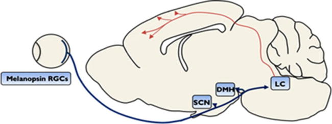
The photic regulation of arousal and mood (PRAM) pathway. A midsaggital section of a rat brain illustrating loci involved in PRAM-associated depressive-like behavior. Structures of PRAM pathway are indicated by blue boxes. SCN, suprachiasmatic nucleus; DMH, dorsomedial hypothalamus; LC, locus coeruleus. Melanopsin-containing retinal ganglion cells (RGCs) are putative the origin of the PRAM pathway, as they project directly onto SCN, their major target, which then projects indirectly onto LC via DMH. Dysregulation of the PRAM pathway may lead to the decreased noradrenergic-LC signaling (red arrows) that is associated with depression
4 MELANOPSIN-EXPRESSING, INTRINSICALLY PHOTOSENSITIVE RETINAL GANGLION CELLS: A DIRECT ROUTE TO THE PRAM PATHWAY?
In recent years there has been great interest in a subset of ∼4–5% of retinal ganglion cells (RGCs) that express the photopigment melanopsin (Schmidt, Chen, & Hattar, 2011). These specialized cells intrinsically respond to light independently of traditional photoreceptor signaling, have robust effects on the regulation of circadian rhythms, and are a prominent candidate cell-type for the nonimage forming functions of light such as mood regulation. Known as intrinsically photosensitive RGCs (ipRGCs), these cells project directly to their major target, SCN (Baver, Pickard, Sollars, & Pickard, 2008; Guler et al., 2008; Hattar et al., 2006; Sollars et al., 2003; Hattar et al., 2002; Provencio et al., 2000), and thus are likely to directly influence PRAM pathway function. Indeed, human fMRI studies indicate that 480 nm blue light, which maximally activates ipRGCs, increases the bilateral activity in the area consistent with LC (Vandewalle et al., 2007). Further, a number of studies show striking effects of melanopsin on mood. For example, in humans, two gene-association studies demonstrated a link between melanopsin and SAD (Roecklein et al., 2009), and between melanopsin and seasonal alterations in sleep timing (Roecklein et al., 2012). Four hundred eighty nanometers blue light, which maximally activates ipRGCs, is superior to green light in modulating emotional brain responses (Vandewalle et al., 2010). Further, depression-like behavior in mice induced by aberrant light cycle conditions is dependent on melanopsin-containing cells (LeGates et al., 2012). In this way, melanopsin-containing ipRGCs represent an attractive candidate cell type involved in the photic regulation of circadian entrainment, arousal, and regulation of mood.
5 A CHEMOGENETIC APPROACH TO TARGETING MELANOPSIN IPRGCS FOR THE TREATMENT OF DEPRESSION
DREADDs are a powerful new chemogenetic tool that is transforming neuropsychiatric research. Allowing for the direct and selective control of neuronal populations, DREADDs modulate neuronal function by targeting specific intracellular signaling pathways. DREADDs are modified human GPCRs that are solely activated by a specific orthogonal ligand, clozapine-N-oxide (CNO), which itself is otherwise pharmacologically inert (Armbruster, Li, Pausch, Herlitze, & Roth, 2007). Three DREADDs commonly used are hM3Dq, hM4Di, and rM3Ds that activate Gq, Gi, and Gs signaling, respectively. Effectively, DREADDs create a powerful “lock and key” approach for targeted neuronal regulation (for example, see Mahler et al., 2014; Vazey and Aston-Jones (2014)). Different from other genetic approaches, such as optogenetics, DREADDs are based upon human proteins and therefore have minimal potential for immunogenic reactivity and require no optical fiber implant, and thus arguably provide greater translational potential for psychiatric disorders. However, despite their translational appeal, current technology requires the direct injection of a viral-vector into the brain, creating multiple surgical risks and potential prohibitive expense for the patient. The eye presents an alternate route of direct administration of the viral-vector, as the retina is the only part of the central nervous system that can be accessed without the risk of invasive brain surgery.
Intravitreal injection (IVI), which bypasses the blood–retinal barrier, allows for highly targeted vector delivery. With the increased use of injectable drugs for the treatment of blinding diseases, IVIs have become one of the most common ophthalmic procedures (Ho & Kuo, 2007). IVIs are rapid, the overall risk for serious complications is low, and only local anesthesia is required. As such, it is feasible to conceive of a therapy whereby DREADDs are transfected specifically into melanopsin ipRGCs via IVI, allowing for regulation of the PRAM pathway with local (eye drops) or systemic (oral dosing) CNO. Clearly further preclinical work is required to demonstrate the therapeutic utility of such an approach, a line of work our laboratory is currently actively pursuing.
6 SUMMARY
The PRAM pathway presents a novel circuit for the regulation of mood in both humans and other species. Evidence indicates that melanopsin-containing ipRGCs are the origin of the PRAM pathway, as they project directly onto SCN, their major target, which then projects indirectly onto LC via DMH. Because access to the retina is simple and carries little risk, IVI-injected DREADDs provide an exciting opportunity for manipulating the PRAM pathway in humans, without the need for brain surgery. While further research is required to understand the precise mechanism(s) undying the PRAM pathway-mediated regulation of mood, we have nonetheless demonstrated a potentially critical role for this pathway in such processes. The recent development of DREADDs, allowing for reversible control of the PRAM pathway, creates exciting new directions for the treatment of depression in humans.
Acknowledgments
Supported by USPHS grant R01MH092868 (GAJ) and by the National Health and Medical Research Council of Australia: 1128089 (HEB), 1072706 (MHJ).
Grant sponsor: USPHS; Grant number: R01MH092868; Grant sponsor: National Health and Medical Research Council of Australia; Grant numbers: 1128089 and 1072706.
Footnotes
Dr. James has no conflict of interest to report. Both Dr. Bowrey and Dr. Aston-Jones are listed as inventors on a pending patent entitled “compositions and methods for treating neuropsychiatric disorders.”
References
- Agren H. Symptom patterns in unipolar and bipolar depression correlating with monoamine metabolites in the cerebrospinal fluid: II. Suicide. Psychiatry Research. 1980;3(2):225–236. doi: 10.1016/0165-1781(80)90039-6. [DOI] [PubMed] [Google Scholar]
- Agren H. Depressive symptom patterns and urinary MHPG excretion. Psychiatry Research. 1982;6(2):185–196. doi: 10.1016/0165-1781(82)90006-3. [DOI] [PubMed] [Google Scholar]
- Allard JS, Tizabi Y, Shaffery JP, Trouth CO, Manaye K. Stereological analysis of the hypothalamic hypocretin/orexin neurons in an animal model of depression. Neuropeptides. 2004;38(5):311–315. doi: 10.1016/j.npep.2004.06.004. [DOI] [PubMed] [Google Scholar]
- American Psychiatric Association. Diagnostic and statistical manual of mental disorders (DSM-5) Arlington, VA: American Psychiatric Pub; 2013. [Google Scholar]
- Antle MC, Silver R. Orchestrating time: Arrangements of the brain circadian clock. Trends in Neuroscience (TINS) 2005;28(3):145–151. doi: 10.1016/j.tins.2005.01.003. https://doi.org/10.1016/j.tins.2005.01.003. [DOI] [PubMed] [Google Scholar]
- Arango V, Underwood MD, Mann JJ. Fewer pigmented locus coeruleus neurons in suicide victims: Preliminary results. Biological Psychiatry. 1996;39(2):112–120. doi: 10.1016/0006-3223(95)00107-7. https://doi.org/10.1016/0006-3223(95)00107-7. [DOI] [PubMed] [Google Scholar]
- Armbruster BN, Li X, Pausch MH, Herlitze S, Roth BL. Evolving the lock to fit the key to create a family of G protein-coupled receptors potently activated by an inert ligand. Proceedings of the National Academy of Sciences of the United States of America. 2007;104(12):5163–5168. doi: 10.1073/pnas.0700293104. https://doi.org/10.1073/pnas.0700293104. [DOI] [PMC free article] [PubMed] [Google Scholar]
- Aston-Jones G, Bloom FE. Activity of norepinephrine-containing locus coeruleus neurons in behaving rats anticipates fluctuations in the sleep-waking cycle. Journal of Neuroscience. 1981;1(8):876–886. doi: 10.1523/JNEUROSCI.01-08-00876.1981. [DOI] [PMC free article] [PubMed] [Google Scholar]
- Aston-Jones G, Chen S, Zhu Y, Oshinsky ML. A neural circuit for circadian regulation of arousal. Nature Neuroscience. 2001;4(7):732–738. doi: 10.1038/89522. https://doi.org/10.1038/89522. [DOI] [PubMed] [Google Scholar]
- Avery DH, Wildschiødtz G, Rafaelsen OJ. Nocturnal temperature in affective disorder. Journal of Affective Disorders. 1982;4(1):61–71. doi: 10.1016/0165-0327(82)90020-9. [DOI] [PubMed] [Google Scholar]
- Baver SB, Pickard GE, Sollars PJ, Pickard GE. Two types of melanopsin retinal ganglion cell differentially innervate the hypothalamic suprachiasmatic nucleus and the olivary pretectal nucleus. European Journal of Neuroscience. 2008;27(7):1763–1770. doi: 10.1111/j.1460-9568.2008.06149.x. https://doi.org/10.1111/j.1460-9568.2008.06149.x. [DOI] [PubMed] [Google Scholar]
- Bernard R, Kerman IA, Thompson RC, Jones EG, Bunney WE, Barchas JD, Watson SJ. Altered expression of glutamate signaling, growth factor, and glia genes in the locus coeruleus of patients with major depression. Molecular Psychiatry. 2011;16(6):634–646. doi: 10.1038/mp.2010.44. https://doi.org/10.1038/mp.2010.44. [DOI] [PMC free article] [PubMed] [Google Scholar]
- Bernstein HG, Heinemann A, Krell D, Dobrowolny H, Bielau H, Keilhoff G, Bogerts B. Hypothalamic nitric oxide synthase in affective disorder: Focus on the suprachiasmatic nucleus. Cellular and Molecular Biology. 2005;51(3):279–284. [PubMed] [Google Scholar]
- Berridge CW, Waterhouse BD. The locus coeruleus-noradrenergic system: Modulation of behavioral state and state-dependent cognitive processes. Brain Research Brain Research Reviews. 2003;42(1):33–84. doi: 10.1016/s0165-0173(03)00143-7. [DOI] [PubMed] [Google Scholar]
- Brundin L, Björkqvist M, Petersén Å, Träskman-Bendz L. Reduced orexin levels in the cerebrospinal fluid of suicidal patients with major depressive disorder. European Neuropsychopharmacology. 2007;17(9):573–579. doi: 10.1016/j.euroneuro.2007.01.005. [DOI] [PubMed] [Google Scholar]
- Buijs RM. Intra-and extrahypothalamic vasopressin and oxytocin pathways in the rat. Cell and Tissue Research. 1978;192(3):423–435. doi: 10.1007/BF00212323. [DOI] [PubMed] [Google Scholar]
- Chan-Palay V, Asan E. Quantitation of catecholamine neurons in the locus coeruleus in human brains of normal young and older adults and in depression. Journal of Comparative Neurology. 1989;287(3):357–372. doi: 10.1002/cne.902870307. https://doi.org/10.1002/cne.902870307. [DOI] [PubMed] [Google Scholar]
- Chrapko W, Jurasz P, Radomski MW, Archer SL, Newman SC, Baker G, Le Melledo JM. Alteration of decreased plasma NO metabolites and platelet NO synthase activity by paroxetine in depressed patients. Neuropsychopharmacology. 2006;31(6):1286–1293. doi: 10.1038/sj.npp.1300961. https://doi.org/10.1038/sj.npp.1300961. [DOI] [PubMed] [Google Scholar]
- Christoph GR, Leonzio RJ, Wilcox KS. Stimulation of the lateral habenula inhibits dopamine-containing neurons in the substantia nigra and ventral tegmental area of the rat. Journal of Neuroscience. 1986;6(3):613–619. doi: 10.1523/JNEUROSCI.06-03-00613.1986. [DOI] [PMC free article] [PubMed] [Google Scholar]
- Cornwell-Jones CA, Palfai T, Krasenbaum D, Byer E, Jr, Clark R, Kinnard K. Housing influences exploration and social interaction of control and DSP-4-treated rats. Physiology & Behavior. 1992;52(2):271–276. doi: 10.1016/0031-9384(92)90270-c. [DOI] [PubMed] [Google Scholar]
- Delini-Stula A, Mogilnicka E, Hunn C, Dooley DJ. Novelty-oriented behavior in the rat after selective damage of locus coeruleus projections by DSP-4, a new noradrenergic neurotoxin. Pharmacology, Biochemistry and Behavior. 1984;20(4):613–618. doi: 10.1016/0091-3057(84)90312-5. [DOI] [PubMed] [Google Scholar]
- Demyttenaere K, Bruffaerts R, Posada-Villa J, Gasquet I, Kovess V, Lepine JP, Chatterji S. Prevalence, severity, and unmet need for treatment of mental disorders in the World Health Organization World Mental Health Surveys. Journal of the American Medical Association. 2004;291(21):2581–2590. doi: 10.1001/jama.291.21.2581. https://doi.org/10.1001/jama.291.21.2581. [DOI] [PubMed] [Google Scholar]
- Duncan WC. Circadian rhythms and the pharmacology of affective illness. Pharmacology & Therapeutics. 1996;71(3):253–312. doi: 10.1016/s0163-7258(96)00092-7. [DOI] [PubMed] [Google Scholar]
- Einat H, Kronfeld-Schor N, Eilam D. Sand rats see the light: Short photoperiod induces a depression-like response in a diurnal rodent. Behavioural Brain Research. 2006;173(1):153–157. doi: 10.1016/j.bbr.2006.06.006. https://doi.org/10.1016/j.bbr.2006.06.006. [DOI] [PubMed] [Google Scholar]
- Fava M, Rush AJ, Alpert JE, Balasubramani GK, Wisniewski SR, Carmin CN, Trivedi MH. Difference in treatment outcome in outpatients with anxious versus nonanxious depression: A STAR*D report. American Journal of Psychiatry. 2008;165(3):342–351. doi: 10.1176/appi.ajp.2007.06111868. https://doi.org/10.1176/appi.ajp.2007.06111868. [DOI] [PubMed] [Google Scholar]
- Fried EI. Problematic assumptions have slowed down depression research: Why symptoms, not syndromes are the way forward. Frontiers in Psychology. 2015;6(309) doi: 10.3389/fpsyg.2015.00309. https://doi.org/10.3389/fpsyg.2015.00309. [DOI] [PMC free article] [PubMed] [Google Scholar]
- Gompf HS, Aston-Jones G. Role of orexin input in the diurnal rhythm of locus coeruleus impulse activity. Brain Research. 2008;1224:43–52. doi: 10.1016/j.brainres.2008.05.060. [DOI] [PMC free article] [PubMed] [Google Scholar]
- Gonzalez MM, Aston-Jones G. Circadian regulation of arousal: Role of the noradrenergic locus coeruleus system and light exposure. Sleep. 2006;29(10):1327–1336. doi: 10.1093/sleep/29.10.1327. [DOI] [PubMed] [Google Scholar]
- Gonzalez MM, Aston-Jones G. Light deprivation damages monoamine neurons and produces a depressive behavioral phenotype in rats. Proceedings of the National Academy of Sciences of the United States of America. 2008;105(12):4898–4903. doi: 10.1073/pnas.0703615105. https://doi.org/10.1073/pnas.0703615105. [DOI] [PMC free article] [PubMed] [Google Scholar]
- Gooley JJ, Lu J, Chou TC, Scammell TE, Saper CB. Melanopsin in cells of origin of the retinohypothalamic tract. Nature Neuroscience. 2001;4(12):1165–1165. doi: 10.1038/nn768. [DOI] [PubMed] [Google Scholar]
- Guler AD, Ecker JL, Lall GS, Haq S, Altimus CM, Liao HW, Hattar S. Melanopsin cells are the principal conduits for rod-cone input to non-image-forming vision. Nature. 2008;453(7191):102–105. doi: 10.1038/nature06829. https://doi.org/10.1038/nature06829. [DOI] [PMC free article] [PubMed] [Google Scholar]
- Hammar A, Ardal G. Cognitive functioning in major depression—A summary. Frontiers in Human Neuroscience. 2009;3:26. doi: 10.3389/neuro.09.026.2009. https://doi.org/10.3389/neuro.09.026.2009. [DOI] [PMC free article] [PubMed] [Google Scholar]
- Hannibal J, Hindersson P, Knudsen SM, Georg B, Fahrenkrug J. The photopigment melanopsin is exclusively present in pituitary adenylate cyclase-activating polypeptide-containing retinal ganglion cells of the retinohypothalamic tract. Journal of Neuroscience. 2002;22(RC191):1–7. doi: 10.1523/JNEUROSCI.22-01-j0002.2002. [DOI] [PMC free article] [PubMed] [Google Scholar]
- Harro J, Oreland L, Vasar E, Bradwejn J. Impaired exploratory behaviour after DSP-4 treatment in rats: Implications for the increased anxiety after noradrenergic denervation. European Neuropsychopharmacology. 1995;5(4):447–455. doi: 10.1016/0924-977x(95)00015-h. [DOI] [PubMed] [Google Scholar]
- Harro J, Pahkla R, Modiri AR, Harro M, Kask A, Oreland L. Dose-dependent effects of noradrenergic denervation by DSP-4 treatment on forced swimming and beta-adrenoceptor binding in the rat. Journal of Neural Transmission (Vienna) 1999;106(7–8):619–629. doi: 10.1007/s007020050184. https://doi.org/10.1007/s007020050184. [DOI] [PubMed] [Google Scholar]
- Hattar S, Kumar M, Park A, Tong P, Tung J, Yau KW, Berson DM. Central projections of melanopsin-expressing retinal ganglion cells in the mouse. Journal of Comparative Neurology. 2006;497(3):326–349. doi: 10.1002/cne.20970. https://doi.org/10.1002/cne.20970. [DOI] [PMC free article] [PubMed] [Google Scholar]
- Hattar S, Liao HW, Takao M, Berson DM, Yau KW. Melanopsin-containing retinal ganglion cells: Architecture, projections, and intrinsic photosensitivity. Science. 2002;295(5557):1065–1070. doi: 10.1126/science.1069609. https://doi.org/10.1126/science.1069609. [DOI] [PMC free article] [PubMed] [Google Scholar]
- Herkenham M, Nauta WJ. Efferent connections of the habenular nuclei in the rat. Journal of Comparative Neurology. 1979;187(1):19–47. doi: 10.1002/cne.901870103. [DOI] [PubMed] [Google Scholar]
- Ho QT, Kuo CJ. Vascular endothelial growth factor: Biology and therapeutic applications. International Journal of Biochemistry & Cell Biology. 2007;39(7–8):1349–1357. doi: 10.1016/j.biocel.2007.04.010. [DOI] [PMC free article] [PubMed] [Google Scholar]
- James MH, Campbell EJ, Dayas CV. Role of the orexin/hypocretin system in stress-related psychiatric disorders. Current Topics in Behavioral Neurosciences. 2016:1–23. doi: 10.1007/7854_2016_56. https://doi.org/10.1007/7854_2016_56. [DOI] [PubMed]
- James MH, Campbell EJ, Walker FR, Smith DW, Richardson HN, Hodgson DM, Dayas CV. Exercise reverses the effects of early life stress on orexin cell reactivity in male but not female rats. Frontiers in Behavioral Neuroscience. 2014;8:244. doi: 10.3389/fnbeh.2014.00244. https://doi.org/10.3389/fnbeh.2014.00244. [DOI] [PMC free article] [PubMed] [Google Scholar]
- Ji H, Shepard PD. Lateral habenula stimulation inhibits rat midbrain dopamine neurons through a GABAA receptor-mediated mechanism. Journal of Neuroscience. 2007;27(26):6923–6930. doi: 10.1523/JNEUROSCI.0958-07.2007. [DOI] [PMC free article] [PubMed] [Google Scholar]
- Jiang WG, Li SX, Liu JF, Sun Y, Zhou SJ, Zhu WL, Lu L. Hippocampal CLOCK protein participates in the persistence of depressive-like behavior induced by chronic unpredictable stress. Psychopharmacology. 2013;227(1):79–92. doi: 10.1007/s00213-012-2941-4. https://doi.org/10.1007/s00213-012-2941-4. [DOI] [PubMed] [Google Scholar]
- Jiang WG, Li SX, Zhou SJ, Sun Y, Shi J, Lu L. Chronic unpredictable stress induces a reversible change of PER2 rhythm in the suprachiasmatic nucleus. Brain Research. 2011;1399:25–32. doi: 10.1016/j.brainres.2011.05.001. https://doi.org/10.1016/j.brainres.2011.05.001. [DOI] [PubMed] [Google Scholar]
- Kessler RC, Berglund P, Demler O, Jin R, Merikangas KR, Walters EE. Lifetime preva and age-of-onset distributions of DSM-IV disorders in the National Comorbidity Survey Replication. Archives of General Psychiatry. 2005;62(6):593–602. doi: 10.1001/archpsyc.62.6.593. https://doi.org/10.1001/archpsyc.62.6.593. [DOI] [PubMed] [Google Scholar]
- Khan A, Khan S, Brown WA. Are placebo controls necessary to test new antidepressants and anxiolytics? International Journal of Neuropsychopharmacology. 2002;5(3):193–197. doi: 10.1017/S1461145702002912. https://doi.org/10.1017/S1461145702002912. [DOI] [PubMed] [Google Scholar]
- Kitayama I, Nakamura S, Yaga T, Murase S, Nomura J, Kayahara T, Nakano K. Degeneration of locus coeruleus axons in stress-induced depression model. Brain Research Bulletin. 1994;35(5–6):573–580. doi: 10.1016/0361-9230(94)90171-6. [DOI] [PubMed] [Google Scholar]
- Koenigsberg HW, Teicher MH, Mitropoulou V, Navalta C, New AS, Trestman R, Siever LJ. 24-h Monitoring of plasma norepinephrine, MHPG, cortisol, growth hormone and prolactin in depression. Journal of Psychiatric Research. 2004;38(5):503–511. doi: 10.1016/j.jpsychires.2004.03.006. [DOI] [PubMed] [Google Scholar]
- Leak RK, Moore RY. Topographic organization of suprachiasmatic nucleus projection neurons. Journal of Comparative Neurology. 2001;433(3):312–334. doi: 10.1002/cne.1142. [DOI] [PubMed] [Google Scholar]
- LeGates TA, Altimus CM, Wang H, Lee HK, Yang S, Zhao H, Hattar S. Aberrant light directly impairs mood and learning through melanopsin-expressing neurons. Nature. 2012;491(7425):594–598. doi: 10.1038/nature11673. [DOI] [PMC free article] [PubMed] [Google Scholar]
- LeGates TA, Fernandez DC, Hattar S. Light as a central modulator of circadian rhythms, sleep and affect. Nature Reviews Neuroscience. 2014;15(7):443–454. doi: 10.1038/nrn3743. [DOI] [PMC free article] [PubMed] [Google Scholar]
- Lépine JP, Briley M. The increasing burden of depression. Neuropsychiatric Disease and Treatment. 2011;7(SUPPL):3–7. doi: 10.2147/NDT.S19617. https://doi.org/10.2147/NDT.S19617. [DOI] [PMC free article] [PubMed] [Google Scholar]
- Lewis CM, Ng MY, Butler AW, Cohen-Woods S, Uher R, Pirlo K, McGuffin P. Genome-wide association study of major recurrent depression in the U.K. population. American Journal of Psychiatry. 2010;167(8):949–957. doi: 10.1176/appi.ajp.2010.09091380. https://doi.org/10.1176/appi.ajp.2010.09091380. [DOI] [PubMed] [Google Scholar]
- Linsell CR, Lightman SL, Mullen PE, Brown MJ, Causon RC. Circadian rhythms of epinephrine and norepinephrine in man. The Journal of Clinical Endocrinology & Metabolism. 1985;60(6):1210–1215. doi: 10.1210/jcem-60-6-1210. [DOI] [PubMed] [Google Scholar]
- Lutter M, Krishnan V, Russo SJ, Jung S, McClung CA, Nestler EJ. Orexin signaling mediates the antidepressant-like effect of calorie restriction. Journal of Neuroscience. 2008;28(12):3071–3075. doi: 10.1523/JNEUROSCI.5584-07.2008. [DOI] [PMC free article] [PubMed] [Google Scholar]
- Mahler SV, Vazey EM, Beckley JT, Keistler CR, McGlinchey EM, Kaufling J, Aston-Jones G. Designer receptors show role for ventral pallidum input to ventral tegmental area in cocaine seeking. Nature Neuroscience. 2014;17(4):577–585. doi: 10.1038/nn.3664. https://doi.org/10.1038/nn.3664. [DOI] [PMC free article] [PubMed] [Google Scholar]
- Moret C, Briley M. The importance of norepinephrine in depression. Neuropsychiatric Disease and Treatment. 2011;7(Suppl. 1):9–13. doi: 10.2147/NDT.S19619. https://doi.org/10.2147/ndt.s19619. [DOI] [PMC free article] [PubMed] [Google Scholar]
- Nemeroff CB. The role of corticotropin-releasing factor in the pathogenesis of major depression. Pharmacopsychiatry. 1988;21(2):76–82. doi: 10.1055/s-2007-1014652. https://doi.org/10.1055/s-2007-1014652. [DOI] [PubMed] [Google Scholar]
- Nocjar C, Zhang J, Feng P, Panksepp J. The social defeat animal model of depression shows diminished levels of orexin in mesocortical regions of the dopamine system, and of dynorphin and orexin in the hypothalamus. Neuroscience. 2012;218:138–153. doi: 10.1016/j.neuroscience.2012.05.033. [DOI] [PubMed] [Google Scholar]
- Ordway GA, LKlimek VA, Mann JJ. Neurocircuitry of Mood Disorders. In: Davis KL, Charney D, Coyle JT, Nemeroff C, editors. Neuropsychopharmacology: The Fifth Generation of Progress (1051–1064) Philadelphia: Lippincott Williams & Wilkins; 2002. [Google Scholar]
- Panda S, Antoch MP, Miller BH, Su AI, Schook AB, Straume M, Hogenesch JB. Coordinated transcription of key pathways in the mouse by the circadian clock. Cell. 2002;109(3):307–320. doi: 10.1016/s0092-8674(02)00722-5. [DOI] [PubMed] [Google Scholar]
- Papakostas GI, Thase ME, Fava M, Nelson JC, Shelton RC. Are antidepressant drugs that combine serotonergic and noradrenergic mechanisms of action more effective than the selective serotonin reuptake inhibitors in treating major depressive disorder? A meta-analysis of studies of newer agents. Biological Psychiatry. 2007;62(11):1217–1227. doi: 10.1016/j.biopsych.2007.03.027. https://doi.org/10.1016/j.biopsych.2007.03.027. [DOI] [PubMed] [Google Scholar]
- Pearson KA, Stephen A, Beck SG, Valentino RJ. Identifying genes in monoamine nuclei that may determine stress vulnerability and depressive behavior in Wistar-Kyoto rats. Neuropsychopharmacology. 2006;31(11):2449–2461. doi: 10.1038/sj.npp.1301100. https://doi.org/10.1038/sj.npp.1301100. [DOI] [PMC free article] [PubMed] [Google Scholar]
- Perlis ML, Giles DE, Buysse DJ, Tu X, Kupfer DJ. Self-reported sleep disturbance as a prodromal symptom in recurrent depression. Journal of Affective Disorders. 1997;42(2–3):209–212. doi: 10.1016/s0165-0327(96)01411-5. [DOI] [PubMed] [Google Scholar]
- Pigott HE, Leventhal AM, Alter GS, Boren JJ. Efficacy and effectiveness of antidepressants: Current status of research. Psychotherapy and Psychosomatics. 2010;79(5):267–279. doi: 10.1159/000318293. https://doi.org/10.1159/000318293. [DOI] [PubMed] [Google Scholar]
- Posmontier B. Sleep quality in women with and without postpartum depression. Journal of Obstetric, Gynecologic, and Neonatal Nursing. 2008;37(6):722–735. doi: 10.1111/j.1552-6909.2008.00298.x. quiz 735–727. https://doi.org/10.1111/j.1552-6909.2008.00298.x. [DOI] [PMC free article] [PubMed] [Google Scholar]
- Prange AJ., Jr The pharmacology and biochemistry of depression. Diseases of the Nervous System. 1964;25:217–221. [PubMed] [Google Scholar]
- Provencio I, Rodriguez IR, Jiang G, Hayes WP, Moreira EF, Rollag MD. A novel human opsin in the inner retina. Journal of Neuroscience. 2000;20(2):600–605. doi: 10.1523/JNEUROSCI.20-02-00600.2000. [DOI] [PMC free article] [PubMed] [Google Scholar]
- Ripke S, Wray NR, Lewis CM, Hamilton SP, Weissman MM, Breen G, Sullivan PF. A mega-analysis of genome-wide association studies for major depressive disorder. Molecular Psychiatry. 2013;18(4):497–511. doi: 10.1038/mp.2012.21. https://doi.org/10.1038/mp.2012.21. [DOI] [PMC free article] [PubMed] [Google Scholar]
- Roecklein KA, Rohan KJ, Duncan WC, Rollag MD, Rosenthal NE, Lipsky RH, Provencio I. A missense variant (P10L) of the melanopsin (OPN4) gene in seasonal affective disorder. Journal of Affective Disorders. 2009;114(1–3):279–285. doi: 10.1016/j.jad.2008.08.005. https://doi.org/10.1016/j.jad.2008.08.005. [DOI] [PMC free article] [PubMed] [Google Scholar]
- Roecklein KA, Wong PM, Franzen PL, Hasler BP, Wood-Vasey WM, Nimgaonkar VL, Manuck SB. Melanopsin gene variations interact with season to predict sleep onset and chronotype. Chronobiology International. 2012;29(8):1036–1047. doi: 10.3109/07420528.2012.706766. https://doi.org/10.3109/07420528.2012.706766. [DOI] [PMC free article] [PubMed] [Google Scholar]
- Rosenthal NE, Sack DA, Gillin JC, Lewy AJ, Goodwin FK, Davenport Y, Wehr TA. Seasonal affective disorder. A description of the syndrome and preliminary findings with light therapy. Archives of General Psychiatry. 1984;41(1):72–80. doi: 10.1001/archpsyc.1984.01790120076010. [DOI] [PubMed] [Google Scholar]
- Rotter A, Asemann R, Decker A, Kornhuber J, Biermann T. Orexin expression and promoter-methylation in peripheral blood of patients suffering from major depressive disorder. Journal of Affective Disorders. 2011;131(1):186–192. doi: 10.1016/j.jad.2010.12.004. [DOI] [PubMed] [Google Scholar]
- Rubin KH, Burgess K. Social withdrawal. In: Vasey MW, Dadds MR, editors. The developmental psychopathology of anxiety. Oxford, UK: Oxford University Press; 2001. pp. 407–434. [Google Scholar]
- Ruhe HG, Mason NS, Schene AH. Mood is indirectly related to serotonin, norepinephrine and dopamine levels in humans: A meta-analysis of monoamine depletion studies. Molecular Psychiatry. 2007;12(4):331–359. doi: 10.1038/sj.mp.4001949. https://doi.org/10.1038/sj.mp.4001949. [DOI] [PubMed] [Google Scholar]
- Salomon RM, Ripley B, Kennedy JS, Johnson B, Schmidt D, Zeitzer JM, Mignot E. Diurnal variation of cerebrospinal fluid hypocretin-1 (Orexin-A) levels in control and depressed subjects. Biological Psychiatry. 2003;54(2):96–104. doi: 10.1016/s0006-3223(02)01740-7. [DOI] [PubMed] [Google Scholar]
- Sands SA, Strong R, Corbitt J, Morilak DA. Effects of acute restraint stress on tyrosine hydroxylase mRNA expression in locus coeruleus of Wistar and Wistar-Kyoto rats. Brain Research Molecular Brain Research. 2000;75(1):1–7. doi: 10.1016/s0169-328x(99)00255-7. [DOI] [PubMed] [Google Scholar]
- Schildkraut JJ. The catecholamine hypothesis of affective disorders: A review of supporting evidence. American Journal of Psychiatry. 1965;122(5):509–522. doi: 10.1176/ajp.122.5.509. https://doi.org/10.1176/ajp.122.5.509. [DOI] [PubMed] [Google Scholar]
- Schmidt TM, Chen SK, Hattar S. Intrinsically photosensitive retinal ganglion cells: Many subtypes, diverse functions. Trends in Neuroscience (TINS) 2011;34(11):572–580. doi: 10.1016/j.tins.2011.07.001. https://doi.org/10.1016/j.tins.2011.07.001. [DOI] [PMC free article] [PubMed] [Google Scholar]
- Serretti A, Mandelli L, Lattuada E, Smeraldi E. Depressive syndrome in major psychoses: A study on 1351 subjects. Psychiatry Research. 2004;127(1–2):85–99. doi: 10.1016/j.psychres.2003.12.025. https://doi.org/10.1016/j.psychres.2003.12.025. [DOI] [PubMed] [Google Scholar]
- Setterfield M, Walsh M, Frey AL, McCabe C. Increased social anhedonia and reduced helping behaviour in young people with high depressive symptomatology. Journal of Affective Disorders. 2016;205:372–377. doi: 10.1016/j.jad.2016.08.020. https://doi.org/10.1016/j.jad.2016.08.020. [DOI] [PubMed] [Google Scholar]
- Shi J, Potash JB, Knowles JA, Weissman MM, Coryell W, Scheftner WA, Levinson DF. Genome-wide association study of recurrent early-onset major depressive disorder. Molecular Psychiatry. 2011;16(2):193–201. doi: 10.1038/mp.2009.124. https://doi.org/10.1038/mp.2009.124. [DOI] [PMC free article] [PubMed] [Google Scholar]
- Sollars PJ, Smeraski CA, Kaufman JD, Ogilvie MD, Provencio I, Pickard GE. Melanopsin and non-melanopsin expressing retinal ganglion cells innervate the hypothalamic suprachiasmatic nucleus. Visual Neuroscience. 2003;20(6):601–610. doi: 10.1017/s0952523803206027. [DOI] [PubMed] [Google Scholar]
- Sprouse J, Braselton J, Reynolds L. Fluoxetine modulates the circadian biological clock via phase advances of suprachiasmatic nucleus neuronal firing. Biological Psychiatry. 2006;60(8):896–899. doi: 10.1016/j.biopsych.2006.03.003. https://doi.org/10.1016/j.biopsych.2006.03.003. [DOI] [PubMed] [Google Scholar]
- Stewart R, Besset A, Bebbington P, Brugha T, Lindesay J, Jenkins R, Meltzer H. Insomnia comorbidity and impact and hypnotic use by age group in a national survey population aged 16 to 74 years. Sleep. 2006;29(11):1391–1397. doi: 10.1093/sleep/29.11.1391. [DOI] [PubMed] [Google Scholar]
- Taheri S, Gardiner J, Hafizi S, Murphy K, Dakin C, Seal L, Bloom S. Orexin A immunoreactivity and prepro-orexin mRNA in the brain of Zucker and WKY rats. Neuroreport. 2001;12(3):459–464. doi: 10.1097/00001756-200103050-00008. [DOI] [PubMed] [Google Scholar]
- Tansey KE, Guipponi M, Perroud N, Bondolfi G, Domenici E, Evans D, Uher R. Genetic predictors of response to serotonergic and noradrenergic antidepressants in major depressive disorder: A genome-wide analysis of individual-level data and a meta-analysis. PLoS Medicine. 2012;9(10):e1001326. doi: 10.1371/journal.pmed.1001326. https://doi.org/10.1371/journal.pmed.1001326. [DOI] [PMC free article] [PubMed] [Google Scholar]
- Vagell ME, McGinnis MY, Possidente BP, Narasimhan VN, Lumia AR. Olfactory bulbectomy increases basal suprachiasmatic cyclic AMP levels in male rats. Brain Research Bulletin. 1991;27(6):839–842. doi: 10.1016/0361-9230(91)90219-a. [DOI] [PubMed] [Google Scholar]
- van den Buuse M, Lambert G, Fluttert M, Eikelis N. Cardiovascular and behavioural responses to psychological stress in spontaneously hypertensive rats: Effect of treatment with DSP-4. Behavioural Brain Research. 2001;119(2):131–142. doi: 10.1016/s0166-4328(00)00349-1. [DOI] [PubMed] [Google Scholar]
- van Esseveldt KE, Lehman MN, Boer GJ. The suprachiasmatic nucleus and the circadian time-keeping system revisited. Brain Research Brain Research Reviews. 2000;33(1):34–77. doi: 10.1016/s0165-0173(00)00025-4. [DOI] [PubMed] [Google Scholar]
- Vandewalle G, Schmidt C, Albouy G, Sterpenich V, Darsaud A, Rauchs G, Dijk DJ. Brain responses to violet, blue, and green monochromatic light exposures in humans: Prominent role of blue light and the brainstem. PLoS One. 2007;2(11):e1247. doi: 10.1371/journal.pone.0001247. https://doi.org/10.1371/journal.pone.0001247. [DOI] [PMC free article] [PubMed] [Google Scholar]
- Vandewalle G, Schwartz S, Grandjean D, Wuillaume C, Balteau E, Degueldre C, Maquet P. Spectral quality of light modulates emotional brain responses in humans. Proceedings of the National Academy of Sciences of the United States of America. 2010;107(45):19549–19554. doi: 10.1073/pnas.1010180107. https://doi.org/10.1073/pnas.1010180107. [DOI] [PMC free article] [PubMed] [Google Scholar]
- Vazey EM, Aston-Jones G. Designer receptor manipulations reveal a role of the locus coeruleus noradrenergic system in isoflurane general anesthesia. Proceedings of the National Academy of Sciences of the United States of America. 2014;111(10):3859–3864. doi: 10.1073/pnas.1310025111. https://doi.org/10.1073/pnas.1310025111. [DOI] [PMC free article] [PubMed] [Google Scholar]
- Volkow ND. The reality of comorbidity: Depression and drug abuse. Biological Psychiatry. 2004;56(10):714–717. doi: 10.1016/j.biopsych.2004.07.007. https://doi.org/10.1016/j.biopsych.2004.07.007. [DOI] [PubMed] [Google Scholar]
- Waxmonsky J. Assessment and treatment of attention deficit hyperactivity disorder in children with comorbid psychiatric illness. Current Opinion in Pediatrics. 2003;15(5):476–482. doi: 10.1097/00008480-200310000-00006. [DOI] [PubMed] [Google Scholar]
- Wray NR, Pergadia ML, Blackwood DH, Penninx BW, Gordon SD, Nyholt DR, Sullivan PF. Genome-wide association study of major depressive disorder: New results, meta-analysis, and lessons learned. Molecular Psychiatry. 2012;17(1):36–48. doi: 10.1038/mp.2010.109. https://doi.org/10.1038/mp.2010.109. [DOI] [PMC free article] [PubMed] [Google Scholar]
- Wu YH, Ursinus J, Zhou JN, Scheer FA, Ai-Min B, Jockers R, Swaab DF. Alterations of melatonin receptors MT1 and MT2 in the hypothalamic suprachiasmatic nucleus during depression. Journal of Affective Disorders. 2013;148(2–3):357–367. doi: 10.1016/j.jad.2012.12.025. https://doi.org/10.1016/j.jad.2012.12.025. [DOI] [PubMed] [Google Scholar]
- Yeoh JW, Campbell EJ, James MH, Graham BA, Dayas CV. Orexin antagonists for neuropsychiatric disease: progress and potential pitfalls. Front Neurosci. 2014;8:36. doi: 10.3389/fnins.2014.00036. https://doi.org/10.3389/fnins.2014.00036. [DOI] [PMC free article] [PubMed] [Google Scholar]