Abstract
Stenotrophomonas maltophilia is found ubiquitously in the environment and is an important emerging nosocomial pathogen. S. maltophilia has been recently described as an Amoebae-Resistant Bacteria (ARB) that exists as part of the microbiome of various free-living amoebae (FLA) from waters. Co-culture approaches with Vermamoeba vermiformis demonstrated the ability of this bacterium to resist amoebal digestion. In the present study, we assessed the survival and growth of six environmental and one clinical S. maltophilia strains within two amoebal species: Acanthamoeba castellanii and Willaertia magna. We also evaluated bacterial virulence properties using the social amoeba Dictyostelium discoideum. A co-culture approach was carried out over 96 hours and the abundance of S. maltophilia cells was measured using quantitative PCR and culture approach. The presence of bacteria inside the amoeba was confirmed using confocal microscopy. Our results showed that some S. maltophilia strains were able to multiply within both amoebae and exhibited multiplication rates up to 17.5 and 1166 for A. castellanii and W. magna, respectively. In contrast, some strains were unable to multiply in either amoeba. Out of the six environmental S. maltophilia strains tested, one was found to be virulent. Surprisingly, this strain previously isolated from a soil amoeba, Micriamoeba, was unable to infect both amoebal species tested. We further performed an assay with a mutant strain of S. maltophilia BurA1 lacking the efflux pump ebyCAB gene and found the mutant to be more virulent and more efficient for intra-amoebal multiplication. Overall, the results obtained strongly indicated that free-living amoebae could be an important ecological niche for S. maltophilia.
Introduction
Stenotrophomonas maltophilia is a non-fermentative Gram-negative bacterium occurring ubiquitously in various natural and anthropogenic environments [1]. The presence of S. maltophilia has been reported in various water sources such as rivers [2], petroleum reservoir waste water in Iran [3], high altitude lakes, as well as in sediment [4] and deep-sea invertebrates [5]. This species also occurs in various soil types all around the world [6,7] where it is a frequent colonizer of the rhizosphere [8,9]. This bacterium shows plant-growth promoting activity as well as antagonistic properties against bacterial and fungal plant pathogens due to its production of phytohormones [10] and chitinolytic activities [11]. It can also degrade a variety of xenobiotics [12,13] and hydrocarbons [14] with a significant role in bioremediation of polluted sites [15]. This bacterium was also found associated with the gut of a bark beetle where it could be implicated in the oxidation, fermentation, and hydrolysis of cellulose and lignin derived aromatic products [16]. Recently, we showed that S. maltophilia is also part of the microbiome of several free-living amoebal genera from soils collected in Burkina Faso and Vietnam [17]. However, its role in the context of amoebal interactions is poorly known.
S. maltophilia is also described as an important nosocomial pathogen responsible for severe infections such as bacteremia, endocarditis, pneumonia, and urinary tract infections among immunocompromised patients [18]. It can also cause infections in animals such as respiratory infections with chronic coughing in horses, canines, and bovines [19–21]. One of the major features of S. maltophilia is the presence of numerous antibiotic resistance coding genes and efflux pump operons that confer frequent Multi-Drug Resistant (MDR) phenotypes among both clinical and environmental isolates [7]. Its genome is also characterized by the presence of several genes involved in virulence such as hemolysin, protease, phospholipase genes, and the smf1-operon which permits biofilm formation [22]. While all S. maltophilia strains have genes conferring virulence, not all of them are virulent. Indeed, the virulence of 59 strains of S. maltophilia was tested with Dictyostelium discoideum amoebal model and it was observed that environmental isolates were less virulent than clinical strains. Furthermore, this study showed that the virulence differed when the strains were tested with D. discoideum or Acanthamoeba castellanii [23].
Most work studying the interactions between amoebae and bacteria focused on Acanthamoeba sp. and L. pneumophila [24,25]. Only two reports from the literature mentioned a co-culture approach between various amoebal species and S. maltophilia and showed that S. maltophilia was able to resist amoebal digestion and even to grow inside the host [26,27]. As these studies focused on three strains of S. maltophilia (patient’s blood culture, hospital water and intra-amoebal bacteria) the conclusion might not be representative of the interaction between S. maltophilia and free-living amoebae.
In this context, the aim of the present study was to determine the survival and growth of various environmental strains of S. maltophilia within two common environmental amoebal species A. castellanii and Willaertia magna and compare their virulence properties. To achieve this purpose, a co-culture approach with these two amoebae was carried out and the abundance of S. maltophilia cells was measured over time using real time quantitative PCR. In parallel, we confirmed the presence of bacteria inside amoeba using confocal microscopy and the viability and multiplication of intramoebal bacteria using a culture approach. Virulence was assessed using the social amoeba, Dictyostelium discoideum.
Materials and methods
Bacterial strains and growth conditions
Five environmental strains of S. maltophilia from our team’s collection were used in this study (Table 1). Two strains (BurA1 and BurE1) were isolated from bulk soil samples collected in sorghum fields in Burkina Faso [7], one strain (PierC1) was isolated from an agricultural soil contaminated with heavy metals, antibiotics, and xenobiotics in the Pierrelaye plain (France). Two strains (MEEB-Am6.1 and MEEB-Am6.2) were isolated by a culturable method from two different amoebal genera i.e. Micriamoeba and Tetramitus and two different soils, Vietnam and Burkina Faso, respectively [17]. Two reference strains of S. maltophilia were added: the clinical reference strain K279a [28] and the environmental reference strain R551-3 [29].
Table 1. Strains and plasmids used in this study.
| Strains or plasmids | Genotype or properties | References |
|---|---|---|
| S. maltophilia BurA1 | Wild type, soil strain | [7] |
| S. maltophilia BurE1 | ||
| S. maltophilia PierC1 | ||
| S. maltophilia MEEB-Am6.1 | Intra-amoebal bacteria | [17] |
| S. maltophilia MEEB-Am6.2 | ||
| S. maltophilia K279a | Clinical reference strain | [28] |
| S. maltophilia R551.3 | Environmental reference strain | [29] |
| BurA1ΔebyCAB | S. maltophilia BurA1 mutant of ebyCAB operon, ΔebyCAB | This study |
| Escherichia coli DH5a | F- Ф80dlacZΔM15 Δ(lacZYA-argF)U169 deoR recA1 endA1 hsdR17 (rk- mk+) phoA supE44, thi-1 gyrA96 relA1λ- | Invitrogen |
| E. coli S17-1 | λpir-positive mating strain | In the laboratory |
| plasmid pGEMT-easy | Ampr, lacZ | Promega |
| plasmid pEX18Tc | sacB, oriT, Tcr | Promega |
| plasmid pGEM-UD | pGEMT-easy containing the upstream and downstream regions of ebyCAB operon | This study |
| plasmid pEX18-UD | pEX18Tc containing the upstream and downstream regions of ebyCAB operon | This study |
Pseudomonas aeruginosa PT5 [30] and Klebsiella pneumoniae KpGe (Lima et al., 2017; unpublished work) were used as reference strains in virulence assays. One day before each experiment, bacteria were sub-cultured in Luria-Bertani (LB) broth at 28°C, and shaken at 180 r.p.m. overnight.
A BurA1 mutant lacking the previously described efflux pump ebyCAB gene [7] was also used in this study. To construct the BurA1ΔebyCAB mutant, two regions of a 1 kb long fragment located upstream and downstream (UD) from the ebyCAB operon were amplified from the genome of S. maltophilia BurA1 using primers upBurA1-F/upBurA1-R and dwBurA1-F/dwBurA1-R (Table 1). The 952-bp and 1018-bp PCR products were subsequently hybridized using complementary regions introduced in primers. The UD fragment obtained was cloned into the vector pGEM-T (Promega) yielding plasmid pGEM-UD. The plasmid was introduced into Escherichia coli DH5α. The plasmid pGEM-UD was then digested by EcoRI in order to release the UD fragment, which was next cloned into plasmid pEx18-Tc. The plasmid pEx18-UD was introduced into E. coli S17-1 by transformation and mobilized into S. maltophilia BurA1 via conjugation. Transconjugants carrying deleted ebyCAB in the chromosome after double-crossover homologous recombination were obtained by a two-step selection on LB agar containing tetracycline (10 μg/ml) / imipenem (32 μg/mL) and then on LB agar containing 10% (wt/vol) sucrose, yielding the deletion mutants BurA1ΔebyCAB (S1 Fig). The correctness of mutant was confirmed by colony PCR.
Amoebal strains and growth conditions
Two axenic free-living amoebae were used to evaluate survival and growth of S. maltophilia strains: Acanthamoeba castellanii L6a and Willaertia magna C2c (kindly provided by Michel Pélandakis, Microbiology, adaptation, pathogeny laboratory, University Lyon 1). They were grown in proteose peptone-yeast-glucose (PYG90) medium supplemented with fetal calf serum (10%) as monolayers in 75 cm2 tissue culture flasks at 28°C.
The axenic D. discoideum strain AX2 (kindly provided by Anne Vianney, CIRI, University Lyon 1) was used for virulence assays. Amoebal cells were grown in cell culture flasks in HL5 Medium [30] at 22.5°C.
Co-cultures of S. maltophilia and free-living amoebae
For co-culture experiments, amoebae were harvested by tapping flasks and adherent trophozoites were washed twice with Page’s Amoeba Saline buffer (PAS) (2.5 mM KH2PO4, 4 mM MgSO4, 0.5 mM CaCl2, 2.5 mM, NA2HPO4, 0.05 mM (NH4)2FeII(SO4)2) by centrifugation (1000 x g, 10 min). The pellet containing amoebal cells was resuspended in PAS supplemented with glucose and yeast extract (in order to avoid encystement) and the final concentration of cells was adjusted to 1.1 x 10⁵ cells.mL-1. One milliliter of each trophozoite suspension was distributed to each well of a 24-well microplate. Microplates were incubated at 25°C for two hours to allow adhesion of amoebal cells. At the same time, S. maltophilia suspension from LB broth was diluted in PAS buffer at a concentration of 2 x 10⁶ cells.mL-1. Then, 100 μL of bacterial suspension was added to each well containing the amoebal cells (multiplicity of infection 2). Microplates were centrifuged at room temperature (1890 x g, 10 min) to enhance contact between bacteria and trophozoite then were incubated at 25°C for one hour. The PAS was removed, two washing steps in PAS were performed and PAS containing gentamycin (200 μg.mL-1) was added to kill extracellular bacteria by incubating microplates one hour at 25°C. The Minimal Inhibitory Concentration of each S. maltophilia strain was previously determined and revealed they are susceptible to 200 μg.mL-1 gentamycin. PAS containing gentamycin was removed, one washing step was performed and PAS was added and microplates ware incubated at 32°C for 96 hours. Samples were harvested at 0, 24, 48, 72, and 96 hours by scraping wells and cell suspensions were used for DNA extraction and quantitative PCR. At each time sampling was performed in triplicate. Co-culture experiments were performed independently to quantify intra-amoebal S. maltophilia by culture approach. The enumeration of intra-amoebal bacteria was realized in duplicate at each kinetic time (0, 24, 48, 72 and 96 hours) by scraping wells and lysing amoebae by pipetting during 2 min 30 sec with a 25G needle. Recovered S. maltophilia were serial diluted, spotted onto agar plate and enumerated the following day (given as colony-forming-unit per mL).
Real-time quantitative PCR (qPCR)
Samples were taken at different post-infection times and total DNA was extracted using Wizard SV Genomic DNA Purification System (Promega, Charbonnières-les-Bains, France). Abundance of S. maltophilia inside amoebae was quantified in duplicate using qPCR with a set of mono-copy gene-specific primers: smeD3: 5’ -CCAAGAGCCTTTCCGTCAT- 3’ and smeD5: 5’-TCTCGGACTTCAGCGTGAC-3’ [32]. qPCR amplification was performed using CFX-96 Connect (Bio-Rad, Marnes-la-Coquette, France) in a 25 μl volume containing 10 μL of Eva Green PCR Mastermix (Bio-Rad, Marnes-la-Coquette, France), 20 pmol of each primer and 5 μL of DNA template. The amplification conditions were as follows: 98°C for 15 minutes, followed by 45 cycles of 98°C for 10 seconds, 63°C for 20 seconds and 72°C for 15 seconds. Fluorescence was measured at the end of each cycle at 72°C and a melting curve analysis (65–98°C) was performed at the end of the amplification procedure.
Confocal microscopy
In order to visualize potential survival of S. maltophilia in amoebae, confocal microscopy was performed on co-cultures. After the incubation periods (0 to 96 hours), cells within the 24-well microplates were fixed with 4% paraformaldehyde (Electron Microscopy sciences, Hatfield) for 30 minutes. Cells were then washed twice with Phosphate Buffer Saline (PBS: 8 g.L-1 NaCl, 0.2 g.L-1 KCl, 1.44g NA2HPO4, 0.24 g.L-1 KH2PO4) and were permeabilized for 10 minutes with 0.1% Triton. Coverslips were incubated for one hour at room temperature in PBS in wet room with primary rat antisera directed against total proteins of S. maltophilia (Abcam, Cambridge, United Kingdom). After washing twice with PBS, coverslips were incubated in wet room for one hour with second anti-rat antibodies coupled to Alexa Fluor 488 (488- emission 505) (Abcam, Cambridge, United Kingdom) in PBS containing concanavalin A (Cayman Chemical company, Ann Arbor, Michigan) in order to label amoebae red. After three washings with PBS and one with deionized water, the coverslips were mounted onto glass slides using the mounting medium Mowiol (Sigma-Aldrich, MO). The observations were performed on a Zeiss confocal microscope LSM800 (Munich, Germany) using a x63 apochromatic objective (NA 1.4), 0.7 μm optical sections and photos were analyzed using Zen software for microscopy. All experiments were performed in triplicate.
Virulence assays
S. maltophilia virulence was determined as previously described [31] using the social amoeba, D. discoideum. Strains of P. aeruginosa PT5 and K. pneumoniae KpGe were used as negative and positive controls, respectively, for each assay. From the overnight bacterial culture, the optical density (OD) at 600 nm was adjusted to 1.5 by dilution in LB. For co-cultures between bacteria and D. discoideum, Sm Agar (FORMEDIUM, Hustanton, United Kingdom) medium was used. One mL of each bacterial suspension was spread on Sm Agar and plates were allowed to dry for one hour to obtain a dry bacterial layer.
Meanwhile, cells of D. discoideum were washed twice in PAS buffer by centrifugation at 1000 g for 10 minutes. The amoebal suspension was adjusted to 2 x 10⁶ cells.mL-1 and diluted in series to reach a final concentration of 7812 cells.mL-1. Five μL of each serial dilution was spotted on the bacterial lawn. Plates were incubated at 22.5°C for five days and appearance of phagocytic plaques was checked at the end of the incubation time. This assay was performed in triplicate.
In order to interpret the results, we used the categories defined by Adamek et al. (2011) [23]: non virulent (less than 400 amoebae for lysis plaque formation), low-virulent (400–2500 amoebae for lysis plaque formation) and virulent (more than 2500 amoebae).
Statistical analysis
Statistical analysis was carried out using non-parametric Kruskal-Wallis to determine statistical differences between groups and non-parametric Friedman test to determine statistical differences between kinetic times into one specific group.
Results
Internalization and intracellular growth of S. maltophilia strains in A. castellanii L6a
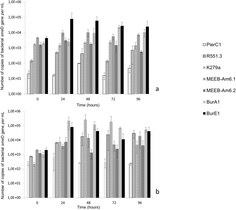
To specifically detect and quantify S. maltophilia cells inside amoeba, we targeted the smeD gene. Based on the results of previous genome sequencing, one copy of the smeD gene was considered to be equivalent to one cell [7].
The beginning of the co-culture experiments (0 h) corresponded to the number of internalized cells. A. castellanii L6a had internalized about 3 x 10³ cells of S. maltophilia strains BurA1, BurE1, MEEB-Am6.1, MEEB-Am6.2 and K279a per mL. It internalized 2 x 101 cells of PierC1 and 1.5 x 10² cells of R551.3 per mL (Fig 1a). The difference between both groups of strains was statistically significant (p < 0.05).
Fig 1. Growth of S. maltophilia strains expressed in number of copies of bacterial smeD gene per mL, in co-culture with amoeba.
a) co-culture with Acanthamoeba castellanii L6a; b) co-culture with Willaertia magna C2c. Means +/- standard deviations from three independent experiments in duplicate are presented.
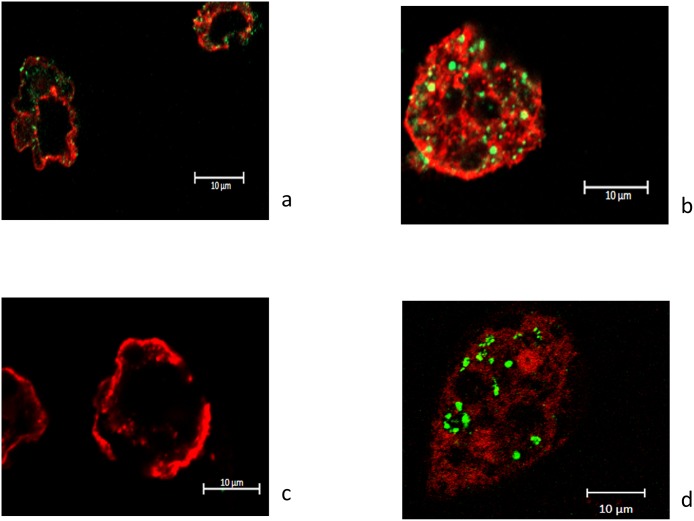
The number of S. maltophilia BurE1 inside A. castellanii increased by about 2 log after 24 hours of co-culture (p < 0.05), and the number of strain BurA1 increased by 1.5 log after 72 hours (p < 0.05). The number of S. maltophilia MEEB-Am6.1 and MEEB-Am6.2 remained stable during the entire course of the co-culture (p < 0.05). With the fluorescent confocal microscopy approach cells of strains BurA1, BurE1 and MEEB-Am6.2 were found in the cytoplasm of A. castellanii L6a (Fig 2a, 2b and 2d).
Fig 2. Fluorescent confocal microscopy images of Acanthamoeba castellanii L6a in co-culture with S. maltophilia strains.
(a) after 48 hours with BurA1, (b) after 24 hours with BurE1, (c) after 24 hours with PierC1 and (d) after 48 hours with MEEB-Am6.2.
At the end of the experiment (96 hours), the number of S. maltophilia BurA1 and BurE1 was always greater than the number of cells at time 0. After 96 hours, cells of strains BurA1, BurE1, were still found inside the amoeba. Regarding the other strains, the number of cells remained relatively constant during the entire course of the experiment (Fig 1a). Using confocal microscopy cells of strains PierC1, MEEB-Am6.1, R551.3 and K279a were not visible in A. castellanii after 24 hours (Fig 2c) or later during the experiment.
Internalization and intracellular growth of S. maltophilia strains in W. magna C2c
At the beginning of the co-culture experiments (0 h), W. magna C2c had internalized about 2 x 10² cells of strains K279 and PierC1 per mL and about 7.5 x 10² cells of strain R551.3 per mL whereas W. magna C2c had internalized about 1 x 10³ to 2 x 10³ cells of strains BurA1, BurE1, MEEB-Am6.1 and MEEB-Am6.2, per mL (Fig 1b). The difference between K279a and PierC1 strains, and the other strains was statistically significant (p < 0.05).
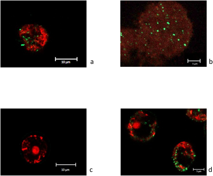
During the co-culture experiment with W. magna C2c, S. maltophilia BurA1, BurE1 and K279a replicated at the highest rates. After 24 hours of co-culture, the number of strains BurA1 and BurE1 increased by about 2.5 log and about 2 log respectively and, after 48 hours of co-incubation the number of strain K279a increased by about 3.5 log (p < 0.05). Fluorescent confocal microscopy experiments confirmed that cells of strains BurA1, BurE1 and K279a were inside the cytoplasm of W. magna C2c (Fig 3).
Fig 3. Fluorescent confocal microscopy images of Willaertia magna C2c in co-culture with S. maltophilia strains.
(a) after 48 hours with BurA1, (b) after 72 hours with BurE1, (c) after 24 hours with PierC1 and after (d) 48 hours with K279a.
Two other strains of S. maltophilia were able to replicate to a lesser extent. After 24 hours of co-incubation, the number of strains MEEB-Am6.1 and MEEB-Am6.2 increased by about 0.5 log and 1 log respectively (p < 0.05). The number of S. maltophilia MEEB-Am6.1 remained stable during the entire course of the experiment whereas those of MEEB-Am6.2 increased by 2 log after 48 hours (p < 0.05). Cells of both strains were detected in the amoeba during the co-culture experiments as seen by confocal microscopy (Fig 3b).
S. maltophilia R551.3 presented a more gradual growth and the number of cells increased by about 1.5 log after 96 hours.
The number of strains PierC1 did not vary during the experiment and microscopy did not allow to detect bacteria in the amoeba regardless of the incubation length (Fig 3c).
Viability of intra-amoebal S. maltophilia
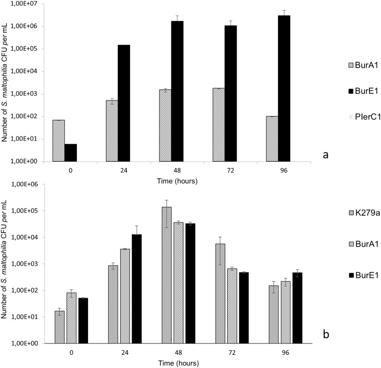
The ability of S. maltophilia strains to multiply inside the amoebae was performed using a culture approach. Representative strains among those showing multiplication properties by qPCR approach were co-cultivated with A. castellanii and W. magna. The number of CFU per mL of co-culture was determined after lysis of amoebae and plating intra-amoebal lysate on agar plates. The number of alive S. maltophilia BurE1, BurA1 and MEEB-Am6.2 strains in A. castellanii demonstrated increase by about 4–5 log, 1.5 log and 2 log respectively during the entire course of co-culture (Fig 4a). The PierC1 strain not able to replicate as quantified by qPCR approach was not detected using the culture approach.
Fig 4. Growth of S. maltophilia strains in co-culture with amoeba, expressed in number of colony forming unit of S. maltophilia per mL.
a) co-culture with Acanthamoeba castellanii L6a; b) co-culture with Willaertia magna C2c. Means +/- standard deviations from two independent experiments in triplicate are presented.
In W. magna the number of S. maltophilia BurE1 and BurA1 increased by about 2.5 log, and K279a increased by 4 log during the entire course of co-culture (p < 0.05) (Fig 4b).
Virulence of S. maltophilia strains
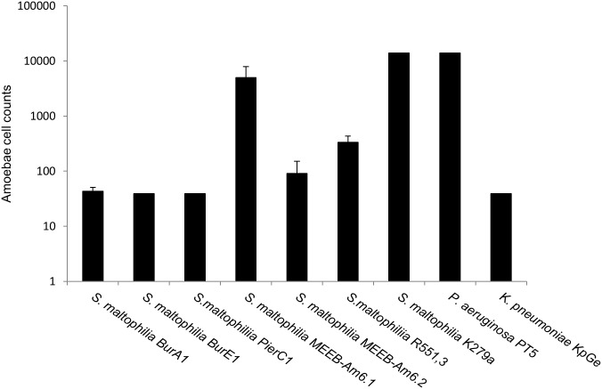
Fig 5 showed the virulence of the various S. maltophilia strains towards D. discoideum. K. pneumoniae KpGe and P. aeruginosa PT5 were used as a positive control for a non-virulent strain and negative control of a virulent strain, respectively.
Fig 5. D. discoideum plate killing assay with seven S. maltophilia strains.
Bars representing the number of amoebae necessary to form a lysis plaque on the bacterial lawn. P. aeruginosa PT5 and K. pneumoniae KpGe were used as negative and positive controls, respectively. Means +/- standard deviations from 3 independent experiments in triplicate are presented.
Five strains of S. maltophilia (BurA1, BurE1, PierC1, R551.3, MEEB-Am6.2) were determined to be non-virulent strains as fewer than 400 amoebae were needed to form lysis plaques such as K. pneumoniae strain. Two strains of S. maltophilia (MEEB-Am6.1, K279A) were characterized as virulent with a similar effect as the control strain P. aeruginosa PT5. None of the S. maltophilia strains tested presented a low-virulent phenotype.
Internalization, intracellular growth and virulence of S. maltophilia BurA1ΔebyCAB
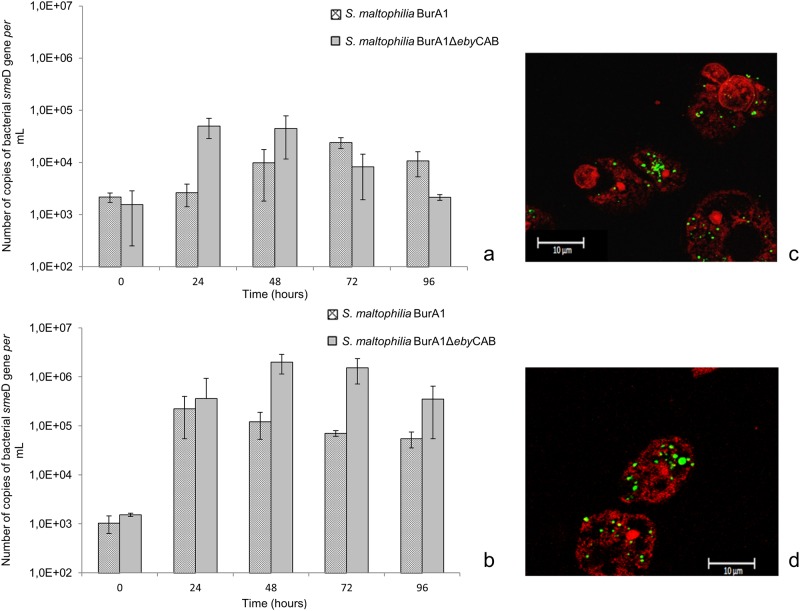
In order to evaluate the role of efflux pumps in the survival and multiplication of environmental S. maltophilia isolates inside amoeba, we chose the model BurA1 and its mutant BurA1ΔebyCAB which lacks the ebyCAB efflux pump gene previously described.
At the beginning of the co-culture experiments (0 h), with both amoebae, the number of internalized S. maltophilia BurA1 and BurA1ΔebyCAB was about 1.5 x 10³ cells per ml. In co-culture with both species of amoebae, S. maltophilia BurA1ΔebyCAB was able to survive and multiply inside amoebae (Figs 6a and 5b). With A. castellanii, the number of S. maltophilia BurA1ΔebyCAB at 24 hours was multiplied by 32 compared to time 0. With W. magna, the population of S. maltophilia BurA1ΔebyCAB at 48 hours was multiplied by a factor of 1307 compared to time 0. Confocal microscopy confirmed the presence of BurA1ΔebyCAB cells inside both amoebae (Fig 6c and 6d).
Fig 6. Growth of S. maltophilia BurA1 and BurA1ΔebyCAB expressed in number of copies of bacterial smeD gene per mL, in co-culture with amoeba and confocal microscopy.
(a) co-culture with A. castellanii L6a; (b) co-culture with W. magna C2c. Means +/- standard deviations from three independent experiments in duplicate are presented; (c) Fluorescent confocal microscopy images of A. castellanii in co-culture with S. maltophilia BurA1ΔebyCAB after 48 hours; (d) Fluorescent confocal microscopy images of W. magna in co-culture with S. maltophilia BurA1ΔebyCAB after 48 hours.
At the end of the experiment with A. castellanii L6a, the number of S. maltophilia BurA1ΔebyCAB cells was lower than with the wild type strain. However, with W. magna, the number of S. maltophilia BurA1ΔebyCAB cells was higher than with S. maltophilia BurA1.
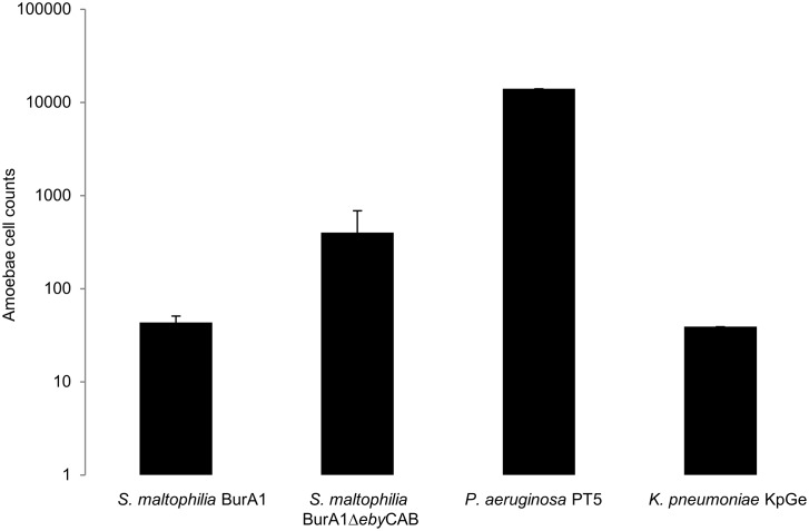
Regarding virulence assays, Fig 7 showed that S. maltophilia BurA1ΔebyCAB was considered to be a low virulence strain because 399 cells of D. discoideum were necessary to form a lysis plaque, whereas the wild type strain was non-virulent because only 43 cells of D. discoideum were necessary to form a lysis plaque.
Fig 7. D. discoideum plate killing assay with two S. maltophilia strains, BurA1 and BurA1ΔebyCAB.
Bars representing the number of amoebae necessary to form a lysis plaque on the bacterial lawn. Pseudomonas aeruginosa PT5 and Klebsiella pneumoniae KpGe are used as negative and positive controls, respectively. Means +/- standard deviations from three independent experiments in triplicate are presented.
Discussion
Free-living amoebae may constitute a host for some bacterial species [33]. Currently, most studies have focused on species known to be endosymbionts of Acanthamoeba, such as the members of the bacterial genera Legionella, Chlamydia or Mycobacterium avium [34]. Other analyses characterizing amoebal microbiomes have shown the presence of various associated bacteria including several human opportunistic pathogens, such as P. aeruginosa and S. maltophilia [17,35,36]. The present study demonstrated that various strains of S. maltophilia regardless of their origin i.e. environmental or clinical, were capable of intra-cellular survival and/or growth inside two different FLA, A. castellanii and W. magna. Recently, Cateau et al. (2014) [26] also reported that both clinical and environmental isolates of S. maltophilia were able to survive and multiply inside V. vermiformis. Our results are the first to compare the behavior of several environmental isolates of S. maltophilia with two amoebal genera and provide insight on whether selectivity towards specific amoebal genera exists or not. We showed that at an early step of the interaction i.e. bacterial internalization (time 0) by the amoeba, differences can be seen in the number of cells internalized from one strain to another. Indeed, with A. castellanii L6a, the numbers of S. maltophilia PierC1 and R551.3 cells internalized is lower than for the other strains. Regarding W. magna C2c, S. maltophilia PierC1 and K29a were the least internalized strains. These differences were confirmed using culture approach.
This observation could be related to differences in the affinity of the amoeba towards S. maltophilia strains or to differences in bacterial strategies to escape phagocytosis [37].
Also, the ability of S. maltophilia to grow inside amoeba is different according to bacterial strains and amoebal genera. For instance, strains BurA1 and BurE1 were able to grow inside A. castellanii L6a, whereas others such as K279a showed the highest growth rate within W. magna C2c. Furthermore, S. maltophilia K279a was found unable to multiply inside Acanthamoeba but increased by 3 log in W. magna. These results clearly showed that the ability of bacteria to survive or multiply in amoeba varies between bacterial strains and that a strain might grow very well in one amoebal species and not in the other. Corsaro et al. (2013) demonstrated that a S. maltophilia strain was able to multiply inside A. castellanii but not in Naegleria lovaniensis [27]. The variability of bacterial proliferation was also reported in a previous study where the multiplication of Legionella pneumophilia differed inside Acanthamoeba, Hartmanella and Willaertia [24]. Interestingly, Dey et al. (2009) [24] showed that W. magna C2c was very resistant towards the proliferation of L. pneumophila Paris but not towards other strains of L. pneumophila. Our study showed that W. magna C2c was more permissive to S. maltophilia strains than A. castellanii L6a.
We noted that after 96 hours of co-culture experiments, no lysis of both amoebae was observed regardless of the strain of S. maltophilia used and no bacterial cells were present in the co-culture medium. These data agree with the partial lysis observed only after 5 day of co-culture between S. maltophilia and V. vermiformis in the study of Evstigneeva et al. (2009) [38]. The incubation period of our co-culture experiment could be increased by a few days in order to determine if S. maltophilia would be able to lyse amoeba and to persist in the environment like the species L. pneumophila [39].
Our study involved two approaches to evaluate bacterial survival in amoebae. The use of qPCR approach was coupled to culture approach and provided data on the viability of S. maltophilia strains inside amoebae. We showed that the qPCR approach and the culture one led to the same trend for S. maltophilia multiplication. To our knowledge our study is the first one that combines both approaches to study bacteria-amoebae interactions as previous reports were based on one or the other approach [24,26,27,40].
In order to survive or multiply inside amoebae, ARB could express virulence genes, such as those reported for L. pneumophila [40] and Chlamydia spp. which possess the type III secretion system as an important virulence feature for adherence and host cell invasion [41]. We used the social amoebae D. discoideum and showed that two out of seven strains of S. maltophilia, MEEB-Am6.1 and K279a, could be considered as virulent strains. This finding is interesting as BurA1 and BurE1 were able to multiply in amoebae and were not virulent, while S. maltophilia MEEB-Am6.1 was virulent but unable to survive and grow in both amoebal species. A similar observation was previously reported from a collection of 59 isolates of S. maltophilia tested in interaction with D. discoideum and A. castellanii [23]. One can hypothesize that the virulence factors of S. maltophilia strains involved in the interaction with D. discoideum might be different from those involved in the interaction with A. castellanii or W. magna. However, it is important to note that our virulence tests were performed at 22.5°C, whereas our co-culture experiments were performed at 32°C. Temperature could be partly responsible for these differences as some bacterial strains express their virulence traits at temperatures higher than 22.5 °C [33].
The genome of S. maltophilia was shown to harbor numerous efflux pumps and these pumps are increasingly recognized as having a role in bacterial physiology and virulence [42]. For instance, the AcrAB-TolC pump of Enterobacter cloacae was found to be involved in virulence [43]. We thus investigated the role of EbyCAB efflux pump previously described [7] and showed that in both amoebae, S. maltophilia BurA1ΔebyCAB multiplied more than the wild type strain and exhibited a higher virulence than the wild type strain. Whether the efflux pump EbyCAB is involved in virulence is still unclear but the expression of this pump seems to decrease the fitness of S. maltophilia BurA1. The exact role of this pump needs to be investigate further as the overexpression of a MDR pump in S. maltophilia was found related to a decrease of fitness and a lower virulence [44].
Conclusion
In conclusion, our results showed for the first time that S. maltophilia isolates with contrasting phenotypes of virulence are able to grow inside two amoebae, A. castellanii and W. magna. These results suggest that in the environment, S. maltophilia could have the potential to infect and proliferate within a large panel of FLA. The fact that this emerging opportunistic pathogen is often found in the amoebal microbiome [35] and that it can multiply in the amoeba support the hypothesis that in the environment, FLA could be a reservoir and vector for the transmission of S. maltophilia. Thus, S. maltophilia could use the amoebae as a “training ground” in order to better resist human macrophages, as demonstrated for L. pneumophila [39], or to increase their virulence and antibiotic resistance properties [45]. In conclusion, FLA constitute an ecological niche for opportunistic bacterial pathogens in which important genetic exchanges between species could occur and contribute to the propagation of antibiotic resistance and virulence genes in the environment [46].
Supporting information
The gene orientation is indicated by arrows. White box: deleted region. Underlined nucleotides represent the nucleotides added to create a complementary region between upstream and downstream fragments.
(TIF)
Data Availability
All relevant data are within the paper and its Supporting Information file.
Funding Statement
The work was supported by “Centre National de la Recherche Scientifique “ in order to perform experiments, and by a research program of the FR BioEnviS. E. Denet, phD Student was supported by the “Ministère de l’Education Nationale, de l’Enseignement Supérieur et de la Recherche.
References
- 1.Ryan RP, Monchy S, Cardinale M, Taghavi S, Crossman L, Avison MB, et al. The versatility and adaptation of bacteria from the genus Stenotrophomonas. Nat Rev Microbiol. 2009;7: 514–525. doi: 10.1038/nrmicro2163 [DOI] [PubMed] [Google Scholar]
- 2.Tacão M, Correia A, Henriques IS. Low prevalence of carbapenem-resistant bacteria in river water: resistance is mostly related to intrinsic mechanisms. Microb Drug Resist. 2015;21: 497–506. doi: 10.1089/mdr.2015.0072 [DOI] [PubMed] [Google Scholar]
- 3.Hassanshahian M, Ahmadinejad M, Tebyanian H, Kariminik A. Isolation and characterization of alkane degrading bacteria from petroleum reservoir waste water in Iran (Kerman and Tehran provenances). Mar Pollut Bull. 2013;73: 300–305. doi: 10.1016/j.marpolbul.2013.05.002 [DOI] [PubMed] [Google Scholar]
- 4.Dungan RS, Yates SR, Frankenberger WT. Transformations of selenate and selenite by Stenotrophomonas maltophilia isolated from a seleniferous agricultural drainage pond sediment. Environ Microbiol. 2003;5: 287–295. [DOI] [PubMed] [Google Scholar]
- 5.Romanenko LA, Uchino M, Tanaka N, Frolova GM, Slinkina NN, Mikhailov VV. Occurrence and antagonistic potential of Stenotrophomonas strains isolated from deep-sea invertebrates. Arch Microbiol. 2008;189: 337–344. doi: 10.1007/s00203-007-0324-8 [DOI] [PubMed] [Google Scholar]
- 6.Pinot C, Deredjian A, Nazaret S, Brothier E, Cournoyer B, Segonds C, et al. Identification of Stenotrophomonas maltophilia strains isolated from environmental and clinical samples: a rapid and efficient procedure J Appl Microbiol. 2011;111: 1185–1193. doi: 10.1111/j.1365-2672.2011.05120.x [DOI] [PubMed] [Google Scholar]
- 7.Youenou B, Favre-Bonté S, Bodilis J, Brothier E, Dubost A, Muller D, et al. Comparative genomics of environmental and clinical Stenotrophomonas maltophilia strains with different antibiotic resistance profiles. Genome Biol Evol. 2015;7: 2484–2505. doi: 10.1093/gbe/evv161 [DOI] [PMC free article] [PubMed] [Google Scholar]
- 8.Berg G, Roskot N, Smalla K. Genotypic and phenotypic relationships between clinical and environmental isolates of Stenotrophomonas maltophilia. J Clin Microbiol. 1999;37: 3594–3600. [DOI] [PMC free article] [PubMed] [Google Scholar]
- 9.Berg G, Eberl L, Hartmann A. The rhizosphere as a reservoir for opportunistic human pathogenic bacteria. Environ Microbiol. 2005;7: 1673–1685. doi: 10.1111/j.1462-2920.2005.00891.x [DOI] [PubMed] [Google Scholar]
- 10.Peralta KD, Araya T, Valenzuela S, Sossa K, Martínez M, Peña-Cortés H, et al. Production of phytohormones, siderophores and population fluctuation of two root-promoting rhizobacteria in Eucalyptus globulus cuttings. World J Microbiol Biotechnol. 2012;28: 2003–2014. doi: 10.1007/s11274-012-1003-8 [DOI] [PubMed] [Google Scholar]
- 11.Zhang Z, Yuen GY, Sarath G, Penheiter AR. Chitinases from the plant disease biocontrol agent, Stenotrophomonas maltophilia C3. Phytopathology. 2001;91: 204–211. doi: 10.1094/PHYTO.2001.91.2.204 [DOI] [PubMed] [Google Scholar]
- 12.Dubey KK, Fulekar MH. Chlorpyrifos bioremediation in Pennisetum rhizosphere by a novel potential degrader Stenotrophomonas maltophilia MHF ENV20. World J Microbiol Biotechnol. 2012;28: 1715–1725. doi: 10.1007/s11274-011-0982-1 [DOI] [PubMed] [Google Scholar]
- 13.Somaraja PK, Gayathri D, Ramaiah N. Molecular Characterization of 2-Chlorobiphenyl Degrading Stenotrophomonas maltophilia GS-103. Bull Environ Contam Toxicol. 2013;91: 148–153. doi: 10.1007/s00128-013-1044-1 [DOI] [PubMed] [Google Scholar]
- 14.Chen S, Yin H, Ye J, Peng H, Zhang N, He B. Effect of copper(II) on biodegradation of benzo[a]pyrene by Stenotrophomonas maltophilia. Chemosphere. 2013;90: 1811–1820. doi: 10.1016/j.chemosphere.2012.09.009 [DOI] [PubMed] [Google Scholar]
- 15.Antonioli P, Lampis S, Chesini I, Vallini G, Rinalducci S, Zolla L, et al. Stenotrophomonas maltophilia SeITE02, a New Bacterial Strain Suitable for Bioremediation of Selenite-Contaminated Environmental Matrices. Appl Environ Microbiol. 2007;73: 6854–6863. doi: 10.1128/AEM.00957-07 [DOI] [PMC free article] [PubMed] [Google Scholar]
- 16.Morales-Jiménez J, Zúñiga G, Ramírez-Saad HC, Hernández-Rodríguez C. Gut-Associated Bacteria Throughout the Life Cycle of the Bark Beetle Dendroctonus rhizophagus (Curculionidae: Scolytinae) and Their Cellulolytic Activities. Microb Ecol. 2012;64: 268–278. doi: 10.1007/s00248-011-9999-0 [DOI] [PubMed] [Google Scholar]
- 17.Denet E, Coupat-Goutaland B, Nazaret S, Pélandakis M, Favre-Bonté S. Diversity of free-living amoebae in soils and their associated human opportunistic bacteria. Parasitol Res. 2017;116: 3151–3162. doi: 10.1007/s00436-017-5632-6 [DOI] [PubMed] [Google Scholar]
- 18.Denton M, Kerr KG. Microbiological and clinical aspects of infection associated with Stenotrophomonas maltophilia. Clin Microbiol Rev. 1998;11: 57–80. [DOI] [PMC free article] [PubMed] [Google Scholar]
- 19.Albini S, Abril C, Franchini M, Hüssy D, Filioussis G. Stenotrophomonas maltophilia isolated from the airways of animals with chronic respiratory disease. Schweiz Arch Für Tierheilkd. 2009;151: 323–328. doi: 10.1024/0036-7281.151.7.323 [DOI] [PubMed] [Google Scholar]
- 20.Ohnishi M, Sawada T, Marumo K, Harada K, Hirose K, Shimizu A, et al. Antimicrobial susceptibility and genetic relatedness of bovine Stenotrophomonas maltophilia isolates from a mastitis outbreak. Lett Appl Microbiol. 2012;54: 572–576. doi: 10.1111/j.1472-765X.2012.03246.x [DOI] [PubMed] [Google Scholar]
- 21.Winther L, Andersen RM, Baptiste KE, Aalbæk B, Guardabassi L. Association of Stenotrophomonas maltophilia infection with lower airway disease in the horse: A retrospective case series. Vet J. 2010;186: 358–363. doi: 10.1016/j.tvjl.2009.08.026 [DOI] [PubMed] [Google Scholar]
- 22.Adamek M, Linke B, Schwartz T. Virulence genes in clinical and environmental Stenotrophomas maltophilia isolates: A genome sequencing and gene expression approach. Microb Pathog. 2014;67–68: 20–30. doi: 10.1016/j.micpath.2014.02.001 [DOI] [PubMed] [Google Scholar]
- 23.Adamek M, Overhage J, Bathe S, Winter J, Fischer R, Schwartz T. Genotyping of environmental and clinical Stenotrophomonas maltophilia isolates and their pathogenic potential. Chakravortty D, editor. PLoS ONE. 2011;6: e27615 doi: 10.1371/journal.pone.0027615 [DOI] [PMC free article] [PubMed] [Google Scholar]
- 24.Dey R, Bodennec J, Mameri MO, Pernin P. Free-living freshwater amoebae differ in their susceptibility to the pathogenic bacterium Legionella pneumophila. FEMS Microbiol Lett. 2009;290: 10–17. doi: 10.1111/j.1574-6968.2008.01387.x [DOI] [PubMed] [Google Scholar]
- 25.Dupuy M, Binet M, Bouteleux C, Herbelin P, Soreau S, Héchard Y. Permissiveness of freshly isolated environmental strains of amoebae for growth of Legionella pneumophila. Descoteaux A, editor. FEMS Microbiol Lett. 2016;363: fnw022. doi: 10.1093/femsle/fnw022 [DOI] [PubMed] [Google Scholar]
- 26.Cateau E, Maisonneuve E, Peguilhan S, Quellard N, Hechard Y, Rodier M-H. Stenotrophomonas maltophilia and Vermamoeba vermiformis relationships: Bacterial multiplication and protection in amoebal-derived structures. Res Microbiol. 2014;165: 847–851. doi: 10.1016/j.resmic.2014.10.004 [DOI] [PubMed] [Google Scholar]
- 27.Corsaro D, Müller K-D, Michel R. Molecular characterization and ultrastructure of a new amoeba endoparasite belonging to the Stenotrophomonas maltophilia complex. Exp Parasitol. 2013;133: 383–390. doi: 10.1016/j.exppara.2012.12.016 [DOI] [PubMed] [Google Scholar]
- 28.Crossman LC, Gould VC, Dow JM, Vernikos GS, Okazaki A, Sebaihia M, et al. The complete genome, comparative and functional analysis of Stenotrophomonas maltophilia reveals an organism heavily shielded by drug resistance determinants. Genome Biol. 2008;9: 1. [DOI] [PMC free article] [PubMed] [Google Scholar]
- 29.Taghavi S, Garafola C, Monchy S, Newman L, Hoffman A, Weyens N, et al. Genome survey and characterization of endophytic bacteria exhibiting a beneficial effect on growth and development of poplar trees. Appl Environ Microbiol. 2009;75: 748–757. doi: 10.1128/AEM.02239-08 [DOI] [PMC free article] [PubMed] [Google Scholar]
- 30.Favre-Bonte S. Biofilm formation by Pseudomonas aeruginosa: role of the C4-HSL cell-to-cell signal and inhibition by azithromycin. J Antimicrob Chemother. 2003;52: 598–604. doi: 10.1093/jac/dkg397 [DOI] [PubMed] [Google Scholar]
- 31.Froquet R, Lelong E, Marchetti A, Cosson P. Dictyostelium discoideum: a model host to measure bacterial virulence. Nat Protoc. 2008;4: 25–30. doi: 10.1038/nprot.2008.212 [DOI] [PubMed] [Google Scholar]
- 32.Alonso A, Martinez JL. Expression of multidrug efflux pump SmeDEF by clinical isolates of Stenotrophomonas maltophilia. Antimicrob Agents Chemother. 2001;45: 1879–1881. doi: 10.1128/AAC.45.6.1879-1881.2001 [DOI] [PMC free article] [PubMed] [Google Scholar]
- 33.Steinert M. Pathogen–host interactions in Dictyostelium, Legionella, Mycobacterium and other pathogens. Semin Cell Dev Biol. 2011;22: 70–76. doi: 10.1016/j.semcdb.2010.11.003 [DOI] [PubMed] [Google Scholar]
- 34.Guimaraes AJ, Gomes KX, Cortines JR, Peralta JM, Peralta RHS. Acanthamoeba spp. as a universal host for pathogenic microorganisms: One bridge from environment to host virulence. Microbiol Res. 2016;193: 30–38. doi: 10.1016/j.micres.2016.08.001 [DOI] [PubMed] [Google Scholar]
- 35.Pagnier I, Valles C, Raoult D, La Scola B. Isolation of Vermamoeba vermiformis and associated bacteria in hospital water. Microb Pathog. 2015;80: 14–20. doi: 10.1016/j.micpath.2015.02.006 [DOI] [PubMed] [Google Scholar]
- 36.Delafont V, Brouke A, Bouchon D, Moulin L, Héchard Y. Microbiome of free-living amoebae isolated from drinking water. Water Res. 2013;47: 6958–6965. doi: 10.1016/j.watres.2013.07.047 [DOI] [PubMed] [Google Scholar]
- 37.Matz C, Kjelleberg S. Off the hook—how bacteria survive protozoan grazing. Trends Microbiol. 2005;13: 302–307. doi: 10.1016/j.tim.2005.05.009 [DOI] [PubMed] [Google Scholar]
- 38.Evstigneeva A, Raoult D, Karpachevskiy L, La Scola B. Amoeba co-culture of soil specimens recovered 33 different bacteria, including four new species and Streptococcus pneumoniae. Microbiology. 2009;155: 657–664. doi: 10.1099/mic.0.022970-0 [DOI] [PubMed] [Google Scholar]
- 39.Greub G, Raoult D. Microorganisms Resistant to Free-Living Amoebae. Clin Microbiol Rev. 2004;17: 413–433. doi: 10.1128/CMR.17.2.413-433.2004 [DOI] [PMC free article] [PubMed] [Google Scholar]
- 40.Messi P, Bargellini A, Anacarso I, Marchesi I, de Niederhäusern S, Bondi M. Protozoa and human macrophages infection by Legionella pneumophila environmental strains belonging to different serogroups. Arch Microbiol. 2013;195: 89–96. doi: 10.1007/s00203-012-0851-9 [DOI] [PubMed] [Google Scholar]
- 41.Siddiqui R, Malik H, Sagheer M, Jung S-Y, Khan NA. The type III secretion system is involved in Escherichia coli K1 interactions with Acanthamoeba. Exp Parasitol. 2011;128: 409–413. doi: 10.1016/j.exppara.2011.05.008 [DOI] [PubMed] [Google Scholar]
- 42.Lin Y-T, Huang Y-W, Chen S-J, Chang C-W, Yang T-C. The SmeYZ Efflux Pump of Stenotrophomonas maltophilia Contributes to Drug Resistance, Virulence-Related Characteristics, and Virulence in Mice. Antimicrob Agents Chemother. 2015;59: 4067–4073. doi: 10.1128/AAC.00372-15 [DOI] [PMC free article] [PubMed] [Google Scholar]
- 43.Perez A, Poza M, Fernandez A, del Carmen Fernandez M, Mallo S, Merino M, et al. Involvement of the AcrAB-TolC Efflux Pump in the Resistance, Fitness, and Virulence of Enterobacter cloacae. Antimicrob Agents Chemother. 2012;56: 2084–2090. doi: 10.1128/AAC.05509-11 [DOI] [PMC free article] [PubMed] [Google Scholar]
- 44.Alonso A. Overexpression of the multidrug efflux pump SmeDEF impairs Stenotrophomonas maltophilia physiology. J Antimicrob Chemother. 2004;53: 432–434. doi: 10.1093/jac/dkh074 [DOI] [PubMed] [Google Scholar]
- 45.Loret J-F, Greub G. Free-living amoebae: Biological by-passes in water treatment. Int J Hyg Environ Health. 2010;213: 167–175. doi: 10.1016/j.ijheh.2010.03.004 [DOI] [PubMed] [Google Scholar]
- 46.Moliner C, Fournier P-E, Raoult D. Genome analysis of microorganisms living in amoebae reveals a melting pot of evolution. FEMS Microbiol Rev. 2010;34: 281–294. doi: 10.1111/j.1574-6976.2010.00209.x [DOI] [PubMed] [Google Scholar]
Associated Data
This section collects any data citations, data availability statements, or supplementary materials included in this article.
Supplementary Materials
The gene orientation is indicated by arrows. White box: deleted region. Underlined nucleotides represent the nucleotides added to create a complementary region between upstream and downstream fragments.
(TIF)
Data Availability Statement
All relevant data are within the paper and its Supporting Information file.


