Daily torpor in the mouse is a comprehensive physiological and biochemical suite of changes in response to a caloric deficit and a cool ambient temperature. The well-orchestrated torpor response is truly an integrative one – it is likely controlled by the brain, involves changes in body position, altered autonomic influence, has a strong circadian component, and involves altered expression/activity of genes in body tissues. Unraveling the neural circuitry for torpor induction, maintenance, and arousal is of great interest for both basic science and potential applications of targeted temperature management. The current article is a comment on our recent publication concerning the differentiation between hypothermia and natural torpor in the mouse.1
For ecophysiologists and natural historians, describing torpor in terms of body temperature and metabolic rate is appropriate and suffices well. However, pharmacological studies are now performed on the standard laboratory mouse. If a drug causes cardiac output to plummet, or interrupts the electron transport chain, one would expect a drop in metabolic rate and subsequent drop in body temperature in a way that is not associated with a natural torpor bout. Because the mouse tolerates bouts of hypothermia quite well, a fall in body temperature in response to a drug is not sufficient to conclude activation of torpor-inducing pathways. There is so much more to torpor than hypometabolism and low body temperature. Defining torpor this way simply ignores the integrative nature of this complex response to caloric deprivation in the mouse.
The literature base concerning the neural circuitry for temperature regulation is vast. In fact, journals like this one devote a great deal of space for discussion of just this topic. While the role of some regions of the brain (e.g., the hypothalamus, the pre-optic area) are known to be important in the regulation of entry and exit into torpor,2 the understanding of neural mechanisms in torpor is only in its infancy. Hence, given the lack of understanding of the neural pathways involved with torpor, the principle question we addressed is how one determines whether hypothermia from administration of a compound to a mouse represents a natural torpor bout.1 We decided to examine robust outputs and consequences of torpor with a battery of tests/measurements. These tests are by no means comprehensive, yet they contain diversity in the approach to examining hypothermia.
We focused on adenosine, an intriguing candidate as a mediator of torpor. Adenosine signals metabolic stress by acting on the inhibitory A1 adenosine receptor to reduce metabolic rate. Central (i.e., intracerebroventricular or ICV) activation of this receptor with cyclohexyladenosine (CHA) induces hypothermia and hypometabolism in arctic ground squirrels, but only during the hibernating season suggesting a seasonal regulation of sensitivity to adenosine.3 Similarly, CHA into the rat, an animal that does not normally engage torpor pathways, induces hypothermia, low heart rate, skipped heart beats, reduced shivering, reduces CO2 production, and lowers brown fat temperature.4 Further, blocking adenosine action centrally can both prevent torpor and induce arousal in hamsters.5 Therefore, we wanted to address the hypothesis that central injection of an adenosine A1 receptor agonist activates a pattern of physiological and biochemical changes that are indistinguishable from torpor.
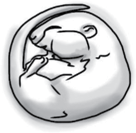
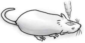
Our first observation was that of an altered body position of the mouse. With central delivery of CHA into the mouse, the mice seemed to stop in their tracks in a splayed position whereas natural torpor resulted in the commonly seen reduction in surface area by assuming a curled position. In common with the arctic ground squirrel and rat, central delivery of CHA into the mouse induced hypothermia. Similar to the response in the rat, this compound induced bradycardia, an increase in heart rate variability, and a lack of shivering. However, it seems that the A1 adenosine receptor agonist does not fully capture the autonomic changes during natural torpor in the mouse (Table 1). While both situations show bradycardia, the HR drop in torpor was much steeper and to a slower rate than that with CHA. The HR and body temperature rise during arousal from CHA-induced hypothermia was similarly blunted. Administration of CHA induced infrequent but excessively long asystoles, often >1 s, whereas natural torpor showed frequent and short-duration skipped beats. The body temperature of the mouse treated with CHA fell five times as fast as during natural torpor, a likely result of the lack of tail vasoconstriction and failure to reduce their surface area to volume ratio. All of these, except body position, suggest that the A1 adenosine receptor agonist did not capture the autonomic balance that drives physiologic changes in natural torpor. Further, mice in natural torpor shiver (i.e., thermoregulate) which was not seen in mice given CHA. Finally, the heart and liver massively increased expression of c-Fos with CHA, possibly a sign that these tissues went cold without proper preparation.
Table 1.
Physiological and biochemical changes in mus musculus in natural torpor and adenosine-induced hypothermia.
Natural torpor
|
Adenosine-induced hypothermia
|
|
|---|---|---|
| Heart rate during entrance to hypothermia | 600 bpm → 100 bpm in 1 h | 600 bpm → 200 bpm in 8 h |
| Heart rate during arousal from hypothermia | 100 bpm → 600 bpm in 30 min | 200 bpm → 600 bpm in 90 min |
| Asystoles | Periodic and frequent skipped beats | Infrequent asystoles with extended interbeat intervals |
| Rate of heat loss during entrance | 0.2°C per minute | 5× faster |
| Rate of heat gain during arousal | 0.4°C per minute | 2× slower |
| Circulating lactate | Low | Low |
| Shivering | Periodically | None |
| c-Fos expression in the heart and liver | No change from normal | 80–100 fold induction |
Special thanks to Francesca Barradale for her drawings used in the table.
In total, our data suggest a different physiologic status when comparing adenosine-induced hypothermia and natural torpor. Since delivery of a small amount of the compound to the brain can evoke changes that lower metabolic rate and heart rate, we suspect that adenosine might be part of the signaling pathway for torpor. However, ICV administration of CHA does not seem to capture fully the repression of the sympathetic nervous system, and/or activation of the parasympathetic nervous system during entrance and maintenance of torpor. It seemed that CHA mimicked more a state of anesthesia than it did a state of torpor. Further interventions that possibly include manipulating adenosine signaling can be used to fully elucidate the neural basis of natural torpor.
References
- [1].Vicent MA, Borre ED, Swoap SJ. Central activation of the A1 adenosine receptor in fed mice recapitulates only some of the attributes of daily torpor. J Comp Physiol B. 2017;187:835-845; PMID:28378088. doi: 10.1007/s00360-017-1084-7. [DOI] [PMC free article] [PubMed] [Google Scholar]
- [2].Heller HC. Hibernation – Neural Aspects. Annu Rev Physiol. 1979;41:305-321; PMID:373593. doi: 10.1146/annurev.ph.41.030179.001513. [DOI] [PubMed] [Google Scholar]
- [3].Jinka TR, Ø Tøien, Drew KL. Season primes the brain in an arctic hibernator to facilitate entrance into torpor mediated by adenosine A1 receptors. J Neurosci. 2011;31:10752-10758. PMID:21795527; doi: 10.1523/JNEUROSCI.1240-11.2011. [DOI] [PMC free article] [PubMed] [Google Scholar]
- [4].Tupone D, Madden CJ, Morrison SF. Central activation of the A1 Adenosine Receptor (A1AR) induces a hypothermic, torpor-like state in the rat. J Neurosci. 2013;33:14512-14525. PMID:24005302; doi: 10.1523/JNEUROSCI.1980-13.2013. [DOI] [PMC free article] [PubMed] [Google Scholar]
- [5].Tamura Y, Shintani M, Nakamura A, Monden M, Shiomi H. Phase-specific central regulatory systems of hibernation in Syrian hamsters. Brain Res. 2005;1045:88-96. PMID:15910766; doi: 10.1016/j.brainres.2005.03.029. [DOI] [PubMed] [Google Scholar]


