Abstract
Context:
Ankle sprains are one of the most common injuries in the physically active population. Previous researchers have shown that supporting the ankle with taping or bracing is effective in preventing ankle sprains. However, no authors have compared the effects of self-adherent tape and lace-up ankle braces on ankle range of motion (ROM) and dynamic balance in collegiate football players.
Objective:
To examine the effectiveness of self-adherent tape and lace-up ankle braces in reducing ankle ROM and improving dynamic balance before and after a typical collegiate football practice.
Design:
Crossover study.
Setting:
Collegiate athletic training room.
Patients or Other Participants:
Twenty-nine National Collegiate Athletic Association Division I football athletes (age = 19.2 ± 1.14 years, height = 187.52 ± 20.54 cm, mass = 106.44 ± 20.54 kg).
Intervention(s):
Each participant wore each prophylactic ankle support during a single practice, self-adherent tape on 1 leg and lace-up ankle brace on the other. Range of motion and dynamic balance were assessed 3 times for each leg throughout the testing session (baseline, prepractice, postpractice).
Main Outcome Measure(s):
Ankle ROM for inversion, eversion, dorsiflexion, and plantar flexion were measured at baseline, immediately after donning the brace or tape, and immediately after a collegiate practice. The Y-Balance Test was used to assess dynamic balance at these same time points.
Results:
Both interventions were effective in reducing ROM in all directions compared with baseline; however, dynamic balance did not differ between the tape and brace conditions.
Conclusions:
Both the self-adherent tape and lace-up ankle brace provided equal ROM restriction before and after exercise, with no change in dynamic balance.
Key Words: ankle support, prophylactic bracing, Y-Balance Test
Key Points
A lace-up ankle brace and self-adherent tape restricted ankle range of motion similarly.
Although both the self-adherent tape and lace-up ankle brace reduced range of motion, neither affected dynamic balance.
Ankle-ligament injuries account for 15% of all athletic injuries, and 85% of these involve the lateral ankle-ligament complex.1 The authors of a recent systematic review2 reported approximately 11.5 ankle sprains per 1000 athlete-exposures across all sports. Athletic movements such as planting, cutting, and jumping often force the ankle into plantar flexion and inversion, the common mechanism for lateral ankle sprain. Ankle sprains range from microtears to complete rupture of the involved ligaments, which can result in long-term pain and dysfunction.
The high incidence and lingering symptoms of ankle sprains have led sports medicine professionals to use taping and bracing as an injury-prevention measure. In 1 study,3 the incidence of ankle sprain decreased from 32.8 to 14.7 per 1000 exposures with the use of prophylactic ankle taping. White cloth tape has traditionally been used in sports medicine settings to decrease ankle range of motion (ROM) and provide external support. Numerous investigators4−8 have shown that white tape loosens with activity, potentially reducing its effectiveness by allowing increased ROM.
Aside from restricting ankle ROM, prophylactic ankle support has been suggested to improve balance and proprioception9,10; however, the scientific data are scarce. The proposed mechanism for this benefit is stimulation of the cutaneous mechanoreceptors and an improved proprioceptive feedback loop.11 Previous researchers12,13 demonstrated that ankle prophylactic devices did not impede dynamic balance. Poor dynamic balance is associated with an elevated risk of lower extremity noncontact injury in collegiate football athletes.14,15 Plisky et al16 observed an elevated risk of lower extremity injury with poor performance on dynamic balance testing. The relationship between the type of ankle support and dynamic balance has not been established in American football athletes.
A new taping product (PowerTape; Andover Healthcare, Salisbury, MA) was recently developed to address the support flaws of current prophylactic ankle devices. Self-adherent tape and prewrap (PowerFlex; Andover Healthcare) are highly moldable and purported to maintain restriction throughout activity.17 One major feature of this new product is the absence of adhesive materials on the tape; instead, the tape adheres to itself, which is proposed to decrease skin irritation because glue is not used to bind the tape to the skin. In addition, the self-adhesive tape and prewrap are claimed to be water resistant, which should decrease the extent to which perspiration affects the performance of the tape. Lastly, the self-adherent prewrap, unlike traditional foam prewrap, is reported to add tensile resistance to the completed ankle taping.17 No published studies have compared self-adherent tape with ankle braces and studies of the benefits of self-adherent tape were not conducted for more than 30 minutes of exercise. It is important to identify the amount of support provided by these 2 prophylactic devices before and after sport-specific activity to aid in the prevention of ankle injury.
The purpose of our study was to examine the effectiveness of self-adherent tape and lace-up ankle braces on ankle ROM and dynamic balance before and after a typical collegiate football practice.
METHODS
Participants
Participants were recruited from a midwestern National Collegiate Athletic Association Division I Football Championship Division team. All recruits regularly wore prophylactic ankle support when engaging in athletic activities and none had a history of ankle or foot surgery. Exclusion criteria were a history of ankle or foot injury within the last 6 months, diagnosis of chronic ankle instability, or surgery to the lower extremity within the last 12 months. Participants were also excluded from the study if they had a diagnosed metabolic disorder or were not currently active in athletics due to another injury or illness. A total of 41 individuals volunteered to participate in the research study. After application of the inclusion and exclusion criteria, 29 individuals (age = 19.2 ± 1.14 years, height = 187.52 ± 20.54 cm, mass = 106.44 ± 20.54 kg) were deemed eligible. The university's institutional review board approved the study.
Procedures
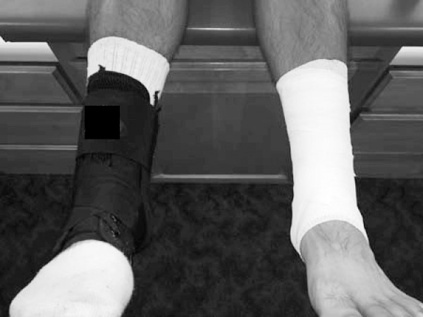
Each volunteer reported to the athletic training room before and after football practice for 1 testing session, signed an informed consent form, and completed the preparticipation questionnaire. Range of motion and dynamic balance were assessed 3 times on each leg throughout the testing session: baseline (before the tape or brace was applied), pretest (immediately after the tape or brace was applied), and posttest (immediately after football practice). Each participant wore each prophylactic ankle support during a single practice: lace-up ankle brace on 1 leg and self-adherent tape on the other. Limb dominance was assessed by asking participants which leg they would use to kick a ball. The tape condition was applied to the randomized condition (dominant or nondominant) and the lace-up brace was applied to the opposite limb (Figure 1). Limb dominance was equally represented in each condition.
Figure 1. .

Bracing and taping conditions.
Range-of-Motion Testing
Each participant was positioned supine on an examination table with the knee fully extended and ankle unsupported off the end of the table. Active ankle ROM in 4 directions (inversion, eversion, dorsiflexion, and plantar flexion) was assessed using a handheld goniometer. The participant was instructed to move in the direction of measurement as far as possible while a trained research assistant performed the goniometer measurements. During inversion and eversion ROM measurements, the fulcrum of the goniometer was centered over the talocrural joint while the stationary arm was centered over the long axis of the tibia and the movement arm was centered over the second metatarsal.18 During dorsiflexion and plantar-flexion ROM measurement, the fulcrum of the goniometer was centered over the lateral malleolus while the stationary arm was aligned with the long axis of the fibula and the movement arm was parallel to the fifth metatarsal.18 All ROM measurements were assessed bilaterally by the same trained research assistant. Before data collection began, intrarater reliability for all ROM measures was established on both ankles of 10 healthy participants with measurements separated by 24 hours. Reliability of the ROM measures was excellent, with intraclass correlation coefficient values ranging from 0.96 to 0.98.
Dynamic Balance
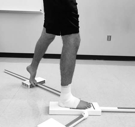
After ROM data collection, the participant's dynamic balance was assessed using the Lower Quarter Y-Balance Test (Figure 2). The Y-Balance Test assesses an individual's dynamic balance in single-legged stance while he or she reaches in 3 directions (anterior, posteromedial, and posterolateral) with the contralateral limb.15 Before beginning the Y-Balance Test, we measured lower extremity limb length using a cloth tape measure to normalize reach distance. The participant was positioned supine, and the researcher performed a Weber-Barstow maneuver19 to equalize the pelvis. Bilateral limb length was measured from the inferior aspect of the anterior-superior iliac spine to the most distal aspect of the medial malleolus.15
Figure 2. .

Y-Balance Test.
When performing the Y-Balance Test, the participant was instructed to stand unshod on the center of the stance platform with the most distal aspect of the foot touching the starting line.15,20 The participant then pushed the reach indicator in the red target area as far as possible while maintaining balance on the contralateral limb.15,20,21 Each reach direction was labeled with reference to the stance leg. Per the Y-Balance Test protocol,15,20 the trial was unsuccessful if the participant (1) failed to maintain unilateral stance (fell off the center platform or touched down on the floor with the reach foot), (2) failed to maintain contact with the reach indicator throughout the motion, (3) used the reach indicator for stance support, or (4) failed to return the reach foot to the starting position. If the participant was unable to perform a successful trial in 3 attempts, additional trials were performed until a successful reach was completed (maximum of 6 attempts).15 If the participant could not perform a successful trial in 6 attempts, the direction was recorded as a fail.15 Performance in 3 test trials was recorded in each direction, and the maximal reach in each direction was used for analysis.21
To control for learning effects, participants performed 6 practice trials on each leg in all 3 directions before formal testing began.20,22 Three test trials were performed in the anterior direction with the right leg, followed by 3 test trials in the anterior direction with the left leg.15 The participant then performed alternating test trials in the posteromedial and posterolateral directions to minimize fatigue.15 The researcher was certified through free online training to collect Y-Balance Test data.20 After data collection, the composite score for the Y-Balance Test was calculated for each limb using the equation: (composite reach score = [(maximum anterior reach + maximum posteromedial reach + maximum posterolateral reach)/(3 × limb length)] × 100).15 This composite score was used for data analysis. Previous researchers15 have shown good to excellent reliability of the Y-Balance Test.
Taping and Bracing
Tape was applied using a standard closed–basket-weave technique18 as follows: (1) The ankle was placed in 90° of dorsiflexion. (2) Two heel and lace pads with a small amount of skin lubricant were applied to the ankle, 1 over the Achilles tendon and 1 over the anterior ankle joint. (3) Eco-Flex prewrap (Cramer Products Inc, Gardner, KS) was applied using a full stretch in a circular pattern from the midfoot to the base of the calf. (4) Three anchor strips of self-adherent tape were applied at the base of the gastrocnemius, followed by 3 medial-to-lateral stirrups. (5) Horseshoe strips were then placed from the base of the anchors to the malleolus. (6) Two figure-8 strips and 2 heel locks (1 medial and 1 lateral) were placed continuously. (7) Two anchor strips were placed at the base of the gastrocnemius, and 1 anchor strip was placed at the midfoot. To control for taping variations, the same athletic trainer applied the self-adherent tape to all participants.
Participants were given a new lace-up ankle brace (model 195T Ultralight; McDavid Inc, Fountain Valley, CA) for testing. Ankle-brace sizing was determined by the self-reported shoe size (small: size 8−9, medium: size 9−11, large: size 11−13, extra large: size 14+) of the participant. The brace was applied by the participant over a single, new, cotton, crewcut sock (model 1602A; Pro Feet, Inc, Burlington, NC) in accordance with the manufacturer's instructions.23 The brace was applied as follows: (1) Lace as tightly as comfortable. (2) Wrap 1 figure-8 strap securely over the top of the foot and up the side of the foot and then attach the hook-and-loop patch; repeat with the opposite strap. (3) Attach the cover strap at the top to secure the ends of the figure-8 straps.23 An athletic trainer supervised the brace application to ensure proper fit.
Each volunteer participated in 1 collegiate football practice (duration = 97.5 ± 17.53 minutes, environmental temperature = 1.61°C ± 5.49°C) over the course of an 8-day testing period. All practices involved the same basic structure and occurred midweek between November 6 and December 4. None of the practices were walkthroughs, and all athletes wore full pads; therefore, all should have applied the same general stress to the tape or brace. At the end of practice, posttest ankle ROM and dynamic balance were assessed with the prophylactic device still on the ankle. A within-subject study design was used in an attempt to standardize the stress applied to each device under the same environmental conditions. Most previous authors have used a standardized exercise protocol to compare prophylactic devices. Because we studied use of the devices in an actual practice, we believed this was the most effective method to ensure the participants placed similar stresses on the conditions. We hypothesized that no differences would exist between the 2 conditions for any of the ROM directions or dynamic balance at either time point.
Statistical Analysis
Four repeated-measures analyses of variance (RM-ANOVAs) were performed to examine the effects of taping or bracing on ROM. The 4 RM-ANOVAs assessed changes in the 4 directions of ROM for the within-subject factors of time (3 levels) and condition (2 levels). One additional RM-ANOVA addressed the effects of taping or bracing on dynamic balance; this RM-ANOVA included the within-subject factors of time (3 levels) and condition (2 levels). Significant differences identified by an RM-ANOVA were further analyzed by examining the mean difference and associated 95% confidence intervals (CIs) to interpret the clinical meaningfulness of the results. To counteract the likelihood of type 1 error, we applied the Bonferroni correction, adjusting the α level of P < .05 to P < .01. Effect sizes were calculated using the Cohen d and categorized as trivial (≤0.20), small (0.21 to 0.49), moderate (0.50 to 0.79), or large (≥0.80). All data were analyzed using SPSS (version 21; IBM Corp, Armonk, NY).
RESULTS
Preliminary analyses of the data revealed no differences in ROM or dynamic balance between participants' dominant and nondominant ankles during the baseline condition. Temperatures and practice lengths for the 8 data-collection periods were similar and are reported in Table 1.
Table 1. .
Environmental Conditions and Practice Length During Data Collection
| Data-Collection Day |
Temperature (°C) |
Dew Point (°C) |
Wind Speed (mph) |
Practice Length (min) |
| 1 | 5.5 | 2.8 | 16 | 110 |
| 2 | 13.28 | 0.0 | 10 | 60 |
| 3 | −2.22 | −7.8 | 13 | 110 |
| 4 | −2.72 | −6.7 | 13 | 110 |
| 5 | −0.5 | −3.3 | 11 | 90 |
| 6 | −2.72 | −3.9 | 14 | 90 |
| 7 | 2.22 | 0.6 | 16 | 100 |
| 8 | 0 | −2.8 | 8 | 110 |
| Mean ± SD | 1.6 ± 5.5 | −2.6 ± 3.6 | 12.6 ± 2.8 | 97.5 ± 17.5 |
Descriptive statistics for ankle ROM and the Y-Balance Test composite scores are presented in Tables 2 and 3. The RM-ANOVA revealed a time effect for all ROM directions. In all cases, ROM was less immediately after the application of either the tape or brace (prepractice). Inversion ROM demonstrated a time effect (F2,27 = 84.53, P = .001), and follow-up testing showed differences in ROM from baseline to prepractice (P = .001; effect size = 1.50; 95% CI = 0.92, 2.08), baseline to postpractice (P = .001; effect size = 0.91; 95% CI = 0.37, 1.45), and prepractice to postpractice (P = .001; effect size = 0.83; 95% CI = 0.29, 1.36). Eversion ROM displayed a time effect (F2,27 = 30.49, P = .001) and follow-up testing indicated differences in ROM from baseline to prepractice (P = .001; effect size = 0.88; 95% CI = 0.34, 1.41) and prepractice to postpractice (P = .001; effect size = 0.85; 95% CI = 0.31, 1.38). Dorsiflexion ROM exhibited a time effect (F2,27 = 64.17, P = .001), and follow-up testing revealed differences in ROM from baseline to prepractice (P = .001; effect size = 1.41; 95% CI = 0.84, 1.99), baseline to postpractice (P = .001; effect size = 0.68; 95% CI = 0.15, 1.20), and prepractice to postpractice (P = .001; effect size = 1.02; 95% CI = 0.47, 1.57). Finally, plantar-flexion ROM showed a time effect (F2,27 = 93.35, P = .001), and follow-up testing demonstrated differences in ROM from baseline to prepractice (P = .001; effect size = 1.17; 95% CI = 0.61, 1.72) and prepractice to postpractice (P = .001; effect size = 0.92; 95% CI = 0.37, 1.46).
Table 2. .
Range-of-Motion and Y-Balance Test Changes by Time and Condition, ° (Mean ± SD)
| Motion |
Time |
Group |
||||
| Baseline |
Pre-Exercise |
Postexercise |
Brace |
Tape |
||
| Inversion | 28.4 ± 7.1 | 18.8 ± 5.6a | 23.0 ± 4.5ab | 22.4 ± 5.3c | 24.4 ± 6.2 | |
| Everson | 6.5 ± 3.3 | 3.9 ± 2.6a | 6.1 ± 2.6b | 5.1 ± 2.7 | 5.9 ± 3.0 | |
| Dorsiflexion | 6.2 ± 4.3 | 0.6 ± .3.6a | 3.8 ± 2.6ab | 3.6 ± 3.3 | 3.4 ± 3.7 | |
| Plantar flexion | 50.4 ± 6.9 | 42.7 ± 6.3a | 48.2 ± 5.7b | 46.2 ± 6.1 | 47.9 ± 6.6 | |
| Composite | 156.9 ± 13.3 | 154.2 ± 14.5 | 157.1 ± 12.6 | 156.1 ± 13.0 | 156.1 ± 14.3 | |
Significant time effect from baseline.
Significant time effect from pre-exercise.
Significant group effect.
Table 3. .
Descriptive Statistics for Ankle Range of Motion, ° (Mean ± SD) and Composite Y-Balance Test Scores (Mean Normalized Reach Distance ± SD) by Group and Time
| Variable |
Baseline |
Pre-Exercise |
Postexercise |
| Inversion | |||
| Brace | 28.01 ± 7.1 | 17.4 ± 4.6 | 21.9 ± 4.1 |
| Tape | 28.7 ± 7.1 | 20.3 ± 6.6 | 24.2 ± 4.9 |
| Eversion | |||
| Brace | 6.4 ± 3.5 | 3.1 ± 2.2 | 5.7 ± 2.3 |
| Tape | 6.7 ± 3.2 | 4.7 ± 3.0 | 6.4 ± 2.8 |
| Dorsiflexion | |||
| Brace | 6.3 ± 4.3 | 0.6 ± 3.2 | 4.0 ± 2.5 |
| Tape | 6.1 ± 4.2 | 0.5 ± 4.0 | 3.7 ± 2.8 |
| Plantar flexion | |||
| Brace | 50.5 ± 7.2 | 41.5 ± 4.9 | 46.6 ± 5.9 |
| Tape | 50.3 ± 6.7 | 43.8 ± 7.6 | 49.8 ± 5.4 |
| Composite Y-Balance Test score | |||
| Brace | 157.0 ± 11.7 | 154.6 ± 14.5 | 156.6 ± 12.7 |
| Tape | 156.8 ± 14.9 | 153.8 ± 15.5 | 157.6 ± 12.5 |
A condition effect was present for inversion with the brace restricting only slightly more motion than the tape (F1,28 = 15.02, P = .001). Follow-up analysis revealed a difference between groups (P = .001; effect size = 0.35; 95% CI = −0.17, 0.87). No condition effects occurred for any other ROM direction.
No significant group × time effects were displayed for inversion (F2,27 = 3.17, P = .06), eversion (F2,27 = 2.37, P = .11), or dorsiflexion (F2,27 = 0.32, P = .97). A significant group × time interaction existed for the plantar-flexion condition: F2,27 = 8.43, P = .001. Although these findings were statistically significant, follow-up analysis revealed that the 95% CIs of the means crossed over one another at each time point. Additionally, the effect sizes were trivial, and the 95% CIs crossed zero for all 3 time points.
The RM-ANOVA for the Y-Balance Test failed to show differences between groups. The Mauchly test indicated that the assumption of sphericity was violated; therefore, the degrees of freedom were corrected using Greenhouse-Geisser estimates of sphericity. No main effect of time for composite balance scores (F1.58,56 = 4.88, P = .02) was present. No main effect occurred for condition (F1,28 = 0.00, P = .99), and no significant condition × time interaction was found (F2,56 = 0.84, P = .43).
DISCUSSION
The purpose of our study was to examine the effectiveness of self-adherent tape and lace-up ankle braces in reducing ankle ROM and improving dynamic balance before and after a collegiate football practice. White cloth tape is commonly used in the athletic setting; however, the potential loosening of the tape during exercise has led some clinicians to switch to self-adherent tape. Our investigation is the first to examine the relationship between these prophylactic devices during a practice in a collegiate football population.
Self-adherent tape and lace-up ankle braces generally provided equal ROM restriction before and after exercise while participants maintained dynamic balance. Plantar-flexion ROM showed slightly greater restriction in the 2 groups. Our results were consistent with those of previously published studies8,24−26 on taping and bracing and indicated that both prophylactic devices immediately restricted ROM in all directions. Although self-adherent tape has not previously been compared with lace-up ankle braces, our ROM results were similar to those of some investigators8,24 who compared white athletic tape and various types of ankle braces. In contrast, other researchers4,5,27 have shown greater restriction of motion with ankle braces than with white athletic tape. Two studies have been conducted to examine ankle ROM differences between self-adherent tape and white athletic tape before and after exercise. Knight et al28 observed no difference in ankle-inversion restriction before or after exercise. In contrast, Purcell et al6 noted greater ROM restriction in the inversion-to-eversion range with self-adherent tape versus white athletic tape after exercise.
Our findings cannot provide evidence for a reduction in ankle injuries when using these prophylactic devices, but many authors5,25,26,29−31 agreed that such devices may be valuable in preventing ligament injuries. Rovere et al32 conducted a 6-year retrospective research study to examine injury prevention related to ankle braces and tape. Football athletes who wore lace-up ankle braces reduced their risk of injury by half compared with football athletes who wore white athletic tape. McGuine et al29 performed a similar study on high school football players and found that lace-up ankle braces reduced the incidence of lateral ankle sprains by 61%. Fumich et al33 reported a 50% decrease in the restrictive properties of white athletic tape after a 2.5-hour football practice compared with before practice. Various types of ankle braces have also been used to prevent ankle sprains. Paris et al4 compared a lace-up brace and subtalar brace with traditional taping; both braces provided greater support than tape throughout activity. Although we failed to demonstrate a difference between products, the 2 supports performed similarly over the duration of a football practice. In contrast to many previous studies on tape, self-adherent tape appeared to provide as much restriction as a new lace-up ankle brace and should be considered a viable option for athletes.
Injury-prevention strategies reach far beyond prophylactic ankle protection; the National Athletic Trainers' Association position statement on ankle injury31 focused on using balance and neuromuscular control as a prevention strategy for ankle sprains. Dynamic balance is needed for all athletic activities, and dysfunctional dynamic balance was associated with an elevated risk of lower extremity injury in collegiate football athletes.14,15 Our results showed that lace-up ankle bracing and self-adherent tape had no effect on dynamic balance, which is consistent with the findings of other researchers12,13 who studied healthy patient populations.
With the cost of health care on the rise, preventing ankle sprains is critical to reduce the potential for long-term disability. As the number of ankle sprains increased in each ankle, Yeung et al34 found an increase in residual symptoms, such as pain, weakness, crepitus, instability, swelling, and stiffness. Residual symptoms were experienced by 9.4% of patients whose ankles had a history of 1 sprain and 37.9% of patients whose ankles had a history of 5 or more sprains.34 Staples35 reported that only 58.7% of patients with grade 3 ankle sprains were completely recovered after 10 years. Hansen et al36 observed that 20.8% of patients who sustained an ankle sprain experienced residual symptoms 3 to 3.6 years postinjury.
Even with the high incidence of ankle injuries, the cost of prophylactic materials can play a large role in use of these products. A lace-up brace (which cost approximately $36.00 in 2015) provides a cost-effective alternative to self-adherent tape (which cost approximately $2.10 per application in 2015) and does not require trained personnel to apply.
LIMITATIONS
As with all research, several minor limitations existed in this study. First, this investigation was limited to football athletes who wore prophylactic ankle supports on a daily basis. Thus, these results may not be generalizable to other athletic populations or to athletes who are first-time wearers of ankle protection. Second, we chose to compare the 2 prophylactic products during 1 in-season football practice, and the brace used was new. It is still unclear when an ankle brace loses its effectiveness over time.
Future research needs to be conducted via a prospective multiseason study to determine the long-term effects of prophylactic ankle devices and the incidence of injury. We showed equal restrictions in ankle ROM with the lace-up ankle braces and the self-adherent tape, but this does not necessarily translate to prevention of injury. Future investigators also should assess the mechanical properties of lace-up ankle braces and self-adherent tape in a laboratory setting. The manufacturer of the self-adherent tape suggested this product is waterproof and contained less than 1% stretch,6 but these values have not been verified in the laboratory setting. Also, no published research has been conducted on the material breakdown of lace-up ankle braces to establish guidelines for replacement. Self-adherent tape should be compared with other types of ankle braces to determine the best ankle-protection device.
CONCLUSIONS
Both the lace-up ankle brace and self-adherent tape provided similar ROM restrictions before and after exercise, with no change in dynamic balance. The decreased ROM provided by the prophylactic devices allows the ankle to maintain neutral positioning, thereby decreasing the chance of ligament injury. Using these devices to restrict ankle ROM would be an effective choice in the collegiate football setting.
REFERENCES
- 1. Garrick JG. . The frequency of injury, mechanism of injury, and epidemiology of ankle sprains. Am J Sports Med. 1977; 5 6: 241– 242. [DOI] [PubMed] [Google Scholar]
- 2. Doherty C, Delahunt E, Caulfield B, Hertel J, Ryan J, Bleakley C. . The incidence and prevalence of ankle sprain injury: a systematic review and meta-analysis of prospective epidemiological studies. Sports Med. 2014; 44 1: 123– 140. [DOI] [PubMed] [Google Scholar]
- 3. Garrick JG, Requa RK. . Role of external support in the prevention of ankle sprains. Med Sci Sports. 1973; 5 3: 200– 203. [PubMed] [Google Scholar]
- 4. Paris DL, Kokkaliaris J, Vardaxis V. . Ankle ranges of motion during extended activity periods while taped and braced. J Athl Train. 1995; 30 3: 223– 228. [PMC free article] [PubMed] [Google Scholar]
- 5. Gross MT, Bradshaw MK, Ventry LC, Weller KH. . Comparison of support provided by ankle taping and semirigid orthosis. J Orthop Sports Phys Ther. 1987; 9 1: 33– 39. [DOI] [PubMed] [Google Scholar]
- 6. Purcell SB, Schuckman BE, Docherty CL, Schrader J, Poppy W. . Differences in ankle range of motion before and after exercise in 2 tape conditions. Am J Sports Med. 2009; 37 2: 383– 389. [DOI] [PubMed] [Google Scholar]
- 7. Greene TA, Hillman SK. . Comparison of support provided by a semirigid orthosis and adhesive ankle taping before, during, and after exercise. Am J Sports Med. 1990; 18 5: 498– 506. [DOI] [PubMed] [Google Scholar]
- 8. Metcalfe RC, Schlabach GA, Looney MA, Renehan EJ. . A comparison of moleskin tape, linen tape, and lace-up brace on joint restriction and movement performance. J Athl Train. 1997; 32 2: 136– 140. [PMC free article] [PubMed] [Google Scholar]
- 9. Feuerbach JW, Grabiner MD, Koh TJ, Weiker GG. . Effect of an ankle orthosis and ankle ligament anesthesia on ankle joint proprioception. Am J Sports Med. 1994; 22 2: 223– 229. [DOI] [PubMed] [Google Scholar]
- 10. Karlsson J, Andreasson GO. . The effect of external ankle support in chronic lateral ankle joint instability. An electromyographic study. Am J Sports Med. 1992; 20 3: 257– 261. [DOI] [PubMed] [Google Scholar]
- 11. Simoneau GG, Degner RM, Kramper CA, Kittleson KH. . Changes in ankle joint proprioception resulting from strips of athletic tape applied over the skin. J Athl Train. 1997; 32 2: 141– 147. [PMC free article] [PubMed] [Google Scholar]
- 12. Barkoukis V, Sykaras E, Costa F, Tsorbatzoudis H. . Effectiveness of taping and bracing in balance. Percept Mot Skills. 2002; 94 2: 566– 574. [DOI] [PubMed] [Google Scholar]
- 13. Hardy L, Huxel K, Brucker J, Nesser T. . Prophylactic ankle braces and star excursion balance measures in healthy volunteers. J Athl Train. 2008; 43 4: 347– 351. [DOI] [PMC free article] [PubMed] [Google Scholar]
- 14. de Noronha M, Franca LC, Haupenthal A, Nunes GS. . Intrinsic predictive factors for ankle sprain in active university students: a prospective study. Scand J Med Sci Sports. 2013; 23 5: 541– 547. [DOI] [PubMed] [Google Scholar]
- 15. Plisky PJ, Gorman PP, Butler RJ, Kiesel KB, Underwood FB, Elkins B. . The reliability of an instrumented device for measuring components of the Star Excursion Balance Test. N Am J Sports Phys Ther. 2009; 4 2: 92– 99. [PMC free article] [PubMed] [Google Scholar]
- 16. Plisky PJ, Rauh MJ, Kaminski TW, Underwood FB. . Star Excursion Balance Test as a predictor of lower extremity injury in high school basketball players. J Orthop Sports Phys Ther. 2006; 36 12: 911– 919. [DOI] [PubMed] [Google Scholar]
- 17. Cohesive bandages. Andover Healthcare Web site. http://www.andovercoated.com/sports/cohesive_bandages.html. Accessed July 14, 2017.
- 18. Starkey C, Ryan JL. . Evaluation of Orthopedic and Athletic Injuries. 2nd ed. Philadelphia, PA: F.A. Davis; 2002. [Google Scholar]
- 19. McFadden D, Seidenberg P. . Physical examination of the hip and pelvis. : Seidenberg P, Bowen J, . The Hip and Pelvis in Sports Medicine and Primary Care. New York, NY: Springer; 2010: 9– 36. [Google Scholar]
- 20. Y-Balance Test. Functional Movement Systems Web site. https://www.functionalmovement.com/system/ybt. Accessed July 14, 2017.
- 21. Butler RJ, Lehr ME, Fink ML, Kiesel KB, Plisky PJ. . Dynamic balance performance and noncontact lower extremity injury in college football players: an initial study. Sports Health. 2013; 5 5: 417– 422. [DOI] [PMC free article] [PubMed] [Google Scholar]
- 22. Hertel J, Miller SJ, Denegar CR. . Intratester and intertester reliability during the star excursion balance tests. J Sport Rehabil. 2000; 9 2: 104– 116. [Google Scholar]
- 23. 195 ankle brace. McDavid USA Web site. http://www.mcdavidusa.com/catalogsearch/result/?q=195+ankle+brace. Accessed July 14, 2017.
- 24. Myburgh KH, Vaughan CL, Isaacs SK. . The effects of ankle guards and taping on joint motion before, during, and after a squash match. Am J Sports Med. 1984; 12 6: 441– 446. [DOI] [PubMed] [Google Scholar]
- 25. Pederson TS, Ricard MD, Merrill G, Schulthies SS, Allsen PE. . The effects of spatting and ankle taping on inversion before and after exercise. J Athl Train. 1997; 32 1: 29– 33. [PMC free article] [PubMed] [Google Scholar]
- 26. Ricard MD, Sherwood SM, Schulthies SS, Knight KL. . Effects of tape and exercise on dynamic ankle inversion. J Athl Train. 2000; 35 1: 31– 37. [PMC free article] [PubMed] [Google Scholar]
- 27. Greene TA, Wight CR. . A comparative support evaluation of three ankle orthoses before, during, and after exercise. J Orthop Sports Phys Ther. 1990; 11 10: 453– 466. [DOI] [PubMed] [Google Scholar]
- 28. Knight B, Oney J, Miller MG, Gyorkos A. . Comparison of self-adherent and cloth tape on dynamic ankle inversion before and after exercise. Athl Train Sports Health Care. 2012; 4 2: 87– 93. [Google Scholar]
- 29. McGuine TA, Hetzel S, Wilson J, Brooks A. . The effect of lace-up ankle braces on injury rates in high school football players. Am J Sports Med. 2012; 40 1: 49– 57. [DOI] [PMC free article] [PubMed] [Google Scholar]
- 30. Dizon JM, Reyes JJ. . A systematic review on the effectiveness of external ankle supports in the prevention of inversion ankle sprains among elite and recreational players. J Sci Med Sport. 2010; 13 3: 309– 317. [DOI] [PubMed] [Google Scholar]
- 31. Kaminski TW, Hertel J, Amendola N, et al. National Athletic Trainers' Association position statement: conservative management and prevention of ankle sprains in athletes. J Athl Train. 2013; 48 4: 528– 545. [DOI] [PMC free article] [PubMed] [Google Scholar]
- 32. Rovere GD, Clarke TJ, Yates CS, Burley K. . Retrospective comparison of taping and ankle stabilizers in preventing ankle injuries. Am J Sports Med. 1988; 16 3: 228– 233. [DOI] [PubMed] [Google Scholar]
- 33. Fumich RM, Ellison AE, Guerin GJ, Grace PD. . The measured effect of taping on combined foot and ankle motion before and after exercise. Am J Sports Med. 1981; 9 3: 165– 170. [DOI] [PubMed] [Google Scholar]
- 34. Yeung MS, Chan KM, So CH, Yuan WY. . An epidemiological survey on ankle sprain. Br J Sports Med. 1994; 28 2: 112– 116. [DOI] [PMC free article] [PubMed] [Google Scholar]
- 35. Staples OS. . Result study of ruptures of lateral ligaments of the ankle. Clin Orthop Relat Res. 1972; 85: 50– 58. [DOI] [PubMed] [Google Scholar]
- 36. Hansen H, Damholt V, Termansen NB. . Clinical and social status following injury to the lateral ligaments of the ankle. Follow-up of 144 patients treated conservatively. Acta Orthop Scand. 1979; 50 6 pt 1: 699– 704. [DOI] [PubMed] [Google Scholar]


