Abstract
Neural efficiency is proposed as one of the neural mechanisms underlying elite athletic performances. Previous sports studies examined neural efficiency using tasks that involve motor functions. In this study we investigate the extent of neural efficiency beyond motor tasks by using a mental subtraction task. A group of elite karate athletes are compared to a matched group of non-athletes. Electroencephalogram is used to measure cognitive dynamics during resting and increased mental workload periods. Mainly posterior alpha band power of the karate players was found to be higher than control subjects under both tasks. Moreover, event related synchronization/desynchronization has been computed to investigate the neural efficiency hypothesis among subjects. Finally, this study is the first study to examine neural efficiency related to a cognitive task, not a motor task, in elite karate players using ERD/ERS analysis. The results suggest that the effect of neural efficiency in the brain is global rather than local and thus might be contributing to the elite athletic performances. Also the results are in line with the neural efficiency hypothesis tested for motor performance studies.
Keywords: Elite athletes, Karate, EEG, Neural efficiency, Event related synchronization (ERS), Mental subtraction
Introduction
Recent evidence is converging to underscore the important role of brain functions in elite athletic performance (for a review: Yarrow et al. 2009). Specifically, the hypothesis of neural efficiency has been proposed as one of the neural mechanisms underlying elite performance. The hypothesis was first proposed by (Haier et al. 1992) after a series of experiments using Positron Emission Tomography (PET) scans showed that brains of individuals with higher intelligence utilized less glucose during abstract reasoning and attention tasks. These results were interpreted as expert brains utilizing less energy than novice brains, as a means of efficient resources distribution, leading to better performance.
Later investigations used other neuroimaging tools such as functional MRI (fMRI) and Electroencephalography (EEG). FMRI captures activity in brain areas that receive more blood during a cognitive task using the Blood Oxygenation Level Dependent (BOLD) signal (Huettel et al. 2008).
In a study using fMRI, stronger activations in a working memory task were suggestive of decreased efficiency at processing information (Rypma and D’Esposito 1999). Another study by (Reichle et al. 2000) found decreased activations in Broca’s area, related with speech production, in subjects with better verbal skills.
EEG records the electrical oscillations produced by the brain and they are classified into different bands. Initially, alpha (8–13 Hz) and beta (13–30 Hz) rhythms were identified by (Berger 1929). This grouping was later enlarged by the definition of delta (0.5–4 Hz), theta (4–8 Hz) and gamma (> 30 Hz) (Niedermeyer 1993).
A relevant index that has been widely used in studies regarding neural efficiency is the Event Related Desynchronization/Synchronization (ERD/ERS) which calculates the difference in band powers between two conditions as a percentage of its power in the first condition (Pfurtscheller and Aranibar 1979). Recently ERS/ERD approach was utilized to relate the aesthetic preference of the volunteers using ERS/ERD metrics computed for traditional EEG bands with the behavioral responses when 3D stimuli were presented. Alpha, theta and delta ERS/ERD values of frontal electrodes were found to discriminate the liking status from disliking status (Chew et al. 2016).
Using EEG, lower alpha ERD were reported by Grabner et al. (2004) in higher IQ individuals during a working memory task. In addition to these studies, Micheloyannis et al. (2006) investigate the neural efficiency hypothesis using a set of experiments that require the activation of the working memory between two group of subjects whose education levels differ as high (university graduates) and less. In that study, theta and gamma band metrics computed as small world organization parameters were shown to differ between the groups under the same mental workload scenarios. An ERS/ERD research has been conducted to analyze the activation of the neural sources when the subjects are faced with novel problems. In that research, people with mathematically gifted brains were compared with the control group and bilateral superior frontal, right inferior frontal, right lateral central, and right temporal regions were found to be related with the neural efficiency hypothesis (Zhang et al. 2015). As a working memory task, mathematical subtraction task was used to check the effect of high and low dose of chemotherapy to central nervous system. No change has been observed in groups both under resting and under mental workload conditions (Maschio et al. 2016).
Experiments investigating neural efficiency have also been conducted in the sports field. Baumeister et al. (2008) reported higher theta powers in frontal and higher parietal alpha powers for expert golfers using ongoing EEG measurements collected during golf putting when compared with novices. Babiloni et al. (2008) found lower alpha ERDs in expert golfers when the stroke is accurate than when it is missed. Babiloni et al. (2009) also reported lower alpha ERD amplitudes in elite gymnasts while judging sports actions. In shooters, alpha ERD values were lower in experts than novices (Haufler et al. 2000).
Yet the concept of neural efficiency remains in debate. In a review by Neubauer and Fink (2009), they compared conflicting fMRI and EEG studies of neural efficiency and concluded that efficiency is observable in up to moderately difficult tasks and mainly in frontal brain areas. In a recent article by Poldrack (2014), he questions the concept of neural efficiency and puts forward other explanations for reduced fMRI activations such as different neural pathways implicated for the given task.
Moreover, most non-sports studies examine efficiency by using tasks related to the expertise of the participants. Similarly sports related studies examine it using motor tasks. There are no studies examining efficiency in cognitive tasks not related to the skill of expertise.
It can be thought that neural efficiency effects might extend beyond the current evidence of its involvement in motor related tasks. We will investigate our hypothesis in elite karate players while performing a mental arithmetic task. To our knowledge, neural efficiency was investigated only once in Karate players using a motor-related task. In that study Del Percio et al. (2008) investigated the hypothesis using Event Related Potentials (ERPs) and concluded that neural efficiency depends on several factors such as sports type, brain hemisphere and side of action. While this study compares EEG band powers and ERD/ERS values between elite karate players and non-athletes.
Methods
Participants
Ten elite karate players (ages: 22.90 ± 3.01, 9 males, all right handed) volunteered to take part in the study. All karate players had a minimum training of 7 years (range 7–30) and attained medals in national and international competitions (including: 1st and 3rd ranks in the world championship). The small number of karate players is due to the lack of more elite athletes with these levels of expertise. The karate group was matched to a control group of 10 non-athletes (ages: 22.93 ± 2.61, 9 males, 1 left handed). There was no significant difference between ages (p = 0.94). All non-athletes never practiced karate or any formal sports training nor any period of lengthy (more than 1 month) or regular (on a weekly basis) training (except for 2 subjects who practised basketball and soccer during childhood for a short period 6 and 10 years ago respectively). All participants were undergraduates and did not suffer from any previous head injuries, neurological or psychiatric disorders, were non smokers and only social drinkers. Written informed consent for the experiment was provided by all participants. The study was approved by Ethical Review Board of the Medical Faculty, Marmara University (approval number: 2013-0191).
Experimental design
The paradigm consists of two blocks, each for 2 min, while recording EEG from the participants. Both blocks were performed with eyes closed. During the first block, subjects were asked to do nothing and keep their eyes closed. The second block consisted of a mental subtraction task. Subjects were asked to silently (mentally) count backwards from 600 subtracting 3 each time while keeping their eyes closed (600, 597, 594 etc.). None of the subjects reached zero during the experimental block. Following the experiment, subjects gave verbal reports of their mental calculations and none of them reported any loss of concentration during the task. Mean of the last number was 391.5 (SD = 21.27) for karate players and it was 403.1 (SD = 27.41) for control subjects. There were no statistical difference of last numbers between two groups (p = 0.326).
To make sure that any differences in performing the mental task were not due to previous better arithmetical skills, each participant after the experiment performed an arithmetical quiz of 9 questions (e.g. 70 * 61, 965 + 754), each question limited to 30 s. No significant difference in the scores were observed between the two groups.
EEG data acquisition and analysis
EEG data were collected using a 16 channel V-AMP amplifier (Brain Products TM, Germany) with a sampling rate of 1024 Hz. 16 electrodes were used in this study (Fp1, Fp2, F3, Fz, F4, FCz, T3, Cz, T4, CPz, Pz, P7, P8, C3, C4 and Oz). Online filtering was applied with a low pass filter at 80 Hz, a high pass filter at 0.1 Hz and a notch filter of 50 Hz. Channel impedances were kept below 5 kΩ. One reference electrode was attached to right ear lobule and a ground electrode to left ear lobule.
EEG data was preprocessed using BrainVision Analyzer 2.0 (Brain Products TM, Germany). The average EEG of all channels in each time instance is used to reference the data. Sampling rate was reduced to 256 Hz. Artifacts segments were marked manually for exclusion from further analysis. Epochs having absolute amplitudes larger than 150 μV is marked and removed from further analysis. In addition to this, a gradient threshold of 50 μV is searched for each channel data using a matlab script. None of the epochs had marked as artefact with respect to gradient threshold. For the karate players, during resting eyes closed task, 5 epochs on average has been removed from further analysis which corresponds to 5 s. A few of the epochs were consecutively marked. The average number of artefact epochs was 6.5 for the task period. On the other hand for control subjects, average number of discarded epoch was 5.8 during rest and 5.4 during the task period.
A further Low pass 40 Hz filter was applied. Data was then exported for analysis in MATLAB using in-house written scripts.
The 16 electrodes were split into 5 group of regions: frontal (F), (Fp1, Fp2, F3, F4, Fz), midline (M) (Fz, FCz, CPz, Cz, Pz), left centro-temporal (LCT) (T3, C3), right centro-temporal (RCT) (T4, C4) and parieto-occipital (PO) (P7, P8, Pz, Oz).
In the concept of this study, delta, theta, alpha and beta band power values of the EEG are computed. For each electrode, the spectral features were computed, and the electrode band power values were spatially averaged. For this, EEG is epoched with time windows having a duration of 1 s. The epoched signal is assumed as quasi-stationary for this time length (Blanco et al. 1995) which enabled us to compute the power spectrum by the application of Fourier Transform to each epoched window. As the band power distribution over the frequency bands of the individuals may vary, obtained power values were normalized using the total power value for each subject. Thus, the spatially normalized band power values are obtained for each frequency band. The normalization procedure performed in this study yields unitless power values. Finally, the electrodes were grouped into five distinct spatial regions, averaged over the epochs and Event Related Desynchronization/Synchronization (ERD/ERS) values for each power band (beta, alpha, theta, delta) was computed using Eq. 1,
| 1 |
where Pc and Pr stand for the band power values of the counting period and resting period, respectively.
Results
Band power analysis
Repeated measures of ANOVA has been conducted for each frequency band using region and task as within subjects factors while group has been selected as between subjects factor.
Delta band
A repeated measures of ANOVA with a Greenhouse-Geisser correction determined that the mean scores of delta band activity differed statistically significantly between regions (F(2.89, 52.18) = 39.47, p < 0.001). Post-hoc tests using Bonferroni correction revealed that delta power of frontal area was significantly higher than the value of all regions. Midline region delta band power was found to be higher than parietal occipital region. Moreover, delta band of left and right centro temporal regions were higher than parieto occipital. The statistically significant mean difference values between regions are summarized in Table 1 for the main effect of region.
Table 1.
Statistically significant delta band power between regions are given with mean differences and standard deviations
| Regions | Mean difference | SD | Sig. |
|---|---|---|---|
| F–M | 0.051 | 0.01 | < 0.001 |
| F–LCT | 0.050 | 0.011 | < 0.001 |
| F–RCT | 0.044 | 0.009 | < 0.001 |
| F–PO | 0.078 | 0.013 | < 0.001 |
| M–PO | 0.027 | 0.009 | < 0.002 |
| LCT–PO | 0.029 | 0.009 | < 0.003 |
| RCT–PO | 0.034 | 0.012 | < 0.001 |
F, M, LCT, RCT and PO stand for frontal, midline, left centro temporal, right centro temporal and parieto occipital, respectively
The mean scores of delta band was significantly different for the main effect of task (F(1, 18) = 4.58, p < 0.046). Mean difference of delta band power measured during eyes closed task was significantly greater than its value computed for the mental workload task (MD = 0.059, SD = 0.027, p < 0.046). There were no significant differences in the other main and interaction effects.
Theta band
A repeated measures of ANOVA with a Greenhouse-Geisser correction determined that the mean scores of theta band activity differed statistically significantly between regions (F(3.29, 59.30) = 30.35, p < 0.001). Post-hoc tests using Bonferroni correction revealed that theta power values of frontal area is significantly greater than bilateral centro temporal regions and parieto occipital region. Additionally, midline theta power was found to be higher than theta band power of right centro temporal and parieto occipital. Parieto occipital theta band power was also found to be lower than power values of left and right centro temporal regions. The statistically significant mean difference values between regions are summarized in Table 2. Remaining main effects for theta band power could not achieve statistical significance level.
Table 2.
Statistically significant theta band power between regions are given with mean differences and standard deviations
| Regions | Mean difference | SD | Sig. |
|---|---|---|---|
| F–LCT | 0.012 | 0.005 | < 0.001 |
| F–RCT | 0.016 | 0.005 | < 0.001 |
| F–PO | 0.032 | 0.006 | < 0.0001 |
| M–RCT | 0.011 | 0.005 | < 0.047 |
| M–PO | 0.027 | 0.005 | < 0.0001 |
| LCT–PO | 0.020 | 0.005 | < 0.0001 |
| RCT–PO | 0.016 | 0.007 | < 0.004 |
F, M, LCT, RCT and PO stand for frontal, midline, left centro temporal, right centro temporal and parieto occipital, respectively
Alpha band
A repeated measures of ANOVA with a Greenhouse-Geisser correction determined that the mean scores of alpha band activity differed statistically significantly between regions (F(1.85, 33.45) = 37.83, p < 0.0001). Post-hoc tests using Bonferroni correction revealed that alpha power value of parieto occipital region was found to be higher than the alpha band power of the other regions. Similarly, Midline alpha power was found to be higher than all of the regions instead of parieto occipital. In addition to this, left centro temporal alpha band power value was found to be higher than frontal alpha value. Significant differences are listed in Table 3.
Table 3.
Statistically significant alpha band power between regions are given with mean differences and standard deviations
| Regions | Mean difference | SD | Sig. |
|---|---|---|---|
| PO–F | 0.087 | 0.018 | < 0.0001 |
| PO–M | 0.040 | 0.010 | < 0.0001 |
| PO–LCT | 0.065 | 0.014 | < 0.0001 |
| PO–RCT | 0.071 | 0.013 | < 0.0001 |
| M–RCT | 0.031 | 0.007 | < 0.0001 |
| M–LCT | 0.025 | 0.008 | < 0.01 |
| LCT–F | 0.022 | 0.008 | < 0.012 |
| M–F | 0.047 | 0.009 | < 0.0001 |
F, M, LCT, RCT and PO stand for frontal, midline, left centro temporal, right centro temporal and parieto occipital, respectively
The mean scores of alpha band was significantly different for the main effect of task (F(1, 18) = 35.70, p < 0.0001). Mean difference of alpha band power measured during eyes closed task was significantly greater than its value computed for the mental workload task (MD = 0.045, SD = 0.01, p < 0.0001). There was a significant interaction between task and group, (F(1, 18) = 12.27, p < 0.003). This effect tells that the level of the task differed with respect to the group. Karate group had higher alpha (MD = 0.185, SD = 0.05) than control group (MD = 0.180, SD = 0.052) during eyes closed period and also during mental workload period (for karate m = 0.167, SD = 0.04), (for control m = 0.108, SD = 0.03).
A significant interaction effect between region and task is observed (F(2.20, 39.72) = 7.52, p < 0.001). This effect tells us that the alpha band power of spatial regions were different for tasks. Post hoc tests using the Bonferroni correction revealed that, alpha band power of PO region was significantly higher than the other regions values both during the resting state and mental workload paradigm (Table 4).
Table 4.
Alpha band power differences in each task condition are reported
| Regions | Task | Mean difference | SD | Sig. |
|---|---|---|---|---|
| PO–F | Resting | 0.109 | 0.034 | < 0.001 |
| PO–M | Resting | 0.048 | 0.018 | < 0.001 |
| PO–LCT | Resting | 0.079 | 0.030 | < 0.001 |
| PO–RCT | Resting | 0.087 | 0.024 | < 0.001 |
| M–F | Resting | 0.061 | 0.020 | < 0.001 |
| RCT–M | Resting | − 0.039 | 0.010 | < 0.001 |
| LCT–F | Resting | 0.030 | 0.014 | < 0.002 |
| LCT–M | Resting | − 0.031 | 0.015 | < 0.018 |
| PO–F | MW | 0.065 | 0.018 | < 0.001 |
| PO–M | MW | 0.032 | 0.012 | < 0.001 |
| PO–LCT | MW | 0.051 | 0.014 | < 0.001 |
| PO–RCT | MW | 0.055 | 0.015 | < 0.001 |
| M–F | MW | 0.033 | 0.012 | < 0.001 |
| RCT–M | MW | − 0.023 | 0.010 | < 0.001 |
The three-way interaction tells us whether the region × task interaction described above is the same for karate players and athletes. There is a significant three-way region × task × group interaction (F(1, 18) = 7.13, p < 0.015). During the mental workload paradigm, karate players have higher alpha band power than the controls measured from frontal (MD = 0.056, SD = 0.025, p < 0.039), RCT regions (MD = 0.055, SD = 0.026, p < 0.046) and PO (MD = 0.083, SD = 0.037, p < 0.036), respectively.
Beta band
A repeated measures of ANOVA with a Greenhouse-Geisser correction determined that the mean scores of beta band activity differed statistically significantly between regions (F(2.50, 45.04) = 18.50, p < 0.0001). Post-hoc tests using Bonferroni correction revealed that beta power value of frontal region was higher than the beta band power of the other regions. Moreover, midline beta band power was found to be lower than left and right centro temporal regions (Table 5).
Table 5.
Statistically significant beta band power between regions are given with mean differences and standard deviations
| Regions | Mean difference | SD | Sig. |
|---|---|---|---|
| F–M | 0.010 | 0.006 | < 0.042 |
| F–LCT | 0.040 | 0.012 | < 0.0001 |
| F–RCT | 0.044 | 0.011 | < 0.0001 |
| F–PO | 0.023 | 0.009 | < 0.014 |
| M–LCT | 0.030 | 0.009 | < 0.001 |
| M–RCT | 0.034 | 0.011 | < 0.001 |
F, M, LCT, RCT and PO stand for frontal, midline, left centro temporal, right centro temporal and parieto occipital, respectively
The mean scores of beta band was significantly different for the main effect of task (F(1, 18) = 6.81, p < 0.018). Mean difference of beta band power measured during eyes closed task was significantly greater than its value computed for the mental workload task (MD = 0.019, SD = 0.009, p < 0.018).
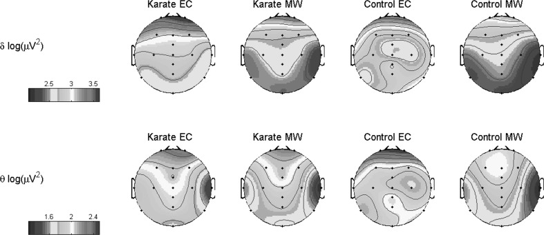
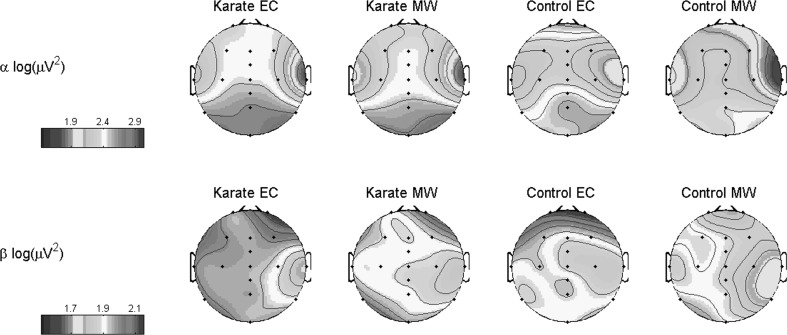
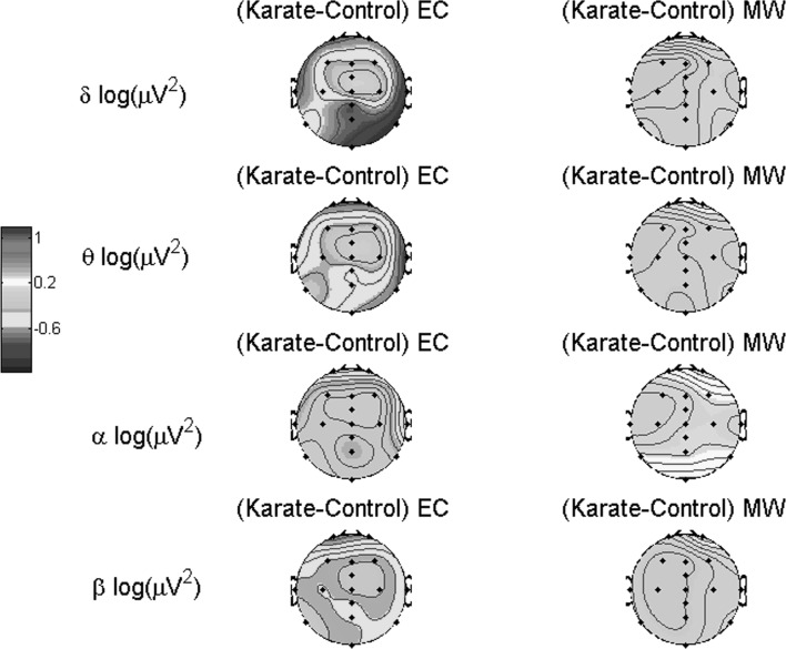
The topographies of each group during each task are shown in Fig. 1 and the differences computed from subtracting the power values of control group from karate group is shown in Figs. 2 and 3.
Fig. 1.
Topographies of logarithmic absolute band power (delta and theta) values for each group and for each task
Fig. 2.
Topographies of logarithmic absolute band power (alpha and beta) values for each group and for each task
Fig. 3.
EEG topographies formed by logarithm of absolute band power differences between groups for each frequency band and task
Event related desyncronization/synchronization (ERD/ERS)
Repeated measures of ANOVA has been conducted using region and frequency band as within subjects factors while group has been selected as between subjects factor. There is no overall effect of either region or frequency, but there is a crossover interaction. Region × frequency interaction was found to be statistically significant (F(4.21, 75.94) = 2.48, p < 0.019). In frontal region theta band power value was found to be higher than delta band value (MD = 0.066, SD = 0.024, p < 0.023).
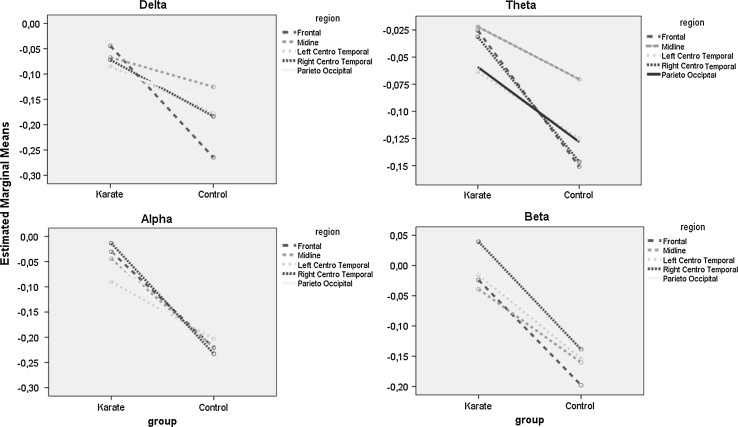
The three-way interaction tells us whether the region × frequency interaction described above is the same for karate players and athletes. There is a significant three-way region × frequency × group interaction (F(1, 18) = 6.61, p < 0.019). In frontal region, alpha and beta ERD/ERS values of karate players were found to be lower than the values of controls (for alpha; MD = 0.19, SD = 0.08, p < 0.009), (for beta; MD = 0.174, SD = 0.088, p < 0.029). Similarly, in midline region, alpha and beta ERD/ERS values were found to be lower for karate players when compared with controls (for alpha; MD = 0.176, SD = 0.071, p < 0.004), (for beta; MD = 0.121, SE = 0.066, p < 0.043). In RCT region, alpha and beta ERS/ERD were lower in karate players (for alpha; MD = 0.219, SD = 0.072, p < 0.001), (for beta; MD = 0.178, SD = 0.081, p < 0.031). In parietal occipital region, alpha ERD/ERS was found to be significantly lower for karate players than the controls (MD = 0.22, SD = 0.056, p < 0.0001). All of the mean differences were computed as subtraction of the ERD/ERS value of karate players from the value corresponding to the values of control subjects. The ERD/ERS values are summarized in Fig. 4.
Fig. 4.
Estimated marginal mean amplitudes of ERD/ERS for each group (Karate, Control) in each task (resting, counting) for each region (Mid: Midline, Lt CT: Left centro-temporal, Rt CT: Right centro-temporal, PO: Parieto-occipital)
Discussion
This study investigates the involvement of neural efficiency in a cognitive task in elite athletes using EEG. A group of elite karate group was compared to a control group of non-atheletes. Participants performed a mental subtraction task.
Delta band power values computed during the resting and mental workload paradigm exhibited higher values in frontal regions when compared to other spatial areas which are in line with the findings of Chen et al. (2008). Rather than the eye blinking or eye movement, cerebral metabolic activity can be thought to be associated with this topographical distribution of delta band (Boord et al. 2007). As expected, when the subjects performed counting process, a reduction in delta band power was observed, as a result of increased mental workload. For the absolute power values of theta band, anterior regions had higher values than the posterior regions while for alpha band posterior regions had higher values than the frontal sites. During the eyes closed resting periods, default mode of the brain is activated. When the mental workload increased, posterior alpha band power significantly decreased. This can be explained by the increased number of desynchronized neurons in network that tries to solve the given problem. Moreover, in our study, posterior alpha band power of the karate players was found to be higher than control subjects under both tasks. A similar increment in absolute alpha band power was observed during the golf performance of expert golf players (Baumeister et al. 2008) and during the imagination of a novel dance scenario by the professional dancers (Fink et al. 2009). According the neural efficiency hypothesis, it can be assumed that less number of resources are allocated for performance of a task. Micheloyannis et al. (2006) showed that reduced values regarding to small world network parameters were exhibited when high educated people were compared with the less educated ones under the same mental workload levels. In addition to this, in a motor task, during upright standing position, alpha inhibition of elite athletes was shown to be less than the control subjects (Del Percio et al. 2009). Our findings about the high alpha power value of elite karate players suggest that increased number of synchronized neurons can be recruited by them when compared with the control subjects. Moreover, in karate players, alpha blockage or alpha inhibition has been shown to be present as a marker of neural efficiency hypothesis, in the concept of this study. Similar to dynamics of alpha band power, beta band power was found to be higher for eyes closed condition than the mental workload. In a recent study, beta band desynchronization was also presented in a motor imagery task which is thought to be a reflection of neuroplastisity (Di Nota et al. 2017).
Finally, this study is the first study to examine neural efficiency related to a cognitive task, not a motor task, in elite karate players using ERD/ERS analysis. The results suggest that the effect of neural efficiency in the brain is global rather than local and thus might be contributing to the elite athletic performances. It can be concluded that the increase in the efficiency of the brain may cause an increase in the sports performance that is based on enhanced motor functions for elite athletes.
Acknowledgements
This work was supported by Research Fund of the Marmara University. Project No: SAG-A-100713-0296.
Contributor Information
Adil Deniz Duru, Email: deniz.duru@marmara.edu.tr.
Moataz Assem, Email: moataz.assem@boun.edu.tr.
References
- Babiloni C, Del Percio C, Iacoboni M, Infarinato F, Lizio R, Marzano N, Crespi G, Dassu F, Pirritano M, Gallamini M, Eusebi F. Golf putt outcomes are predicted by sensorimotor cerebral EEG rhythms. J Physiol. 2008;586(1):131–139. doi: 10.1113/jphysiol.2007.141630. [DOI] [PMC free article] [PubMed] [Google Scholar]
- Babiloni C, Del Percio C, Rossini PM, Marzano N, Iacoboni M, Infarinato F, Eusebi F, et al. Judgment of actions in experts: a high-resolution EEG study in elite athletes. Neuroimage. 2009;45(2):512–521. doi: 10.1016/j.neuroimage.2008.11.035. [DOI] [PubMed] [Google Scholar]
- Baumeister J, Reinecke K, Liesen H, Weiss M. Cortical activity of skilled performance in a complex sports related motor task. Eur J Appl Physiol. 2008;104(4):625–631. doi: 10.1007/s00421-008-0811-x. [DOI] [PubMed] [Google Scholar]
- Berger H. Uber das Elektrenkephalogramm des Menschen. Arch Psychiatr Nervenkrankh. 1929;87:527–570. doi: 10.1007/BF01797193. [DOI] [Google Scholar]
- Blanco S, García H, Quian Quiroga R, Romanelli L, Rosso OA. Stationarity of the EEG series. IEEE Eng Med Biol. 1995;14:395–399. doi: 10.1109/51.395321. [DOI] [Google Scholar]
- Boord PR, Rennie CJ, Williams LM. Integrating “brain” and “body” measures: correlations between EEG and metabolic changes over the human lifespan. J Integr Neurosci. 2007;6:205–218. doi: 10.1142/S0219635207001416. [DOI] [PubMed] [Google Scholar]
- Chen ACN, Feng W, Zhao H, Yin Y, Wang P. EEG default mode network in the human brain: spectral regional field powers. Neuroimage. 2008;41:561–574. doi: 10.1016/j.neuroimage.2007.12.064. [DOI] [PubMed] [Google Scholar]
- Chew LH, Teo J, Mountstephens J. Aesthetic preference recognition of 3D shapes using EEG. Cognit Neurodyn. 2016;10(2):165–173. doi: 10.1007/s11571-015-9363-z. [DOI] [PMC free article] [PubMed] [Google Scholar]
- Del Percio C, Rossini PM, Marzano N, Iacoboni M, Infarinato F, Aschieri P, Eusebi F, et al. Is there a “neural efficiency” in athletes? A high-resolution EEG study. Neuroimage. 2008;42(4):1544–1553. doi: 10.1016/j.neuroimage.2008.05.061. [DOI] [PubMed] [Google Scholar]
- Del Percio C, Babiloni C, Marzano N, Iacoboni M, Infarinato F, Vecchio F, Lizio R, Aschieri P, Fiore A, Toràn G, Gallamini M, Baratto M, Eusebi F. Neural efficiency of athletes’ brain for upright standing: a high-resolution EEG study. Brain Res Bull. 2009;79(3–4):193–200. doi: 10.1016/j.brainresbull.2009.02.001. [DOI] [PubMed] [Google Scholar]
- Di Nota PM, Chartrand JM, Levkov GR, Montefusco-Siegmund R, DeSouza JFX (2017) Experience-dependent modulation of alpha and beta during action observation and motor imagery. BMC Neurosci 18–28 [DOI] [PMC free article] [PubMed]
- Fink Andreas, Graif Barbara, Neubauer Aljoscha C. Brain correlates underlying creative thinking: EEG alpha activity in professional vs. novice dancers. Neuroimage. 2009;46:854–862. doi: 10.1016/j.neuroimage.2009.02.036. [DOI] [PubMed] [Google Scholar]
- Grabner RH, Fink A, Stipacek A, Neuper C, Neubauer AC. Intelligence and working memory systems: evidence of neural efficiency in alpha band ERD. Cognit Brain Res. 2004;20(2):212–225. doi: 10.1016/j.cogbrainres.2004.02.010. [DOI] [PubMed] [Google Scholar]
- Haier RJ, Siegel B, Tang C, Abel L, Buchsbaum MS. Intelligence and changes in regional cerebral glucose metabolic rate following learning. Intelligence. 1992;16(3–4):415–426. doi: 10.1016/0160-2896(92)90018-M. [DOI] [Google Scholar]
- Haufler AJ, Spalding TW, Santa Maria DL, Hatfield BD. Neuro-cognitive activity during a self-paced visuospatial task: comparative EEG profiles in marksmen and novice shooters. Biol Psychol. 2000;53(2–3):131–160. doi: 10.1016/S0301-0511(00)00047-8. [DOI] [PubMed] [Google Scholar]
- Huettel SA, Song AW, McCarthy G. Functional magnetic resonance imaging. 2. Sunderland: Sinauer Associates; 2008. [Google Scholar]
- Maschio M, Marchesi F, Dispenza S, Dinapoli L, Sperati F, Petreri G, Gumenyuk S, Dessanti ML, Zarabla A, Cantelmi T, Mengarelli A. Effect of high dose cytosine arabinoside on quantitative EEG in patients with acute myeloid leukemia. Cognit Neurodyn. 2016;10(2):185–188. doi: 10.1007/s11571-015-9370-0. [DOI] [PMC free article] [PubMed] [Google Scholar]
- Micheloyannis S, Pachou E, Stam CJ, Vourkas M, Erimaki S, Tsirka V. Using graph theoretical analysis of multi channel EEG to evaluate the neural efficiency hypothesis. Neurosci Lett. 2006;402(3):273–277. doi: 10.1016/j.neulet.2006.04.006. [DOI] [PubMed] [Google Scholar]
- Neubauer AC, Fink A. Intelligence and neural efficiency. Neurosci Biobehav Rev. 2009;33(7):1004–1023. doi: 10.1016/j.neubiorev.2009.04.001. [DOI] [PubMed] [Google Scholar]
- Niedermeyer E. The normal EEG of the waking adult. In: Niedermeyer E, Lopes da Silva FH, editors. Electroencephalography: basic principles, clinical applications and related fields. 3. Baltimore: Williams and Wilkins; 1993. pp. 131–152. [Google Scholar]
- Pfurtscheller G, Aranibar A. Evaluation of event-related desynchronization (ERD) preceding and following voluntary self-paced movement. Electroencephalogr Clin Neurophysiol. 1979;46(2):138–146. doi: 10.1016/0013-4694(79)90063-4. [DOI] [PubMed] [Google Scholar]
- Poldrack RA. Is “efficiency” a useful concept in cognitive neuroscience? Dev Cognit Neurosci. 2014;11:12–17. doi: 10.1016/j.dcn.2014.06.001. [DOI] [PMC free article] [PubMed] [Google Scholar]
- Reichle ED, Carpenter P, Just MA. The neural bases of strategy and skill in sentence-picture verification. Cognit Psychol. 2000;40(4):261–295. doi: 10.1006/cogp.2000.0733. [DOI] [PubMed] [Google Scholar]
- Rypma B, D’Esposito M. The roles of prefrontal brain regions in components of working memory: effects of memory load and individual differences. Proc Natl Acad Sci. 1999;96(May):6558–6563. doi: 10.1073/pnas.96.11.6558. [DOI] [PMC free article] [PubMed] [Google Scholar]
- Yarrow K, Brown P, Krakauer JW. Inside the brain of an elite athlete: the neural processes that support high achievement in sports. Nat Rev Neurosci. 2009;10(8):585–596. doi: 10.1038/nrn2672. [DOI] [PubMed] [Google Scholar]
- Zhang L, Gan JQ, Wang H. Localization of neural efficiency of the mathematically gifted brain through a feature subset selection method. Cognit Neurodyn. 2015;9(5):495–508. doi: 10.1007/s11571-015-9345-1. [DOI] [PMC free article] [PubMed] [Google Scholar]


