Abstract
Laboratory studies suggested that general anesthetics induce neuroapoptosis and inhibit neurogenesis in developing brains of animals. Minocycline exerts neuroprotection against a wide range of toxic insults in neurodegenerative diseases models. Here, we investigate whether minocycline can alleviate neurogenetic damage and improve cognition following midazolam exposure in neonatal rats. Postnatal 7 days rats were divided randomly into three groups: control group (C), midazolam group (M), and minocycline pretreatment group (MP). After exposure to midazolam, the cell proliferation in the subventricular zone (SVZ) and the subgranular zone (SGZ) of the hippocampus in pups was analyzed by bromodeoxyuridine immunochemistry at 7 days after the administration of anesthesia. Cognitive function was assessed using the Morris water-maze test at 35 days after midazolam exposure. Compared with the control, midazolam reduced cell proliferation both in the SVZ and in the SGZ of the hippocampus of neonatal rats, and decreased spatial learning and memory ability of rats in adulthood significantly. Pretreatment with minocycline increased cell proliferation both in the SVZ and in the SGZ of the hippocampus and improved spatial learning and memory ability compared with midazolam, but it did not mitigate the changes to the normal levels compared with the controls. Our results indicated that pretreatment with minocycline can alleviate midazolam-induced damage in neural stem cell proliferation of neonatal rats and improve spatial learning and memory ability of rats in adulthood.
Keywords: midazolam, minocycline, Morris water maze, neurodegenerative, neuroprotective
Introduction
Although developmental neurotoxicity of anesthesia agents has received attention from anesthetists for decades, it is still common practice to expose infants or toddlers to general anesthesia in pediatric surgical practice. Studies suggest that almost all the general anesthetics could induce structural and functional changes in the brain of neonatal animals (reviewed in the study by Lin et al. 1). Moreover, large-scale clinical studies also indicated that learning disabilities and behavioral disturbances in some children are correlated with surgery under anesthesia before 4 years of age, especially in children undergoing multiple surgeries 2.
One important phase of brain development for mammals is called brain growth spurt (BGS), in which neural stem cells proliferate, differentiate, and migrate abundantly in the brain. The characteristics of BGS make the brain of neonatal animals more susceptible than the mature brain to exogenous insults. Midazolam, a γ-aminobutyric acid A receptor agonist, is used commonly for the induction and maintenance of anesthesia, as well as to promote sedation in the ICU. Despite its growing popularity, researches showed that its neurodegenerative 3 and neuroapoptotic effects 4 may contribute toward the learning and memory deficits in later life after exposure to the immature brains 5. Therefore, it is necessary to seek neuroprotective strategies when using midazolam as an anesthetic during this period.
The mechanisms of midazolam’s developmental neurotoxicity are not fully understood 6. Some studies indicated that activating γ-aminobutyric acid A receptors could result in depolarization and excitatory toxicity in neurons during BGS 7. Boscolo et al. 8 speculated that impairment of mitochondrial integrity and accumulation of reactive oxygen species could contribute toward the neuronal loss and cognitive dysfunction caused by midazolam, isoflurane, and nitrous oxide anesthesia. Others report that midazolam can block voltage-dependent calcium channels in neurons, which is related to caspase-8-independent apoptosis 4. All these changes were associated with the structural remolding of the hippocampus and the prevention of memory formation following midazolam exposure 9. However, whether exposure to midazolam impairs neurogenesis in neonatal brain needs to be elucidated.
Minocycline, as a long-acting tetracycline agent, is considered to be neuroprotective and anti-inflammatory. Several studies have reported that minocycline was beneficial in neurological models such as spinal-cord injury 10, traumatic brain injury 11, ischemic stroke 12, and Parkinson’s disease 13. The positive outcome of minocycline is associated with inhibition of microglia activation 14, inhibition of caspase-1 and caspase-3 15, and cytochrome c release 16. Recently, we reported that minocycline attenuated ketamine-induced injury in neural stem cells (NSCs) 17 and restores neurogenesis in the subventricular zone (SVZ) and the subgranular zone (SGZ) of the hippocampus after ketamine exposure in neonatal rats 18. However, whether minocycline can attenuate the developmental neurotoxicity of midazolam remains unclear.
Given that minocycline broadly ameliorates central nervous system injury, in the present study, we investigated whether minocycline could protect the brain from impairment in neurogenesis and cognition after midazolam exposure in neonatal rats.
Methods
Animal models and drugs’ administration
As there were 8–10 pups in every litter, 36 healthy postnatal 7 days (PND7) Sprague-Dawley rat pups weighing 15–30 g from four litters were provided by the Experimental Animal Center of Xi’an Jiaotong University. All procedures were performed in accordance with the National Institutes of Health Sciences and followed the Guide for the Care and Use of Laboratory Animals. Experimental protocols were approved by the Committee of Animal Care and Use Administration of Xi’an Jiaotong University. Experimental rats in every litter were allocated randomly into the control group (C), midazolam group (M), and minocycline pretreatment groups (MP).
Rat litters (including the mother and the pups) were kept in 210-square-inch plastic cages bedded by wood-derived materials. The pups were allowed access to milk freely and the mothers were fed with standard nonmedicated laboratory rodent food supplied by the Experimental Animal Center of Xi’an Jiaotong University. The rats in the minocycline pretreatment group received minocycline (St Louis, Missouri, USA, dissolved in normal saline) 40 mg/kg 30 min before an injection of midazolam intraperitoneally (9 mg/kg, diluted in normal saline, intraperitoneal). The rats of the midazolam group received equivalent volumes of normal saline as minocycline before midazolam exposure. The rats in the control group received equivalent volumes of normal saline as other groups. At ∼50 min later, 40% of the midazolam loading dose was administered to maintain the anesthesia. Our preliminary experiment showed that the maximal liquid volume that PND7 rats can endure is about 100 μl; thus, each rat received a solution less than 100 μl in volume at the end of the anesthesia. During the process of anesthesia, the respiration, the skin color, and body movement of all the experimental subjects were monitored. Furthermore, the infant pulse oximetry probes were attached to the abdomen of the rats at 0, 60, 120, and 180 min to detect the oxygen saturation (SpO2) of the anesthetized pups. After spontaneous recovery from the anesthesia at ∼3 h later, the rats (n=7 in each group) were allowed to remain with their mother until PND21 before separation to cages with a size of 140-square-inch individually. At PND42, the behavioral study was carried out with the Morris water maze.
Histological specimen
The pups (n=5 in each group) received bromodeoxyuridine (BrdU) (50 mg/kg, intraperitoneal injection) every 24 h for 7 consecutive days, which started from the end of the anesthesia, and then they were killed for the detection of neurogenesis in the SVZ and SGZ at PND14. As we described in previous studies 18, after anesthetization with 40 mg/kg sodium pentobarbital, the rats were perfused with 0.9% normal saline, followed by 4% paraformaldehyde transcardially. The brains were removed to 4% paraformaldehyde overnight for postfixation and then a 30% glucose solution overnight for dehydration. Finally, the coronal sections (20 µm) from bregma +0.2 mm to bregma −6.0 mm were collected using a freezing microtome.
BrdU immunohistochemistry
BrdU (Sigma-Aldrich Inc., St Louis, Missouri, USA) is commonly used in the detection of cell proliferation in living tissue for its substitute of thymidine during the S phase of DNA replication. Briefly, five 20 μm coronal sections (spaced ∼100 μm apart) of SVZ and SGZ per animal were collected for immunohistochemistry. After denaturation of the DNA with 2 N HCl for 30 min, the sections were rinsed in 100 mM boric acid (pH 8.5) for 10 min at room temperature, and then incubated in 1% H2O2 in PBS for 20 min following blocking solution (4% goat serum and 0.3% Triton X-100 in PBS) for 1 h at room temperature before treatment with the anti-BrdU antibody (1 : 1000, ab8152; Abcam, Cambridge, UK) overnight at 4°C. The sections that were incubated with PBS without the primary antibody were used as negative controls. After rinsing with 10 mM PBS (pH 7.4, 5 min×3 times), the sections were incubated with fluorescein-labeled secondary antibodies (1 : 200, A0521; Beyotime, Shanghai, China) for 2 h at room temperature. The sections were rinsed as above, and then the immune reactive cells were visualized by fluorescence microscopy (BX51; Olympus, Tokyo, Japan) with five randomly selected fields 50×50 µm in size; then, the Image Pro Plus (Systat Software, San Jose, California, USA) (v-5.02) was used to count and calculate the numbers of BrdU-positive cells per mm2 in SVZ and SGZ by a blinded observer. The purpose of this method is to obtain the mean value of each animal for comparison instead of the actual amount of cell proliferation.
Morris water-maze test
At PND42 (first training day), the rats were trained in the Morris water maze to test their learning and memory ability. The Morris water-maze test was performed as described by Sase et al. 19, with minor modifications. It was a black pool filled with 20±1°C water to a depth of 25 cm. The maze was divided geographically into four equal quadrants: N, E, S, and W. A hidden circular platform (11 cm in diameter) was located in the center of the southwest quadrant and submerged 1.5 cm beneath the surface of the water. A charge-coupled device camera was mounted above so that the animal’s motion, for example, the latency to find the platform during the training session, the distance traveled as well as the swimming speed could be recorded automatically and sent to a computerized system (EthoVision, 3.1 version; Noldus, Wageningen, the Netherlands). If the rats failed to find the platform after 120 s, they were placed manually on it for 30 s. After 5 consecutive days of navigation trial, a probe trial was conducted where the rats would swim for 120 s without a platform and the time spent in the target zone was measured. Finally, these data were analyzed by an independent investigator who was blinded to the treatment.
Statistical analysis
Statistical analysis was carried out using the Sigmaplot 12.0 (Systat Software). In detail, a one-way analysis of variance was used to determine differences in neurogenesis between the three groups, whereas to determine differences between groups on Morris water-maze performance, a two-way repeated-measures analysis of variance was used. All the data are presented as mean±SEM. A P value of less than 0.05 was considered significant and Graph Pad Prism Software (Graph Pad Software, San Diego, California, USA) was used for graphical presentation.
Results
Breathing indicators
During anesthesia, the rats were completely anesthetized and did not show voluntary movement and apparent changes in skin color or respiratory rate. There was no significant difference among all groups in oxygen saturation (SpO2) at 0, 60, 120, and 180 min after the anesthesia (data not show).
Cell proliferation in the subventricular zone and subgranular zone of the hippocampus
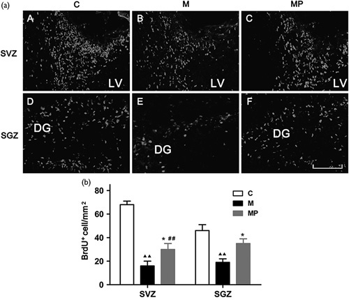
As shown in Fig. 1, BrdU-positive cells were distributed in SVZ and SGZ among all groups. The number of BrdU-positive cells in the midazolam group decreased significantly compared with the control group both in SVZ and in SGZ [F(2, 12)=43.44, P<0.01, in SVZ and F(2, 12)=9.964, P<0.01 in SGZ], respectively. The number of BrdU-positive cells in the minocycline pretreatment group was significantly higher than that of the midazolam group both in SVZ and in SGZ [F(2, 12)=43.44, P=0.032 in SVZ and F(2, 12)=9.964, P=0.044 in SGZ], respectively. However, it was less than that of the control group in SVZ [F(2, 12)=43.44, P<0.01], but not SGZ [F(2, 12)=9.964, P=0.093].
Fig. 1.

(a) Bromodeoxyuridine (BrdU)-positive cells in the subventricular zone (SVZ) and the subgranular zone (SGZ). (A-F). Representative images of BrdU- positive cells in the SVZ and SGZ PND 14 in different groups. LV, lateral ventricle. DG, dentate gyrus. Scale bar=100μm. (b) Quantitative analysis of BrdU-positive cells. Values are presented as mean±SEM. ▲▲P<0.01 Midazolam group compared with the control group. *P<0.05 Minocycline pretreatment group compared with the midazolam group. ##P<0.01 Minocycline pretreatment group compared with the control group. n=5 in each group.
Spatial learning ability
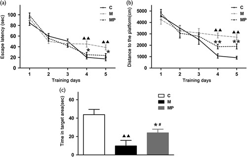
As indicated in Fig. 2a, all animals showed a progressive decline in the escape latency, but further analysis suggested that the rats in the midazolam group took longer to find the hidden platform on the fourth and fifth training day compared with the control group [F(2, 18)=8.581, P<0.01, on the fourth training day and F(2, 18)=7.206, P<0.01 on the fifth training day]. Moreover, the latency of the minocycline pretreatment group decreased significantly compared with that of the midazolam group [F(2, 18)=8.581, P=0.011 on the fourth training day and F(2, 18)=7.206, P=0.035 on the fifth training day]. In addition, there was no difference between the control and the minocycline pretreatment group for latency on the fourth (F(2, 18)=8.581, P=0.478) and fifth training days [F(2, 18)=7.206, P=0.296]. A similar tendency to change could be seen in the swimming distance, which reflects the moving paths of rats to reach the platform (Fig. 2b) [F(2, 18)=19.653, P<0.01 on the fourth training day and F(2, 18)=21.980, P<0.01 on the fifth training day between the control and the midazolam groups; F(2, 18)=19.653, P<0.01 on the fourth training day and F(2, 18)=21.980, P<0.01 on the fifth training day between the midazolam and minocycline pretreatment groups], but there was a significant difference between the control and the minocycline pretreatment group in the swimming path [F(2, 18)=19.653, P=0.011 on the fourth training day and F(2, 18)=21.980, P<0.01 on the fifth training day].
Fig. 2.

The results of the Morris water maze. (a) Latency to reach the hidden platform. The differences between the groups were not significant for the first 3 days (P>0.05), but on the fourth day and fifth day, the subjects showed longer latency in the midazolam group compared with the control group (P<0.01), and rats in the minocycline treatment group spent a shorter time to find the platform than that in the midazolam group (P<0.05), but this was not significantly different compared with the control group (P>0.05). (b) Swimming distance to reach the hidden platform. A tendency similar to latency can be observed. (c) Comparison of the time spent in the effective area. Rats in the midazolam group spent less time in the target region than that in the control group (P<0.01), and the time in the effective areas was significantly longer than that of the midazolam group after minocycline pretreatment (P<0.05). However, there was a significant difference between the control and the minocycline pretreatment group (P<0.05). Values are presented as mean±SEM. ▲▲P<0.01 Midazolam group compared with the control group. *P<0.05, **P<0.01 Minocycline pretreatment group compared with the midazolam group. #P<0.05 Minocycline pretreatment group compared with the control group. n=7 in each group.
In the probe trial, the platform was removed and the animals were allowed to swim for 120 s freely. It was found that the time spent in the target area for the midazolam group was significantly shorter than that in the control group (Fig. 2c) [F(2, 18)=14.755, P<0.01]. However, rats in the minocycline pretreatment group spent a longer time in the effective region than that in the midazolam group (Fig. 2c) [F(2, 18)=14.755, P=0.015], but it was shorter compared with the control group [F(2, 18)=15.755, P=0.027] (Tables 1–4).
Table 1.
Number of BrdU+ cells observed in different groups

Table 4.
Data of time in target area in the Morris water maze between different groups

Table 2.
Data of escape latency in the Morris water maze between different groups

Table 3.
Data of swimming distance to the target in the Morris water maze between different groups

Discussion
Here, we reported that neonatal midazolam exposure decreased cell proliferation in the SVZ and SGZ and impaired cognitive functions of rats in adulthood. However, minocycline pretreatment alleviated the changes induced by midazolam in neonatal rats.
Neurogenesis is composed of NSC proliferation, neuronal differentiation, migration, maturation, and integration into neural networks. In mammals, most neurons are generated before birth, but new neurons are added continuously to certain brain areas throughout life. These neurons are derived from NSCs that are located primarily in two distinct areas of the brain: the SVZ and the SGZ. NSC proliferation is one of the most basic events for neurogenesis. Considering the significance of neurogenesis during the BGS period, we evaluated the cell proliferation using BrdU labeling. Our study showed that midazolam inhibited the cell proliferation in the neurogenetic regions of neonatal rats, indicating that neonatal midazolam exposure might impair neurogenesis. Young et al. 20 reported the deleterious effects of midazolam on apoptotic neurodegeneration in infant rodents after 6 h exposure at a subclinical dose. Boscolo et al. 8 also reported the neurotoxicity of midazolam on the developing brain of PND7 rats. Recently, it was shown that a single maternal clinical dose of midazolam is sufficient to induce significant neuroapoptosis in fetal guinea-pigs 21. Interestingly, we found that minocycline alleviates cellular proliferating changes induced by midazolam in neurogenetic regions of neonatal rats, indicating that minocycline might enhance NSC proliferation following midazolam exposure during the brain development which is benefit for the neurogenesis. The finding about the neuroprotection of minocycline on brain development is in consistency with our recent study, which reported the protective effect of minocycline against ketamine-induced injury in NSCs 17. Whether minocycline can mitigate midazolam-induced neuroapoptosis needs to be determined further.
In humans, BGS begins at the last trimester of pregnancy and ends at 2–3 years. For rodents, BGS peaks around 2 weeks of life. Considering the different lengths of BGS in humans and rodents, it can be argued that 3 h of midazolam exposure might be equal to days to weeks for clinical patients. Actually, sometimes, infants require sedation by midazolam for weeks in the ICU. Moreover, the dose of midazolam in this study is also in the sedation (subanesthetic) range for rat pups. Considering the different duration for BGS between human and rodents, the further study needs to detect the impact of shorter midazolam administration on developing brains which is closer to the clinical practice.
Learning disabilities and behavioral disturbances in some children are correlated with surgery under anesthesia before 4 years of age 2. To evaluate the spatial learning and memory ability of adult rats that were subjected to anesthetic exposure during the neonatal stage, the Morris water maze was used in the present study. The rats took a longer time to find the hidden platform in the latency trial and spent a shorter time in the effective regions in the probe trial after midazolam exposure compared with the controls, indicating that neonatal midazolam exposure induces long-term memory deficits in rodents. These findings are similar to the previous report 22. However, the rats in the minocycline pretreatment group took less time to find the hidden platform in the latency trial and spent more time in the effective regions in the probe trial compared with midazolam exposure, indicating that minocycline ameliorates the long-term memory deficits in rodents who received midazolam in the early stage of life. In terms of assessment of learning and memory ability, the Morris water-maze test used in our study was slightly different from the studies carried out by Sase et al. 19. A pool with a diameter of 150 cm and a height of 60 cm was used in Sase’s study, but a pool with a diameter of 140 cm and a height of 70 cm was used in ours. The rats in Sase’s study were trained for 4 training days, followed by a probe trial with 60 s of free swimming, but the rats in ours were trained for 5 days, followed by a probe trial to swim freely for 120 s. In addition, Sase et al. 19 subjected the rats to an acclimatization training session on the first training day, whereas we did not. Whether these differences impacted the accuracy of cognitive evaluation needs to be determined further.
The study by Timic et al. 22 suggested that midazolam impaired the spatial memory of rats might result from retrograde amnesia, which is the pharmacological property of midazolam. Other studies showed that midazolam-induced excitotoxicity, impaired synaptogenesis, or inhibited long-term potentiation of the hippocampus, which may contribute toward spatial learning and memory dysfunction in adults 23. Whether midazolam-induced damage in neurogenesis contributes toward long-term memory deficits in rodents remains unknown. It was speculated that the progressive decline in cognitive function is a consequence of an early loss or suppression of pools of rapidly dividing, multipotent precursors, which might have created an ongoing deficit in neurogenesis that worsened over time as the gap in NSC numbers between normal controls and exposed animals widened with rapid cell division 24. In our study, midazolam decreased NSC proliferation and impaired cognitive functions, but minocycline pretreatment alleviated the deleterious effects of midazolam on NSCs and neurocognition, strengthening the connection between neurogenesis and cognitive dysfunction in developmental anesthetic neurotoxicity. The present findings indicated that the protective effect of minocycline on NSC proliferation may be one of the potential mechanisms by which it improves behavior performance after neonatal midazolam exposure in rats, but minocycline pretreatment did not alleviate the injury induced by midazolam to the normal level both in the number of BrdU-positive cells in SGZ and the performance in the probe trial.
It is worth emphasizing that there are some limitations in this study. First, NSC differentiation, neuronal apoptosis, or migration in the developing brain were not detected because our main aim was to observe whether clinically relevant doses of midazolam could inhibit NSC proliferation in neonatal rats and cause cognitive deficits in adults. Second, we only measured the time spent in the target region of the probe trial to analyze the spatial learning and memory; our conclusion will be stronger if we can compare more parameters such as target versus nontarget quadrant data. Third, we did not investigate the underlying mechanism for the neuroprotection of minocycline. However, we found that it was related to the PI3K/Akt pathway in our previous study, which exposed PND7 rats to ketamine instead of midazolam 18; further studies are needed to confirm this.
Conclusion
In this study, we found that minocycline pretreatment was associated with more cell proliferation in SVZ and SGZ, as well as better spatial memory and learning ability after midazolam exposure; we speculate that the use of minocycline will be a valuable neuroprotective strategy before anesthesia with midazolam in the future. Further studies should be carried out to confirm this.
Acknowledgements
The authors thank Professor Malgorzata Garstka for reading the manuscript.
This work was supported by the National Natural Science Foundation of China (81071071,81171247), the Key Science and Technology Innovation Team of Shaanxi Province (2014KCT-22), and the Science and Technology Development Project of Shaanxi Province grants (2013KTCL03-09).
Conflicts of interest
There are no conflicts of interest.
Footnotes
Praveen K. Giri and Yang Lu contributed equally to the writing of article.
References
- 1.Lin EP, Soriano SG, Loepke AW. Anesthetic neurotoxicity. Anesthesiol Clin 2014; 32:133–155. [DOI] [PubMed] [Google Scholar]
- 2.Wilder RT, Flick RP, Sprung J, Katusic SK, Barbaresi WJ, Mickelson C, et al. Early exposure to anesthesia and learning disabilities in a population-based birth cohort. Anesthesiology 2009; 110:796–804. [DOI] [PMC free article] [PubMed] [Google Scholar]
- 3.Duerden EG, Guo T, Dodbiba L, Chakravarty MM, Chau V, Poskitt KJ, et al. Midazolam dose correlates with abnormal hippocampal growth and neurodevelopmental outcome in preterm infants. Ann Neurol 2016; 79:548–559. [DOI] [PubMed] [Google Scholar]
- 4.Stevens MF, Werdehausen R, Gaza N, Hermanns H, Kremer D, Bauer I, et al. Midazolam activates the intrinsic pathway of apoptosis independent of benzodiazepine and death receptor signaling. Reg Anesth Pain Med 2011; 36:343–349. [DOI] [PubMed] [Google Scholar]
- 5.Xu B, Yang J, Kang F, Li J. The inflammatory response of two different kinds of anesthetics on vascular cognitive impairment rats and the effect on long term cognitive function. Int J Clin Exp Med 2015; 8:16694–16698. [PMC free article] [PubMed] [Google Scholar]
- 6.Luo J, Guo J, Han D, Li H. Comparison of dexmedetomidine and midazolam on neurotoxicity in neonatal mice. Sheng Wu Yi Xue Gong Cheng Xue Za Zhi 2013; 30:607–610. [PubMed] [Google Scholar]
- 7.Owens DF, Kriegstein AR. Developmental neurotransmitters? Neuron 2002; 36:989–991. [DOI] [PubMed] [Google Scholar]
- 8.Boscolo A, Milanovic D, Starr JA, Sanchez V, Oklopcic A, Moy L, et al. Early exposure to general anesthesia disturbs mitochondrial fission and fusion in the developing rat brain. Anesthesiology 2013; 118:1086–1097. [DOI] [PMC free article] [PubMed] [Google Scholar]
- 9.McEwen BS. Physiology and neurobiology of stress and adaptation: central role of the brain. Physiol Rev 2007; 87:873–904. [DOI] [PubMed] [Google Scholar]
- 10.Papa S, Caron I, Erba E, Panini N, De Paola M, Mariani A, et al. Early modulation of pro-inflammatory microglia by minocycline loaded nanoparticles confers long lasting protection after spinal cord injury. Biomaterials 2016; 75:13–24. [DOI] [PubMed] [Google Scholar]
- 11.Li J, Chen J, Mo H, Chen J, Qian C, Yan F, et al. Minocycline protects against NLRP3 inflammasome-induced inflammation and P53-associated apoptosis in early brain injury after subarachnoid hemorrhage. Mol Neurobiol 2016; 53:2668–2678. [DOI] [PubMed] [Google Scholar]
- 12.Fagan SC, Cronic LE, Hess DC. Minocycline development for acute ischemic stroke. Transl Stroke Res 2011; 2:202–208. [DOI] [PMC free article] [PubMed] [Google Scholar]
- 13.Garrido-Mesa N, Zarzuelo A, Galvez J. Minocycline: far beyond an antibiotic. Br J Pharmacol 2013; 169:337–352. [DOI] [PMC free article] [PubMed] [Google Scholar]
- 14.Wu DC, Jackson-Lewis V, Vila M, Tieu K, Teismann P, Vadseth C, et al. Blockade of microglial activation is neuroprotective in the 1-methyl-4-phenyl-1,2,3,6-tetrahydropyridine mouse model of Parkinson disease. J Neurosci 2002; 22:1763–1771. [DOI] [PMC free article] [PubMed] [Google Scholar]
- 15.Chen M, Ona VO, Li M, Ferrante RJ, Fink KB, Zhu S, et al. Minocycline inhibits caspase-1 and caspase-3 expression and delays mortality in a transgenic mouse model of Huntington disease. Nat Med 2000; 6:797–801. [DOI] [PubMed] [Google Scholar]
- 16.Zhu S, Stavrovskaya IG, Drozda M, Kim BY, Ona V, Li M, et al. Minocycline inhibits cytochrome c release and delays progression of amyotrophic lateral sclerosis in mice. Nature 2002; 417:74–78. [DOI] [PubMed] [Google Scholar]
- 17.Lu Y, Lei S, Wang N, Lu P, Li W, Zheng J, et al. Protective effect of minocycline againstketamine-induced injury in Neural Stem Cell: involvement of PI3K/Akt and Gsk-3 beta pathway. Front Mol Neurosci 2016; 9:135. [DOI] [PMC free article] [PubMed] [Google Scholar]
- 18.Lu Y, Giri PK, Lei S, Zheng J, Li W, Wang N, et al. Pretreatment with minocycline restores neurogenesis in the subventricular zone and subgranular zone of the hippocampus after ketamine exposure in neonatal rats. Neuroscience 2017; 352:144–154. [DOI] [PubMed] [Google Scholar]
- 19.Sase A, Dahanayaka S, Hoger H, Wu G, Lubec G. Changes of hippocampal beta-alanine and citrulline levels are paralleling early and late phase of retrieval in the Morris water maze. Behav Brain Res 2013; 249:104–108. [DOI] [PubMed] [Google Scholar]
- 20.Young C, Jevtovic-Todorovic V, Qin YQ, Tenkova T, Wang H, Labruyere J, et al. Potential of ketamine and midazolam, individually or in combination, to induce apoptotic neurodegeneration in the infant mouse brain. Br J Pharmacol 2005; 146:189–197. [DOI] [PMC free article] [PubMed] [Google Scholar]
- 21.Rizzi S, Carter LB, Ori C, Jevtovic-Todorovic V. Clinical anesthesia causes permanent damage to the fetal guinea pig brain. Brain Pathol 2008; 18:198–210. [DOI] [PMC free article] [PubMed] [Google Scholar]
- 22.Timic T, Joksimovic S, Milic M, Divljakovic J, Batinic B, Savic MM. Midazolam impairs acquisition and retrieval, but not consolidation of reference memory in the Morris water maze. Behav Brain Res 2013; 241:198–205. [DOI] [PubMed] [Google Scholar]
- 23.Tokuda K, O'Dell KA, Izumi Y, Zorumski CF. Midazolam inhibits hippocampal long-term potentiation and learning through dual central and peripheral benzodiazepine receptor activation and neurosteroidogenesis. J Neurosci 2010; 30:16788–16795. [DOI] [PMC free article] [PubMed] [Google Scholar]
- 24.Kang E, Berg DA, Furmanski O, Jackson WM, Ryu YK, Gray CD, et al. Neurogenesis and developmental anesthetic neurotoxicity. Neurotoxicol Teratol 2017; 60:33–39. [DOI] [PMC free article] [PubMed] [Google Scholar]


