Abstract
An examination of neurophysiologic features of median nerve in third trimester of regularly controlled normal risk pregnancies is performed at the Department of Neurophysiology of Primary Health Centre in Tuzla during January / April 2006. Examined group consisted of 40 young females in third trimester of pregnancy, and average age of 25,6 ± 4,9 years. Control group consisted of young healthy females with average age of 31,1 ± 4,4 years. Symptoms and signs of carpal tunnel sy. (CST) had 12 patients, but diagnosis is neurophysiologically confirmed in 9 (75%) patients. In group of pregnant females without symptoms 3 (10,7%) patients showed neurophysiologic evidence of CTS. Sensory velocity of right median nerve was significantly lower in pregnant group of patiens (p=0,002), but area of sensory potentials on both sides were lower in pregnant group (p<0,0001). Area of CMAP of right median nerve was significantly lower in pregnant group (p=0,0003). Significant differences in CTS group compared with control group were in sensory velocities of median nerve (left median nerve p=0,0007, right p<0,0001), and area of SNAP of both sides (left p<0,0001, right p=0,0001), but area of CMAP right (p=0,0003). In CTS group 7 females had unilateral and 5 had bilateral neurophysiological changes. Our conclusion is that neuro-physiological parameters of median nerve in third trimester of pregnancy are changed mainly due to high prevalence of CTS that might disturb quality of life and have psychological and physical implications on future mother. Hence, it is necessary to, continuously, pay enough attention in prevention or treatment of mentioned syndrome in this population group.
Keywords: angiotensin converting enzyme, unilateral nephrectomy, compensatory renal hypertrophy
INTRODUCTION
Although, pregnancy is physiological condition, for some neurological diseases pregnancy presents additional factor of risk, e.g. carpal tunnel syndrome (CTS) which is the most frequent neurophaty occurred because of clamp. Many studies indicating that approximately 34% of pregnant women simptoms of CTS (1). Actual incidence of CTS during pregnancy is ranging from 2 - 25% (2). This syndrome is most frequently diagnosed in second and third trimester (3). Three quarter of women have bilateral symptoms. Half of multiples had similar symptoms during prior pregnancies. Preggies are more often predispositioned for development of CTS in second half of pregnancy, because retention of fluid is often in later stage of pregnancy which causes swelling of tissues. Woman who has to take off their rings because of edema have this syndrome two times more expressed with symptoms (73%), and also woman with CTS symptoms more often have preeclampsia, hypertension and edema (2). Italian study on frequency of carpal tunnel has found clinical signs in 62% of woman while neurophysiological indicators confirmed diagnosis in 43% of preggies. Visible correlation between edema and neurophysiological findings is notable (4). Increase of weight and retention of fluid as a result of decrease of venous circulation and changes in hormonal status, including increased serum of estrogen, aldosteron and cortisol, are increasing the frequency for occurrence of this syndrom. It is suggested that, also, increased level of prolactine could be ethio-logical factor in development of CTS becaue symptoms are getting worse during the night, which is coincidence with diumal level of prolactine (5). Relaxin could also be potential ethiological factor due to his presence in high concentration from 18th week of pregnancy and his reduction 48 hrs after delivery. Presupposition is that this could lead to relaxation of lig. Carpi transversum, which leads to his flatness and compression of n. medianus (6). Objective sensory changes often include tactile discriminations, damage of sensibility for touch and pain with preserved eminence of thenar. As a consequence of compression neural structures, rigidity of fingers is very often. But in progressed stage, atrophy of muscles of thenar (particulary m. abductor pollicis brevis) is also present. By percussion of carpal tunnel area, in 60 % of patients, it is possible to cause Tinel’s sign which is nos specific sign for this syndrome. Flexion of patients hand lasting for one minute (Phalen’s maneuver) or hyperextension of wrist (reversre Phalen’s maneuver) could reproduce symptoms. This test has 80% of specificity but very low sensitivity. Firm pressure over the carpal tunnel for approximately 30 seconds is reproducing the symptoms (so called. Carpal compression test) (7). Sensitivity of this test is 89 % and specificity is 96%. Palpatory diagnosis implies examination of soft tissue in carpal area with sensitivity of 90 % and specificity of 75% or higher (8). Electrophysiological studies, including electromyogra-phy (EMG) and nervous conductive studies (electroneu-rography ENG) are used in combination with specific signs and symptoms and they creating criteria standards for CTS diagnosis and exemption of other neurological diagnosis. The most sensitive electrodiagnostic test for CTS is a study of sensory conduction of n. medianus, and it shows extended distal latence and slowing down the speed of sensory conduction through the wrist in 70 to 90 % of patients. Recording the latence on a short distance in flow of n. medianus from palmar part to the fist and comparing this latence with ulnary nerve on the same distance (conductive studies on palmary nerves) may increase sensitivity of conductive studies of sensory nerves (9). The most sensitive test for an early diagnosis of CTS is determination of sensory speed of conductivity by stimulation of the first finger and registration on the wrist (10). Sensory potential could be absent, and most of the patients with medium to serious nerve damage have prolonged distal motor latence of n. medianus, reduced and expanded M-potential. If motor fibres are caught, result of that is weakness of the fingers and muscular atrophy: lumbrical (I and II), short abductor, opo-nens and short flexor of the thumb. Electrophysiological studies are giving the exact evaluation about how serious nerve damage is. Patient with medium CTS have only a sensory abnormalities. Those with sensory and motor abnormalities have moderate CTS. Evidences of axonal loss (e.g. attenuated or absent sensory or motor response on distal from carpal tunnel or neuropatho-logical abnormality on pin EMG) are classified as a serious CTS. This kind of neurophysiological changes are used for evaluation of eventual modal treatments (9).
GOALS OF RESEARCH
- Compare neurophysiological parameters of n. medianus of preggies in third trimester with control group of healthy woman who are not pregnant.
- Determine frequency of neurophysiological and clinical signs of CTS on preggies in third trimester.
EXAMINEES AND METHODS
Study is performed at the Department of Neurophysiol-ogy of Primary Health Centre in Tuzla during January / April 2006. Control group consisted of young healthy females with average age of 31,1 ± 4,4 (24-40) years. 19 (63,3%) of them had a history of previous labors. Examined group consisted of 40 young females of average age of 25,6 ± 4,9 (17 - 39) years and 26 (65%) of them never had a labor. Average age of pregnancy was 34,65 ± 3,5 (27 - 39) weeks. This is *random pattern* od preggies that were regularly coming for gynecological examinations at Department for medical care and service of healthy and pregnant woman in Medical Centre in Tuzla, and all of them accepted this neurophysiological analyses. First that was taken was medical history, then examination was done and after that neurophysiological processing. Condition for this examination was that neither of examinees had polineuropathy or any other sickness which could influence on speed of nerve conduction. Testing was performed at room temperature and physiological temperature of the skin of examined females in lying position. At the beginning, they were divided on those with and without clinical symptoms (based on torpid fingers at day or night) and signs (Phalen’s and reverse Phalen’s tryout, Tinnel’s sign and tenar atrophy). EMNG machine Medelec Synergy (EMG and EP Systems / OXFORD INSTRUMENTS 2004) was used in measurement of neurographic parameters. Bipolar electrode (so called Large touchproof) for superficial stimulation and registration were used. Sensory conductive velocity of n. medianus was measured by stimulation of the wrist and registration on forefinger, on folding of first joint of finger. Terminal motor latence measured upon stimulation of the wrist was 6 cm proximal on the wrist and by registration on thenar, more preciselly on the belly of m. abductor brevis. Following was analyzed by ENG processing: velocity of sensory conduction with area of action potential of sensory nerve (SNAP), terminal motor latence of n. medianus bilaterally with area of complex motor action potential (CMAP). Areas of SNAP and CMAP were measured from the place of primary deflaction from isoelectric line after stimulative arthefact till final return on isoelectric line. Stimulation while determining motor and sensory response was performed by the end of growth of CMAP and SNAP amplitudes. If person with clinical symptoms and signes did not accomplish neurophysical criteria, she was not placed among persons with CTS. On the other side, if person without clinical symptoms and signes accomplished neurophysical criteria for CTS, she was placed into the group of examinees with this syndrome. Standard statistical parameters used in data analyses are arithmetic mean and standard deviation with usage of T-test. Differences were appreciated for p< 0,05.
RESULTS
NEUROPHYSIOLOGICAL PARAMETERS OF N. MEDIANUS IN CONTROL GROUP
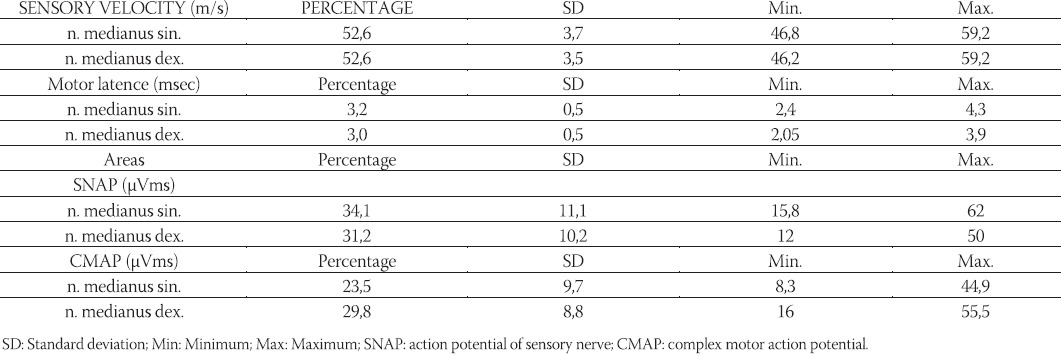
Parameters of neurographyc analyses of both sides of n. medianus are showed in Table 1.
TABLE 1.
Parameters of neurographyc analyses of a medianus of 30 healthy examinees

NEUROPHYSIOLOGICAL PARAMETERS OF N. MEDIANUS IN THIRD TRIMESTER OF PREGNANCY
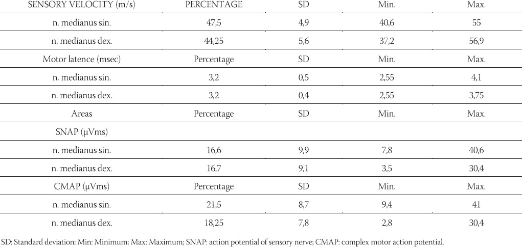
When this study was performed 3 patients were complaining about pain in the neck and 17 patients had periodical pain in lumbar spine. Parameters of neu-rographyc analyses of n. medianus in third trimester of pregnancy are showed in Table 2. Positive signes and simptoms had 12 preggies. From 12 patients with positive simptoms and signes of CTS, 9 (75%) of them had electroneurographically verified CTS. From all patients without simptoms, 3 (10,7%) of them complied criteria for CTS. Hence, 12 patients had CTS; 7 was bilateral and 5 unilateral.
TABLE 2.
Parameters of neurographyc analyses of η medianus in third trimester of pregnancy

NEUROPHYSIOLOGICAL PARAMETERS OF N. MEDIANUS IN THIRD TRIMESTER OF PREGNANCY WITH SYNDROME OF COMPRESSION OF N. MEDIANUS AT THE WRIST
Average age of preggies with verified CTS was 26,8 ± 5,5 (19 - 39) years and 5 of them were with first pregnancy. Average age of pregnancy, in weeks, was 35,3 ± 4,0 (27 - 39). Neurophysiological parameters of patients with CTS are showed in Table 3. Significant differences in parameters of preggies in third trimester by comparison with control group were in velocity of sensoric conduction of n. medianus dex., area of SNAP on both sides and CMAP area on right side (Table 4). Significant differences in parameters of preggies in third trimester with compression of n. medianus at the wrist by comparison with control group were in velocity of sensoric conduction of n. medianus dex., area of SNAP on both sides and CMAP area on right side (Table 4).
TABLE 3.
Parameters of neurographyc analyses of η medianus in third trimester of pregnancy with compression of η medianus at the wrist

TABLE 4.
Signification of differences of neurophysiological parameters in control group of preggies in third trimester and preggies with compression of η medianus at the wrist

DISCUSSION
It is visible that major number of parameters of n. medianus in third trimester f pregnancy is changed: in velocity of sensory conduction of n. medianuss dex., ara of SNAP on both sides and area of CMAP on the right side. Change of those parameters, of course, is referred to patients with CTS. In examined pattern of preggies in third trimester, 30 % of them had CTS. Prevalence of CTS in third trimester of pregnancy in one study was similar to this what we have found in our study and this prevalence was 28% (11). But, clinical finding by itself, without electroneurographical confirmation, was not enough to give final diagnosis of this sy. This sy. was proved even at examinees who had no symptoms, although in minor number (10,7%). This means that, for definitive diagnose of this disease, clinical examination is very important but it is necessary to perform definitive electroneurographical confirmation as well. In one study performed in Iran, 17 % of preggies had CTS, 23,5% of them had bilateral form and 17,5% of them had a serious form of CTS. Prevalence of clinical symptoms was 36% and prevalence of signes (Phalen’s and Tinnel’s) was 26% (12). In our study, 42% woman who were pregnant for the first time had CTS. In Seror’s analyses (1998), in third trimester of pregnancy, symptoms occurred at 12 patients, conductive block, motor or sensory was found at 20/30 patients and serious denervation occurred at 5 patients. Based on our annotation, if CTS in pregnancy is proved, it is possibly that he will be bilateral (58%) than unilateral. Additional anamnestic data showed that from 5 preggies with unilateral CTS even 3 of them had professional risk of getting this disease. According to data from the literature, % of woman are with bilateral symptoms. Half of those who were pregnant before had similar symptoms in previous pregnancies (3,14). According to a study, 50 woman of middle age 30.5 +/- 4.0 with for the first time identified new symptoms of CTS in pregnancy, 24% of them were acctually at first pregnancy. The most frequent appearance of symptoms was in in third trimester (50%) and most of them were bilateral (68% numbness, 67% pain) (13). Conductive velocity of n.medianus (both sensory velocity and motor latence) in examined group was significantly changed in the right hand, not in the left, which is explained based on physiological differences related to dominant and nondominant hand. Dominant hand, the one that was used more often then the other, also has major predisposition for appearance of CTS. It is interesting that non of those patients visited neurologist before and we also have to notice that this study was performed on random pattern of examinees who were coming for regular control examination at gynecologist. According to one study of woman with CTS symptoms in 75% of cases were with disorder of hand and rhythm of sleep and only 46% of those patients mentioned those symptoms to her doctor. Only 16% (35% of those who were complaining about symptoms) were treated and only one half of those cases got better (2). It is ironic that in period when woman needs a healthy hands so she could prepare herself for a labor and care for the baby, woman with CTS often have painful and numb fingers, primarily thumb. Also, additiona problem is the fact that because of the pain pregnant woman cannot sleep and rest properly which causes further exhaustion and violation of quality of life. Our study shows that this problem is not getting enough attention. The fact that non of pregnant patients did not seek for help because of painful and numb hands, and the fact that 3 preggies with unilateral CTS in their anamnesis had painful hands even before pregnancy is telling us that patient as well as medical provider is not informed very well. Adequate, in most cases even very simple treatment or advice (position of the fingers while sleeping, diet, orthrosis) could minimise possibility of clinical occurrence of CTS in population of preggies so that they could easily endure severity of pregnancy and labor as well as breastfeeding period. Based on this we can make conclusion that medical providers are mostly focused on adequate growth and development of the baby, while pregnant woman is placed into a second plan. Her health is actually dictating adequate care of the baby. These problems should be pointed out continuously, prevention modes should be offered and medical providers should be educated (specially those who are working on consultation of pregnant woman) as well as patients how to recognize the symptoms, if it is possible, and how to treat them which is very important for the mother as well as for her descendants.
CONCLUSION
- Significant differences in parameters of preggies in third trimester in relation to control group are in velocity of sensory conduction of n.medianus dex., area of SNAP on both sides and area of CMAP on the right side.
- It is found that 12 preggies in third trimester of pregnancy had positive symptoms and signes on carpal compression of n. medianus (CTS) and 9 (75%) of those CTS cases were electroneurographically verified. Among examinees who had no symptoms, 3 (10,7%) of them fulfilled criteria for CTS. Rate of neurophysiologicaly verified CTS in third trimester of pregnancy was 30%. In most cases (58%) CTS in period of pregnancy is bilateral.
REFERENCES
- 1.McLennan H.G, Oats J.N, Walstab J.E. Survey of hand symptoms in pregnancy. Med. J. Aust. 1987;147:542–544. doi: 10.5694/j.1326-5377.1987.tb133678.x. [DOI] [PubMed] [Google Scholar]
- 2.Voitk A.J, Mueller J.C, Farlinger D.E, Johnston R.U. Carpal tunnel syndrome in pregnancy. Can. Med. Assoc. J. 1983;128:277–281. [PMC free article] [PubMed] [Google Scholar]
- 3.Ekman-Ordeberg G, Salgeback S, Ordeberg G. Carpal tunnel syndrome in pregnancy. A prospective study. Acta Obstet. Gyne-col. Scand. 1987;66:233–235. doi: 10.3109/00016348709020753. [DOI] [PubMed] [Google Scholar]
- 4.Padua L, Aprile I, Caliandro P, Mondelli M, Pasqualetti P, Tonali P.A. Carpal tunnel syndrome in pregnancy: multiperspective follow-up of untreated cases. Neurology. 2002;26(59(10)):1643–1646. doi: 10.1212/01.wnl.0000034764.80136.ef. [DOI] [PubMed] [Google Scholar]
- 5.Snell N.J, Coysh H.L, Snell B.J. Carpal tunnel syndrome presenting in the puerperium. Practitioner. 1980;224(1340):191–193. [PubMed] [Google Scholar]
- 6.Nicholas G.G, Noone R.B, Graham W.p. Carpal tunnel syndrome in pregnancy. Hand. 1971;3:80–83. doi: 10.1016/0072-968x(71)90016-7. [DOI] [PubMed] [Google Scholar]
- 7.Durkan J.A. The carpal-compression test. An instrumented device for diagnosing carpal tunnel syndrome. Orthop Rev. 1994;23(6):522–525. [PubMed] [Google Scholar]
- 8.Sucher B.M. Palpatory diagnosis and manipulative management of carpal tunnel syndrome. J. Am. Osteopath. Assoc. 1994;94(8):647–663. [PubMed] [Google Scholar]
- 9.Stevens J.C. AAEM minimonograph #26: the electrodiagnosis of carpal tunnel syndrome. American Association of Electrodiag-nostic Medicine. Muscle Nerve. 1997;20(12):1477–1486. doi: 10.1002/(sici)1097-4598(199712)20:12<1477::aid-mus1>3.0.co;2-5. [DOI] [PubMed] [Google Scholar]
- 10.Sharma K.R, Rotta F, Romano J, Ayyar D.R. Early diagnosis of carpal tunnel syndrome: Comparison of digit 1 with wrist and distoproximal ratio. Neurology & Clinical Neurophysiology. 2006;2(9):2–10. doi: 10.1162/15268740151079491. [DOI] [PubMed] [Google Scholar]
- 11.Atisook R, Benjapibal M, Sunsaneevithayakul P, Roongpisuthip-ong A. Carpal tunnel syndrome during pregnancy: prevalence and blood level of pyridoxine. J Med. Assoc. Thai. 1995;78(8):410–414. [PubMed] [Google Scholar]
- 12.Bahrami M.H, Rayegani S.M, Fereidouni M, Baghbani M. Prevalence and severity of carpal tunnel syndrome (CTS) during pregnancy. Electromyogr. Clin. Neurophysiol. 2005;45(2):123–125. [PubMed] [Google Scholar]
- 13.Stolp-Smith K.A, Pascoe M.K, Ogburn P.L.J. Carpal tunnel syndrome in pregnancy: frequency, severity, and prognosis. Arch. Phys. Med. Rehabil. 1998;79:1285–1287. doi: 10.1016/s0003-9993(98)90276-3. [DOI] [PubMed] [Google Scholar]
- 14.Massey E.W. Carpal tunnel syndrome in pregnancy. Obstet. Gynecol. 1978;33:145–148. doi: 10.1097/00006254-197803000-00001. [DOI] [PubMed] [Google Scholar]


