Dermsocopy of Sarcoidosis: A Useful Clue to Diagnosis
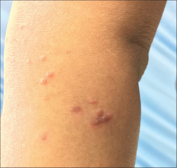
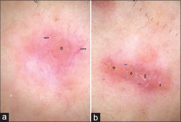
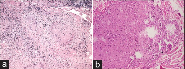
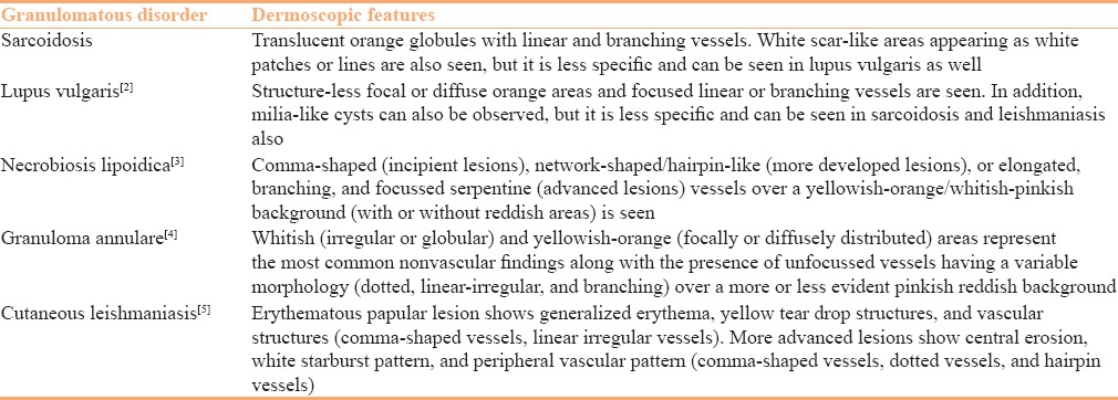
A 28-year-old male presented with a 1-year history of multiple, raised, reddish lesions over the right eye and right forearm. There was no other significant history. On examination, multiple, discrete, shiny, reddish brown papules were present over the right eye and the anteromedial aspect of the right forearm. Few lesions over the forearm were grouped together forming a small plaque [Figure 1]. Diascopy of the lesions showed apple jelly color. A clinical diagnosis of papular sarcoidosis was made. Dermoscopy (DermLite II hybrid m; 3Gen; polarized mode, ×10 magnification) revealed multiple linear and branching vessels over translucent yellowish-orange globular structures. Scar-like depigmented areas were also seen [Figure 2]. Histology was done and showed multiple well-defined granulomas in the dermis consisting of epithelioid cells, histiocytes, few multinucleated giant cells, and lymphocytes, which was consistent with sarcoidosis [Figure 3a and b]. The dermoscopy findings in our case are in line with those previously described by Pellicano et al. in their study.[1] The dermsocopic finding of various granulomatous disorders are summarized in Table 1.
Figure 1.

Multiple, discrete, reddish-brown infiltrated papules over the anteromedial aspect of the forearm
Figure 2.

(a) Multiple linear and branching vessels (seen as arrows) over translucent yellowish-orange globular structures (seen as star). (b) Multiple arborizing vessels (blue arrow) overlying translucent reddish orange background (star) with scar-like depigmented areas (black arrow) (Polarized mode, ×10)
Figure 3.

(a) Multiple well-defined granulomas seen in the dermis having epithelioid histiocytes, multinucleated giant cells, and few lymphocytes (H and E, 10×). (b) Closer view showing epithelioid cell granuloma with histiocytes, epithelioid cells, and scattered lymphocytes (H and E, 40×)
Table 1.
Dermoscopic findings of granulomatous disorders

Financial support and sponsorship
Nil.
Conflicts of interest
There are no conflicts of interest.
References
- 1.Pellicano R, Tiodorovic-Zivkovic D, Gourhant JY, Catricala C, Ferrara G, Caldarola G, et al. Dermoscopy of cutaneous sarcoidosis. Dermatology. 2010;221:51–4. doi: 10.1159/000284584. [DOI] [PubMed] [Google Scholar]
- 2.Brasiello M, Zalaudek I, Ferrara G, Gourhant JY, Capoluongo P, Roma P, et al. Lupus vulgaris: A new look at an old symptom--the lupoma observed with dermoscopy. Dermatology. 2009;218:172–4. doi: 10.1159/000182255. [DOI] [PubMed] [Google Scholar]
- 3.Errichetti E, Stinco G. Dermoscopy in General Dermatology: A Practical Overview. Dermatol Ther (Heidelb) 2016;6:471–507. doi: 10.1007/s13555-016-0141-6. [DOI] [PMC free article] [PubMed] [Google Scholar]
- 4.Errichetti E, Lallas A, Apalla Z, Di Stefani A, Stinco G. Dermoscopy of Granuloma Annulare: A Clinical and Histological Correlation Study. Dermatology. 2017 doi: 10.1159/000454857. [Epub ahead of print] [DOI] [PubMed] [Google Scholar]
- 5.Llambrich A, Zaballos P, Terrasa F, Torne I, Puig S, Malvehy J. Dermoscopy of cutaneous leishmaniasis. Br J Dermatol. 2009;160:756–61. doi: 10.1111/j.1365-2133.2008.08986.x. [DOI] [PubMed] [Google Scholar]


