ABSTRACT
Episcleritis is the inflammation of the thin, loose, highly vascular connective tissue layer that lies between the conjunctiva and sclera. Incidence is less than 1/1000. It is more common in women and those between 40 and 50 years of age. Most cases are idiopathic. It is classified into simple and nodular. Most attacks resolve within 1–3 months. The nodular type tends to be more recurrent and painful. It presents with acute onset of redness, lacrimation, and photophobia. The diagnosis of is essentially clinical, and eye pain or tenderness should raise the concern for scleritis. Ophthalmological referral is recommended to rule out scleritis. Bloodwork to diagnose associated systemic rheumatological disease may be helpful. Cold compresses and artificial tears provide symptomatic relief. Topical nonsteroidal anti-inflammatory drugs (NSAIDs) and steroids are used for persistent symptoms. Rarely, systemic steroids may be necessary. Immunosuppressive treatment to control an underlying autoimmune disorder is the last resort for resistant cases.
KEYWORDS: Episcleritis, scleritis, simple episcleritis, topical steroids, red eye
1. Introduction
Episcleritis is the inflammation of a thin, loose, highly vascular connective tissue layer that lies deep to Tenon capsule and superficial to the sclera between the conjunctive and sclera [1]. From its earliest description, confusion existed between episcleritis and scleritis [2]. Incidence is less than 1/1000. It is more common in women and those between 40 and 50 years of age [3]. Most cases are idiopathic, but it can be associated with connective tissue diseases or caused by an exogenous stimulus. It is usually a mild and self-limiting disease but can be recurrent without any complications.
2. Pathophysiology
The hypothesized pathophysiology is non-granulomatous inflammation of the superficial vascular network of the episclera that leads to vascular dilatation and perivascular infiltration [1].
Watson and Hayreh classified episcleritis into simple and nodular. Simple episcleritis is characterized by intense but non-raised engorgement of the subconjunctival vessels affecting one or more quadrants of one or both eyes. It is more common (70%) than the nodular type (30%). In contrast, in nodular episcleritis, the intense engorgement of the episcleral blood vessels surrounds a localized tender and movable swelling [2].
Most patients have intermittent bouts of moderate or severe inflammation at intervals of 1–3 months, lasting 7–10 days, and occurring much more commonly during the spring and autumn than in the summer or winter [5]. The nodular type tends to be more recurrent and painful [4].
3. Clinical presentation
As a rule, episcleritis presents with acute onset of redness, lacrimation, and photophobia. Generally, it is painless with minor eye tenderness, while scleritis is severely painful.
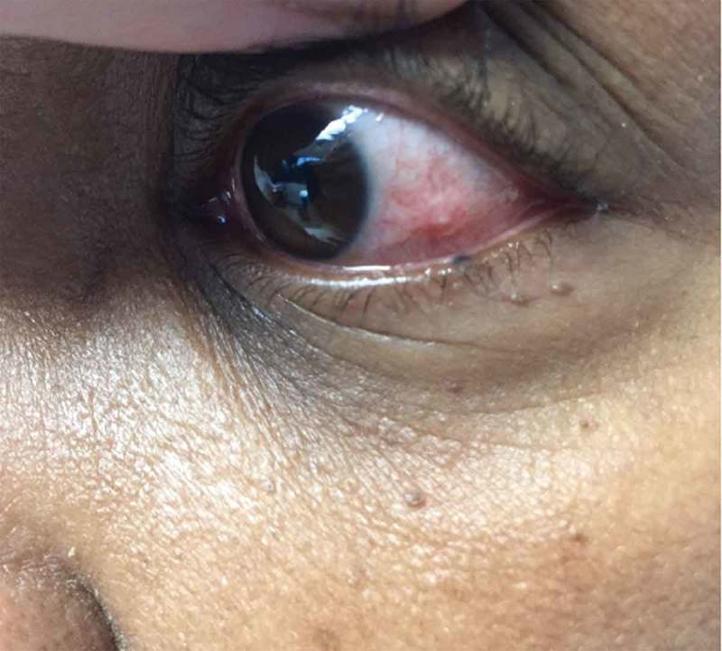
Episcleritis commonly affects a single quadrant in one eye as opposed to scleritis that may involve more than one quadrant (Figure 1).
Figure 1.

Congestion of the left lower quadrant of the left eye consistent with episcleritis.
Bilateral involvement suggests underlying systemic disease [5]. Other symptoms of underlying conditions might be helpful in the diagnosis, e.g. rheumatoid arthritis, scleroderma, systemic lupus erythematosus, and dermatomyositis.
4. Diagnosis
4.1. Clinical signs
The diagnosis of episcleritis is essentially clinically, and there are no published guidelines for diagnosis. Vision usually is unimpaired. If there is impaired vision or severe pain, other diagnoses should be considered. White sclera can be seen between superficial dilated blood vessels. Tenderness on examination usually points to scleritis rather than episcleritis.
Episcleritis usually does not give rise to scleritis. The lone exception is in ocular herpes zoster, which will occasionally present as a self-resolving episcleritis during the vesicular stage of disease and can recur at the same location as scleritis several months later [6].
4.2. Tests
Blood and radiographic tests are seldom helpful and are mainly used to diagnose associated autoimmune conditions. Conjunctival culture or corneal staining are rarely needed. The use of phenylephrine hydrochloride 2.5% drops is helpful in distinguishing episcleritis (blood vessels blanch) from scleritis (does not blanch) [1] but not available at most primary care sites. Scleral biopsy should be done by an ophthalmologist if histologic diagnosis is needed due to failure of therapy.
4.3. Differential diagnosis
Scleritis, conjunctivitis, keratitis, acute anterior uveitis, and acute angle-closure glaucoma.
5. Treatment
Episcleritis is a relatively benign, self-limited condition without long-term complications. Symptomatic relief using cold compresses should be tried, but they do not suppress any inflammation or actual tissue damage. Complete resolution is often achieved without any treatment.
If the episcleritis is secondary to connective tissue disorder, control of the underlying systemic disease is crucial for controlling the ocular complications. Additionally, there is an association between the severity of underlying disease and the severity of any ocular recurrences should they occur.
Topical NSAIDs appear to be ineffective compared to artificial tears in treating the signs or symptoms of idiopathic episcleritis [7]. Systemic NSAIDs can be tried after weighing the cost versus benefits [8]. Topical steroids are very effective treatment to minimize the inflammation. Either prednisolone acetate (0.125% and 1%) or prednisolone sodium phosphate (0.125% and 1%) can be used. Intraocular pressure should be measured every 2–4 weeks for the first 2 months of therapy. Use caution in corneal abrasions, diabetes mellitus, and open-angle glaucoma [9]. Systemic steroids are infrequently used unless needed to control the underlying systemic disease. Treatment must be initiated in consultation with the treating ophthalmologist to exclude other ophthalmological diseases.
In unclear cases, ophthalmological referral is needed to differentiate from scleritis, as this can be a sight threatening condition. There are no specific tests for episcleritis
6. Conclusion
Episcleritis is usually encountered at the primary care settings. It is a benign disease. Primary care physician should be able to reassure their patients. Symptomatic management is the rule of thumb as the disease is a self-limited disease without complications.
Disclosure statement
No potential conflict of interest was reported by the authors.
References
- [1]. Sainz De La Maza M, Molina N, Gonzalez-Gonzalez LA, et al. Clinical characteristics of a large cohort of patients with scleritis and episcleritis. Ophthalmology. 2012;119:43–50. [DOI] [PubMed] [Google Scholar]
- [2]. Watson PG, Hayreh SS.. Scleritis and episcleritis. Br J Ophthalmol. 1976;60(163):LP–191. [DOI] [PMC free article] [PubMed] [Google Scholar]
- [3]. McGavin D. D., Williamson J., Forrester J. V., et al Episcleritis and scleritis. A study of their clinical manifestations and association with rheumatoid arthritis. Br J Ophthalmol. 1976;60:192 LP–226. [DOI] [PMC free article] [PubMed] [Google Scholar]
- [4]. Bone P, Mader R. Kelley’s textbook of rheumatology. 2013. DOI: 10.1016/B978-1-4377-1738-9.00102-X. [DOI] [Google Scholar]
- [5]. Jabs DA, Mudun A, Dunn JP, et al. Episcleritis and scleritis: clinical features and treatment results. Am J Ophthalmol. 2000;130:469–476. [DOI] [PubMed] [Google Scholar]
- [6]. Tuft SJ, Watson PG. Progression of scleral disease. Ophthalmology. 1991;98:467–471. [DOI] [PubMed] [Google Scholar]
- [7]. Williams CPR, Browning AC, Sleep TJ, et al. A randomised, double-blind trial of topical ketorolac vs artificial tears for the treatment of episcleritis. Eye. 2005;19:739–742. [DOI] [PubMed] [Google Scholar]
- [8]. Berchicci L., Miserocchi E, Di Nicola M, et al. Clinical features of patients with episcleritis and scleritis in an Italian tertiary care referral center. Eur J Ophthalmol. 2014;24:293–298. [DOI] [PubMed] [Google Scholar]
- [9]. Leibowitz HM, Hyndiuk RA, Lindsey C, et al. Fluorometholone acetate: clinical evaluation in the treatment of external ocular inflammation. Ann Ophthalmol. 1985;16:1110–1115. [PubMed] [Google Scholar]


