Abstract
Despite the advances in imaging, surgery and radiotherapy, the majority of patients with brainstem gliomas die within 2 years after initial diagnosis. Factors that contribute to the dismal prognosis of these patients include the infiltrative nature and anatomic location in an eloquent area of the brain, which prevents total surgical resection and the presence of the blood-brain barrier (BBB), which reduces the distribution of systemically administered agents. The development of new therapeutic approaches which can circumvent the BBB is a potential path to improve outcomes for these children. Convection-enhanced delivery (CED) and intranasal delivery (IND) are strategies that permit direct drug delivery into the central nervous system (CNS) and are an alternative to intravenous injection (IV). We treated rats bearing human brainstem tumor xenografts with nanoliposomal irinotecan (CPT-11) using CED, IND, and IV. A single treatment of CED irinotecan had a similar effect on overall survival as multiple treatments by IV route. IND CPT-11 showed significantly increased survival of animals with brainstem tumors, and demonstrated the promise of this non-invasive approach of drug delivery bypassing the BBB when combined with nanoliposomal chemotherapy. Our results indicated that using CED and IND of nanoliposomal therapy increase likelihood of practical therapeutic approach for the treatment of brainstem gliomas.
Introduction
Among brain tumors, brainstem tumors are particularly rare, occurring in only several hundred children in the United States each year and only in 1–2% of adults. Despite improvements in survival for hematologic and other solid tumors, the survival for children with brainstem gliomas remains poor with few patients remaining alive two years after diagnosis [1]. The factors that have limited treatment improvement include an infiltrative character in a non-resectable location, an aggressive pattern of growth, and finally, the inability to achieve high tissue drug concentrations due to an intact blood brain barrier (BBB) [2, 3]. A large number of Phase I studies conducted over the past several decades have failed to demonstrate any improvement in survival, or even any response, in the majority of these patients [1, 4]. Indeed, the only modality of therapy that prolongs survival, fractionated radiotherapy, has not changed in several decades. While surgical resection is not possible, biopsies and directed therapies are being explored. In particular, there is growing interest in direct delivery of chemotherapeutic agents.
During the last ten years, there have been improvements in the technology designed to directly delivery of therapeutic agents by convection-enhanced delivery (CED) into the central nervous system (CNS) [5]. CED relies upon continuous infusion with a positive pressure gradient to drive a therapeutic agent through a target volume. CED has shown promising results in animal models and is being evaluated in human clinical trials [6–8]. As expected, there are many variables that may affect the clinical efficacy of CED including volume of distribution, reflux, drug concentration, and delivery time [9, 10].
CED is well suited for the delivery of liposomes and particulate drug carriers which have the potential to provide a sustained level of drug and to reach cellular targets with improved specificity [11, 12]. The theoretical and observed consequence of liposomal delivery is markedly reduced CNS toxicity when compared to CED with free drug. Irinotecan, in particular, shows promise for the treatment of gliomas based on both in vitro and in vivo pre-clinical results with nanoliposomal irinotecan (nal-IRI) [6, 12], as well as demonstrated clinical free activity of free irinotecan [13]. Nal-IRI is a novel highly stabilized formulation of irinotecan that allows for slow and sustained release of the encapsulated irinotecan [14], which was recently approved for the treatment of gemcitabine refractory pancreatic cancer [15], but also has significant activity in treating brain tumors if delivered effectively. The limitations of CED, however, include the need for a surgical procedure, with its accompanying risks and complications, and the exacting technical requirements.
Intranasal delivery (IND) is a promising and practical noninvasive method for delivering therapeutic agents to the brain bypassing BBB using the unique anatomic connections of the olfactory and trigeminal nerves from the nasal mucosa to the CNS [16]. Intranasally administered drugs reach the CNS and/or CSF within minutes of administration by using an extracellular route through perineural and perivascular channels, without binding to any receptor or relying upon axonal transport. There are also the advantages of avoidance of hepatic first-pass elimination, thereby reducing systemic side effects and elimination of surgical risk. In brain tumor models, many anti-cancer agents such as methotrexate [17], 5-fluorouracil [18], and raltitrexed [19], have been delivered successfully to the brain using IND.
In this study, our goal was to compare three delivery routes, intravenous injection (IV), CED, and IND, for nal-IRI in an orthotopic rodent brainstem tumor model.
Materials and Methods
Cell Culture
U-87 MG human glioblastoma cell line was obtained from the Department of Neurological Surgery Tissue Bank at the University of California, San Francisco (UCSF), and was propagated as exponentially growing monolayers in complete medium consisting of Eagle’s minimal essential medium supplemented with 10% fetal bovine serum and non-essential amino acids. A standard U87 MG cell line is maintained at the UCSF Tissue Bank to provide for consistency among institutional investigators. GS2 cell line was obtained from Manfred Westphal, Department of Neurological Surgery, University Hospital Eppendorf, Hamburg, Germany, and maintained as a neurosphere culture, as previously described [20]. DNA fingerprints were obtained to confirm the identity of the cell lines. Cells from pediatric H3K27M diffuse midline gliomas were not used due to their unavailability at the time this study was conducted.
Cell Proliferation Assay
Tumor cells were cultured in the presence of 0, 0.0001, 0.001, 0.01, 0.1, 1, 10, 100, or 1000 nM SN38 (7-Ethyl-10-hydroxycamptothecin, Sigma-Aldrich) for 4 days. Proliferation effect was assessed by counting viable cells. Trypsinized cell suspensions were stained with trypan blue, and viable cells determined by hemocytometer counting. All in vitro assays and analyses were performed with mean and standard deviation (SD) values plotted from triplicate samples.
Animals
Six-week-old male athymic rats (rnu/rnu, homozygous) were purchased from the National Cancer Institute (Frederick, MD). Rats were housed in an animal facility and were maintained in a temperature-controlled and light-controlled environment with an alternating 12-hour light/dark cycle. All protocols were approved by the UCSF Institutional Animal Care and Use Committee.
Surgical Procedure for Implantation of Tumor Cells
Before injecting tumor cells into the brainstem, rats were anesthetized by intraperitoneal injection of 75 mg/kg of ketamine and 7.5 mg/kg of xylazine. Anesthetized rats were then positioned in a stereotactic device (David Kopf Instruments, Tujunga, CA) using ear bars. A burr hole was drilled through the skull 1.0 mm behind the lambda, and 9.6 mm deep from the inner surface of the skull. 1 × 10⁵ tumor cells suspended in 1μl HBSS were injected slowly (over 1 minute) into the pontine tegmentum using a guide-screw system [20]. All procedures were carried out under sterile conditions.
In vivo BLI Monitoring
GS2 cells were transduced with a lentiviral vector containing firefly luciferase (Fluc) under the control of the spleen focus forming virus (SFFV) promoter as previously described [20]. Briefly, lentiviral vectors were generated by transfection of 293T (human embryonal kidney) cells with plasmids encoding the vesicular stomatitis virus G envelope, gag-pol, and Fluc genes [21, 22]. Cells were screened for transfection efficiency by treatment with luciferin (D-luciferin potassium salt, 150mg/kg, Gold Biotechnology, St Louis, MO) in vitro and examination by a Xenogen IVIS Lumina System (Xenogen Corp., Alameda, CA).
In vivo BLI was performed with the Xenogen IVIS Lumina System coupled LivingImage software for data acquisition (Xenogen Corp.). Rodents were anesthetized with 75mg/kg of ketamine and 7.5mg/kg of xylazine and imaged 12 min after intraperitoneal injection of luciferin. Signal intensity was quantified within a region of interest over the head that was defined by the LivingImage software. To facilitate comparison of growth rates, each rat’s luminescence readings were normalized against its own luminescence reading at the day before initiation of therapy, thereby allowing each rat to serve as its own control [20].
Liposomal Agent
Nanoliposomal irinotecan (nal-IRI) is a highly stabilized liposomal formulation containing nano-sized irinotecan crystals complexed with sucrose octasulfate in the liposome interior [14] and was generously provided by Merrimack Pharmaceuticals (Cambridge, MA). The preparation of nanoliposomal nal-IRI used in the experiments that follow had a particle size of 112.0 ±11.6 nm, as determined by dynamic light scattering, and a drug-to-phospholipid (PL) ratio of 754 ± 21 g irinotecan/mol PL. N,NV-bis-octadecyl-4,4,4V,4V-tetramethylindacarbocyanin iodide [DiIC18(3); Molecular Probes, Inc., Eugene, OR] was included in the formulation at a ratio of 0.3 mol % of the total phospholipid for fluorescent labeling. The final concentration of the drug based on irinotecan content was 50 mg irinotecan/ml.
Intra-nasal Delivery
To administer the drugs through the nasal cavity, animals were anesthetized with 2–2.5% isoflurane and placed in an anesthesia chamber. Six microliter (μl) drops of soluble form of therapeutic agents were administered with a small pipette every 2 min into alternate sides of the nasal cavity for a total of 22 min (a total volume of 66 μl) and 3.3 mg irinotecan. This method of administration results in consistent deposition in the olfactory epithelium without respiratory distress [23]. Following IND, the animals remained in a supine position for 15 min in order for absorption to occur through the nasal mucosa.
Convection-enhanced Delivery
CED was performed by micro-infusion of liposomal agents as previously described [24]. Briefly, the infusion system consisted of a fused-silica needle cannula with a 1-mm stepped design continuous with a fused silica tube (Polymicro Technologies, Phoenix, AZ) leading to a 24 gage needle that protruded from the silica guide base. A one mL syringe with silica cannula was loaded with liposomal agents and mounted onto a micro-infusion pump (BeeHive, Bioanalytical Systems, West Lafayette, IN). The syringe with silica cannula was mounted onto a stereotactic holder and guided to targeted region of the brains through a puncture hole made in the skull for tumor cells implantation. The liposomal agent was infused following ascending rates to achieve the 20 μL total infusion volume: 0.1 μL/min (5 minutes) + 0.2 μL/min (5 minutes) + 0.5 μL/min (5 minutes) + 0.8 μL/min (20 minutes). For the 50 mg/mL nal-IRI this translated to a total irinotecan dose of 1 mg per rat. The cannula was removed 2 min following completion of infusion.
Statistical Analysis
The Kaplan–Meier estimator and Prism software were used to generate and analyze survival plots. Differences between survival plots were calculated using a log-rank test. For all other comparisons, a 2-tailed unpaired t-test was used (GraphPad Software).
RESULTS
Toxicity of Liposomal Irinotecan by CED and IND
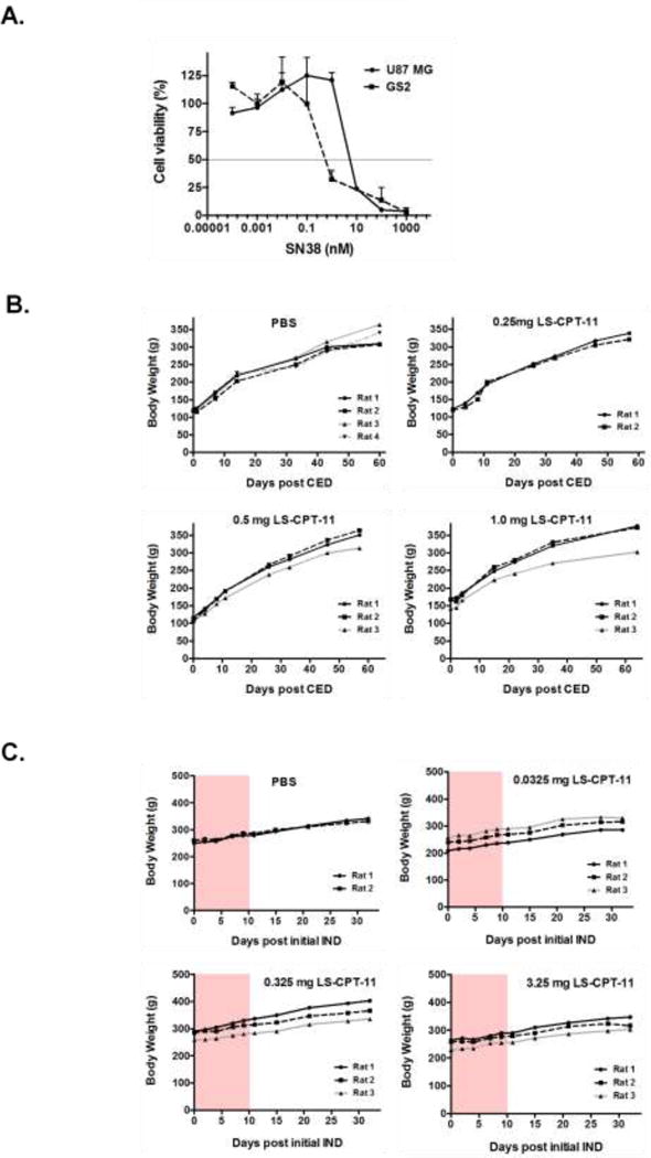
In advance of conducting experiments to evaluate toxicity of nal-IRI in vivo, we examined in vitro response to the active metabolite of irinotecan, SN38, using U-87 MG and GS2 cell lines (Figure 1A). Cell proliferation assay results showed a 50% reduction in U-87 MG and GS2 cell number at 2.42 ± 0.13 and 0.65 ± 0.15 nM of SN38. In order to determine whether the liposomal formulation of irinotecan (nal-IRI) had any overt toxicity by either CED or IND, we treated naïve rats with single CED at dose of 0.25 mg (2 rats), 0.5 mg (3 rats), and 1.0 mg of nal-IRI (3 rats), 20 μl of PBS was used in the control group (4 rats). There was no effect on body weight of the animals that received nal-IRI by CED (Figure 1B), and no animals developed symptoms attributable to either the surgical procedure or drug delivery. Similarly, animals received varying total dose of nal-IRI by IND. We administered a maximum solubility dose (MSD) of nal-IRI (3.25 mg in 65 μl PBS) in 3 rats, 0.1 of MSD (0.325 mg in 65 μl PBS) in 3 rats, and 0.01 of MSD (0.0325 mg in 65 μl PBS) in 3 rats, through the nasal cavity, daily for 10 days. 65 μl of PBS was delivered intranasally in 2 rats as control (Figure 1C; shaded area represents duration of treatment). There was no effect on body weight and no animals experienced symptoms.
Figure 1.

Effect of active metabolite of CPT-11, SN38, on human glioma cell proliferation and toxicity of nanoliposomal CPT-11 (LS-CPT11) by CED and IND in rats. A. Graph showing proliferation response of human glioma cells to increasing concentration of SN38. Values shown are the average (mean ± SD) from triplicate samples for each incubation condition. SN38 shows valuable, but consistently anti-proliferative effect at concentrations of 0.1–100 nM. B. Graphs showing direct injection of either PBS or nanoliposomal CPT-11 by CED demonstrates no obvious toxicity. A maximum volume of 20 μl of infusate volume was used. C. IND with nanoliposomal CPT-11 over a hundred-fold dose range for 10 consecutive days (pink area) did not result in any appreciable toxicity.
Distribution of Liposomes by CED and IND
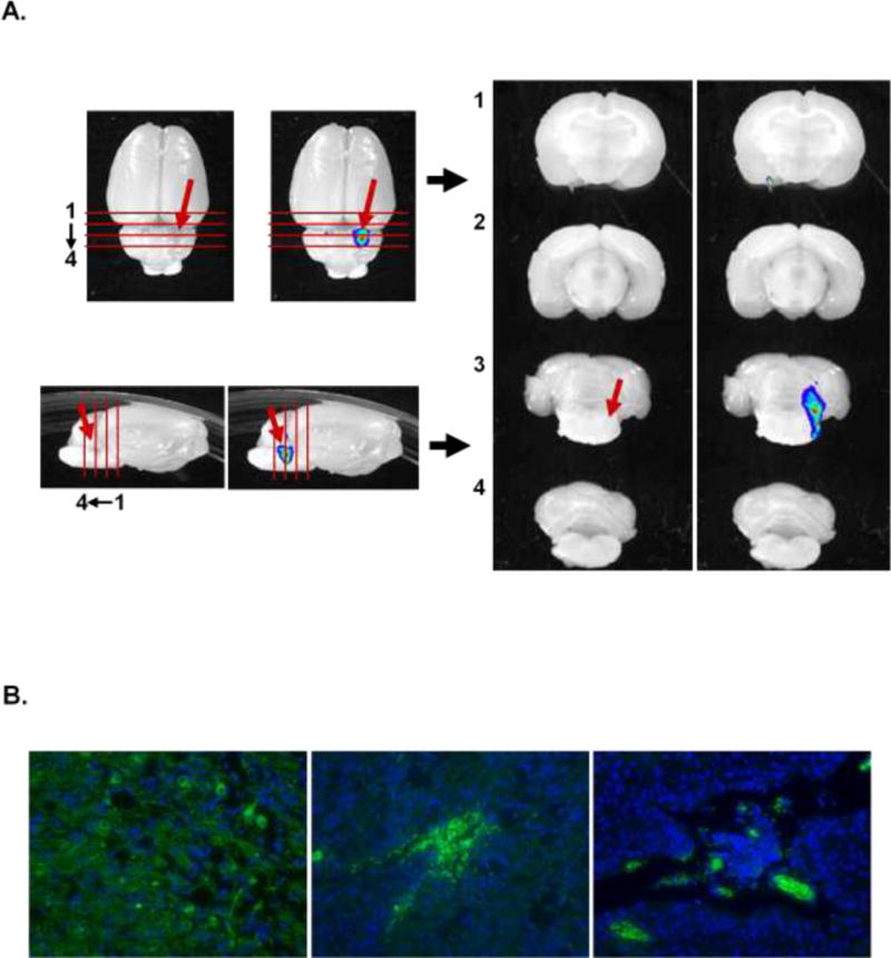
In order to qualitatively confirm the distribution of DiI labeled liposomes (Dil-LS), DiI-LS was infused into the brainstem of naïve rats by CED. Two naïve rats receiving DiI-LS were euthanized with transcardial perfusion at 3 h following CED (Figure 2). Sequential sections were taken of the brainstem and ex vivo fluorescent imaging was performed. Ex vivo fluorescent image was detected in the infusion site over the ipsilateral pons (Figure 2A). The fluorescent microscope images showed diffuse distribution in the pons that indicates parenchymal penetration of liposomes (Figure 2B).
Figure 2.

Ex vivo distribution of fluorescent liposomes using CED in the rat brainstem. A. Ex vivo image demonstrates relatively efficient distribution at the target site (narrows) and for a short distance along injection track. B. Fluorescence microscope indicates diffuse distribution of DiI liposomes in the pons. DNA staining is by DAPI.
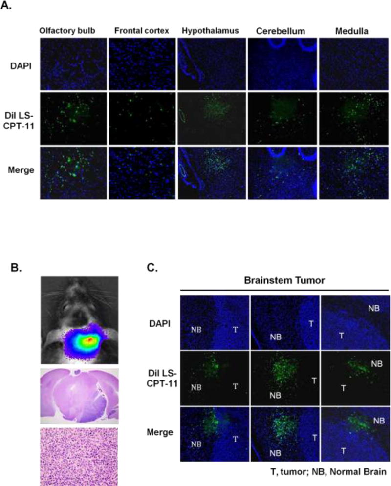
Following intranasal delivery of DiI nal-IRI, animals were euthanized and the brains isolated for subsequent analysis with fluorescent microscopy (Figure 3). There was clear evidence of fluorescent signal within the olfactory bulb, frontal cortex, hypothalamus, cerebellum and medulla (Figure 3A). In tumor-bearing animals (Figure 3B), the fluorescent signal mainly clustered at the tumor/brain interface (Figure 3C).
Figure 3.

Distribution of fluorescently labeled liposomal CPT-11 by IND in normal brain and brainstem tumor. A. Fluorescent signals were detected from olfactory bulb throughout the different brain regions. B. 1 × 10⁵ luciferase-modified GS2 glioblastoma cells were injected into brainstem in athymic rats using an implantable guide-screw system. Bioluminescence imaging (BLI) shows a corresponding signal from the brainstem tumor (upper). Histologic analysis reveals GS2 tumor growth in the pons (middle: 2 × magnification, lower: 40 × magnification). C. Fluorescent labeled liposomal CPT-11 accumulated in the brainstem 6 hours following IND.
In Vivo Efficacy of Liposomal Irinotecan by IV, CED, and IND
We then examined the in vivo efficacy of nal-IRI using three delivery routes: IV, CED, and IND. IV injections were three times of 30 mg/kg of nal-IRI (3 rats) and free irinotecan (3 rats) once a week for three weeks. The IV control group comprised of 3 rats receiving IV PBS. We administered nal-IRI by CED once at 0 mg (3 rats), 0.01 mg (4 rats), 0.1 mg (4 rats), and 1.0 mg (4 rats). Intranasal nal-IRI was administered MSD dose at 3.25 mg/day (6 rats) as well as empty LS (5 rats) for 15 days as total 48.7 mg of irinotecan.
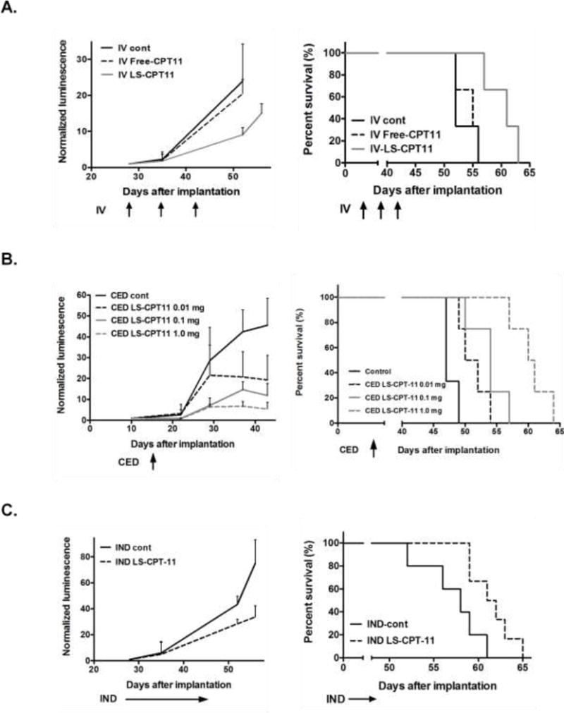
Following three IV doses of nal-IRI, there was a significant increase in survival compared to rats receiving empty liposome controls (empty-LS) - IV control (median survival of IV control 52 d vs. IV-nal-IRI 61 d, significant p = 0.02, Figure 4A). In contrast, there was no difference in survival between control and IV free irinotecan. This was consistent with previous reports utilizing this treatment approach [12].
Figure 4.

A comparison of survival between animals with brainstem tumors treated by IV, IND and CED routes. A. Rats with brainstem tumor were treated with vehicle (PBS), free-CPT11 or liposomal CPT-11 (LS-CPT11) by IV at daily dose of 30mg/kg once a week for three weeks. Bioluminescence values were normalized against bioluminescence values obtained at the beginning of therapy. Growth curve for brainstem tumor shows more growth delay of rats received IV LS-CPT11 in compared to the animals received vehicle control and free-CPT11 (left). Corresponding survival plots for each treatment (right). Statistical analysis was performed using a log-rank test (IV LS-CPT11: p = 0.0224). B. Rats were treated with LS-CPT11 by CED once at 0, 0.01, 0.1, and 1.0 mg. Growth curve for brainstem tumor shows dose dependent inhibition of the brainstem tumor growth by CED of LS-CPT11 (left). Every dose of LS-CPT11 significantly prolonged animal survival in compared to the animal treated with CED of empty LS (right, 0.01 mg: p = 0.0304, 0.1 mg: p = 0.001, 1.0 mg: p = 0.001). C. Rats received IND of LS-CPT11 at daily dose of 3.25 mg for 15 days as total dose of 48.7 mg. IND of LS-CPT11 inhibits growth of brainstem tumor (left). IND of LS-CPT11 significantly prolonged animal survival in compared to the animals received IND of empty-LS (right, p = 0.022).
We then compared the effects of local delivery with CED or IND using the same tumor model. CED of nal-IRI showed dose dependent inhibition of the growth of GS2 brainstem tumor and significant increased survival with median survival of 51 days for 0.01 mg (p = 0.03), 54 days for 0.1 mg (p = 0.01), and 60.5 days for 1.0 mg (p = 0.01) of nal-IRI when compared to CED of empty-LS with median survival of 47 days (Figure 4B). Fifteen-days treatment of IND of nal-IRI also inhibited brainstem tumor growth and increased survival with median survival of 61.5 days for nal-IRI (p = 0.02) compared to IND of empty-LS (median survival of 58 days) (Figure 4C).
DISCUSSION
The development of new strategies to deliver therapeutic agents across the BBB is a priority if the outcomes associated with malignant brain tumors are to improve. Local delivery strategies are being explored for the treatment of brainstem gliomas because of the specific therapeutic challenges regarding their site of origin, infiltrative nature, and presence of an intact BBB. Based on our previous experience with the development of brainstem tumor models [20, 25, 26, 27], we decided to evaluate the efficacy of three delivery strategies that may have clinical relevance.
CED relies on positive pressure infusion of a therapeutic agent into the brain using a MR-guided surgical approach. A previous phase III clinical trial examining the use of an IL-13 antagonist in adult patients with glioblastoma did not demonstrate efficacy but a number of technical limitations were noted [28]. In addition to CED, additional technical refinements can improve the duration of therapeutic agents in the target tissue. Liposomal carriers are used to package drugs that then allow sustained release. A highly stable nanoliposomal formulation of irinotecan (nal-IRI) has shown greater brain and tumor retention for a prolonged period than free irinotecan and effective anti-tumor activity in intracerebral glioblastoma xenografts, when administered by CED [6]. A clinical trial currently underway uses liposome encapsulated irinotecan delivered by CED for patients with recurrent GBM (NCT02022644). This study delivers a variable volume of agent to a defined tumor volume using a MR-guided approach. There is interest in using a similar strategy for children with diffuse midline gliomas with histone H3K27M mutation, especially as the same formulation of liposomal nal-IRI can be adapted for use in children. In order to examine the feasibility of using liposomal nal-IRI in the brainstem, we used an orthotopic xenograft brainstem tumor model to assess the efficacy of nal-IRI delivered by the IV route, IND route, or by CED.
Our results demonstrate that naïve non-tumor bearing animals tolerated our local delivery strategies. Increasing the total drug delivered by CED four-fold did not result in any noticeable animal toxicity. Similarly, treatment by IND over a hundred-fold dose range did not cause any toxicity. Histological examination of the nasal mucosa was not performed as prior studies did not observe any congestion, edema, epithelial sloughing, necrosis or hemorrhage of the nasal mucosa with administration of plain liposomes in human volunteers [29] or calcitonin loaded liposomes in rats [30]. In the CED, reflux of infused nal-IRI to the upper parenchymal brain was observed (Figure 2A). The difficulties associated with accurate brainstem targeting by CED may be due to the high cellular density in the pons. Because of technical limitations regarding survival surgery and repeated treatments, we used only one CED treatment in our experimental model. This single treatment had a qualitatively similar effect on improvement in survival as compared to multiple treatments through either the IV or IND route. The survival benefit was significant but modest with nal-IRI treatment using each delivery method. This could be related to limited conversion rate of irinotecan to SN38 in the brain tumor due to low activity of carboxyl esterase. In addition, the duration of treatment was short for all treatment groups. Our expectation is that greater survival benefits will be observed with repeated treatment and prolonged delivery.
The primary limitation of this model is the use of U87 MG and GS2 GBM cells which show significant difference in genetics, epigenetics, and in vivo growth in compared to pediatric diffuse midline glioma cells with histone H3K27M mutation. At the time that these experiments were planned and conducted, reliable pediatric diffuse midline glioma models were not available. Nevertheless, the use of these adult GBM cells is to compare new drug delivery strategies in appropriate anatomic xenograft models for brainstem glioma which do grow in the pons with a reasonable time period, and that the growth of these tumors can be monitored accurately with bioluminescent imaging [20]. We subsequently successfully developed cell lines derived from a pediatric diffuse midline glioma with histone H3K27M mutation [25, 26]. The delivery methods utilized in this model system can be easily adapted to examine a xenograft model derived from pediatric diffuse midline glioma cells with histone H3K27M mutation. An additional technical limitation of this study is that the therapeutic agent (nal-IRI) cannot be directly visualized using non-invasive techniques. However, our goal with this study was to obtain pre-clinical data using the same agent which is currently being evaluated in human clinical trials (NCT03086616) [31]. In existing and planned human studies, gadolinium is being used as a surrogate marker for drug distribution during CED. Finally, liposomes allow the potential for simultaneous packaging of markers such as gadolinium which can be monitored the distribution of liposomal therapeutics with CED by MRI in rodents [32], non-human primates [33, 34] and in human [31, 35].
The results demonstrate that there is pre-clinical evidence in support of the use of nal-IRI via CED for the treatment of brainstem tumors. There are clearly key differences between a rodent model and the human clinical situation. While the entire tumor volume in rodents can be likely covered with a small volume of infusate, in humans, the target volume in the pons may be as large as 20–30 cc’s. There will be clear risks associated with direct delivery to the brainstem that will need to be incorporated in the clinical trial design. There are additional variables such as total infusate volume, infusion rate, and multiple infusions that will need to be examined directly in human studies.
Acknowledgments
This work was supported by the Pediatric Brain Tumor Foundation Institute Award to the University of California San Francisco, NIH grants RO1NS093079, and by a generous gift from the Segal Family Foundation.
Footnotes
Conflict of interest: None.
References
- 1.Hargrave D, Bartels U, Bouffet E. Diffuse brainstem glioma in children: critical review of clinical trials. Lancet Oncol. 2006;7:241–248. doi: 10.1016/S1470-2045(06)70615-5. [DOI] [PubMed] [Google Scholar]
- 2.Buczkowicz P, Hoeman C, Rakopoulos P, Pajovic S, Letourneau L, Dzamba M, Morrison A, Lewis P, Bouffet E, Bartels U, Zuccaro J, Agnihotri S, Ryall S, Barszczyk M, Chornenkyy Y, Bourgey M, Bourque G, Montpetit A, Cordero F, Castelo-Branco P, Mangerel J, Tabori U, Ho KC, Huang A, Taylor KR, Mackay A, Bendel AE, Nazarian J, Fangusaro JR, Karajannis MA, Zagzag D, Foreman NK, Donson A, Hegert JV, Smith A, Chan J, Lafay-Cousin L, Dunn S, Hukin J, Dunham C, Scheinemann K, Michaud J, Zelcer S, Ramsay D, Cain J, Brennan C, Souweidane MM, Jones C, Allis CD, Brudno M, Becher O, Hawkins C. Genomic analysis of diffuse intrinsic pontine gliomas identifies three molecular subgroups and recurrent activating ACVR1 mutations. Nat Genet. 2014;46:451–456. doi: 10.1038/ng.2936. [DOI] [PMC free article] [PubMed] [Google Scholar]
- 3.Goodwin CR, Xu R, Iyer R, Sankey EW, Liu A, Abu-Bonsrah N, Sarabia-Estrada R, Frazier JL, Sciubba DM, Jallo GI. Local delivery methods of therapeutic agents in the treatment of diffuse intrinsic brainstem gliomas. Clin Neurol Neurosurg. 2016;142:120–127. doi: 10.1016/j.clineuro.2016.01.007. [DOI] [PubMed] [Google Scholar]
- 4.Frazier JL, Lee J, Thomale UW, Noggle JC, Cohen KJ, Jallo GI. Treatment of diffuse intrinsic brainstem gliomas: failed approaches and future strategies. J Neurosurg Pediatr. 2009;3:259–269. doi: 10.3171/2008.11.PEDS08281. [DOI] [PubMed] [Google Scholar]
- 5.Han SJ, Bankiewicz K, Butowski NA, Larson PS, Aghi MK. Interventional MRI-guided catheter placement and real time drug delivery to the central nervous system. Expert Rev Neurother. 2016;16:635–639. doi: 10.1080/14737175.14732016.11175939. [DOI] [PMC free article] [PubMed] [Google Scholar]
- 6.Chen PY, Ozawa T, Drummond DC, Kalra A, Fitzgerald JB, Kirpotin DB, Wei KC, Butowski N, Prados MD, Berger MS, Forsayeth JR, Bankiewicz K, James CD. Comparing routes of delivery for nanoliposomal irinotecan shows superior anti-tumor activity of local administration in treating intracranial glioblastoma xenografts. Neuro Oncol. 2013;15:189–197. doi: 10.1093/neuonc/nos305. [DOI] [PMC free article] [PubMed] [Google Scholar]
- 7.Kawakami K, Kawakami M, Kioi M, Husain Sr, Puri RK, Puri RK. Distribution kinetics of targeted cytotoxin in glioma by bolus or convection-enhanced delivery in a murine model. J Neurosurg. 2004;101:1004–1011. doi: 10.3171/jns.2004.101.6.1004. [DOI] [PubMed] [Google Scholar]
- 8.Saito R, Krauze MT, Noble CO, Drummond DC, Kirpotin DB, Berger MS, Park JW, Bankiewicz KS. Convection-enhanced delivery of Ls-TPT enables an effective, continuous, low-dose chemotherapy against malignant glioma xenograft model. Neuro Oncol. 2006;8:205–214. doi: 10.1215/15228517-2006-001. [DOI] [PMC free article] [PubMed] [Google Scholar]
- 9.Fiandaca MS, Forsayeth JR, Dickinson PJ, Bankiewicz KS. Image-guided convection-enhanced delivery platform in the treatment of neurological diseases. Neurotherapeutics : the journal of the American Society for Experimental NeuroTherapeutics. 2008;5:123–127. doi: 10.1016/j.nurt.2007.10.064. [DOI] [PMC free article] [PubMed] [Google Scholar]
- 10.Jahangiri A, Chin AT, Flanigan PM, Chen R, Bankiewicz K, Aghi MK. Convection-enhanced delivery in glioblastoma: a review of preclinical and clinical studies. J Neurosurg. 2017;126:191–200. doi: 10.3171/2016.1.jns151591. [DOI] [PMC free article] [PubMed] [Google Scholar]
- 11.Noble CO, Krauze MT, Drummond DC, Forsayeth J, Hayes ME, Beyer J, Hadaczek P, Berger MS, Kirpotin DB, Bankiewicz KS, Park JW. Pharmacokinetics, tumor accumulation and antitumor activity of nanoliposomal irinotecan following systemic treatment of intracranial tumors. Nanomedicine (Lond) 2014;9:2099–2108. doi: 10.2217/nnm.13.201. [DOI] [PubMed] [Google Scholar]
- 12.Noble CO, Krauze MT, Drummond DC, Yamashita Y, Saito R, Berger MS, Kirpotin DB, Bankiewicz KS, Park JW. Novel nanoliposomal CPT-11 infused by convection-enhanced delivery in intracranial tumors: pharmacology and efficacy. Cancer Res. 2006;66:2801–2806. doi: 10.1158/0008-5472.can-05-3535. [DOI] [PubMed] [Google Scholar]
- 13.Vredenburgh JJ, Desjardins A, Reardon DA, Friedman HS. Experience with irinotecan for the treatment of malignant glioma. Neuro Oncol. 2009;11:80–91. doi: 10.1215/15228517-2008-075. [DOI] [PMC free article] [PubMed] [Google Scholar]
- 14.Drummond DC, Noble CO, Guo Z, Hong K, Park JW, Kirpotin DB. Development of a highly active nanoliposomal irinotecan using a novel intraliposomal stabilization strategy. Cancer Res. 2006;66:3271–3277. doi: 10.1158/0008-5472.can-05-4007. [DOI] [PubMed] [Google Scholar]
- 15.Wang-Gillam A, Li CP, Bodoky G, Dean A, Shan YS, Jameson G, Macarulla T, Lee KH, Cunningham D, Blanc JF, Hubner RA, Chiu CF, Schwartsmann G, Siveke JT, Braiteh F, Moyo V, Belanger B, Dhindsa N, Bayever E, Von Hoff DD, Chen LT. Nanoliposomal irinotecan with fluorouracil and folinic acid in metastatic pancreatic cancer after previous gemcitabine-based therapy (NAPOLI-1): a global, randomised, open-label, phase 3 trial. Lancet. 2016;387:545–557. doi: 10.1016/s0140-6736(15)00986-1. [DOI] [PubMed] [Google Scholar]
- 16.Dhuria SV, Hanson LR, Frey WH., 2nd Intranasal delivery to the central nervous system: mechanisms and experimental considerations. J Pharm Sci. 2010;99:1654–1673. doi: 10.1002/jps.21924. [DOI] [PubMed] [Google Scholar]
- 17.Shingaki T, Inoue D, Furubayashi T, Sakane T, Katsumi H, Yamamoto A, Yamashita S. Transnasal delivery of methotrexate to brain tumors in rats: a new strategy for brain tumor chemotherapy. Mol Pharm. 2010;7:1561–1568. doi: 10.1021/mp900275s. [DOI] [PubMed] [Google Scholar]
- 18.Sakane T, Yamashita S, Yata N, Sezaki H. Transnasal delivery of 5-fluorouracil to the brain in the rat. J Drug Target. 1999;7:233–240. doi: 10.3109/10611869909085506. [DOI] [PubMed] [Google Scholar]
- 19.Wang D, Gao Y, Yun L. Study on brain targeting of raltitrexed following intranasal administration in rats. Cancer Chemother Pharmacol. 2006;57:97–104. doi: 10.1007/s00280-005-0018-3. [DOI] [PubMed] [Google Scholar]
- 20.Hashizume R, Ozawa T, Dinca EB, Banerjee A, Prados MD, James CD, Gupta N. A human brainstem glioma xenograft model enabled for bioluminescence imaging. J Neurooncol. 2010;96:151–159. doi: 10.1007/s11060-009-9954-9. [DOI] [PMC free article] [PubMed] [Google Scholar]
- 21.Dinca EB, Sarkaria JN, Schroeder MA, Carlson BL, Voicu R, Gupta N, Berger MS, James CD. Bioluminescence monitoring of intracranial glioblastoma xenograft: response to primary and salvage temozolomide therapy. J Neurosurg. 2007;107:610–616. doi: 10.3171/JNS-07/09/0610. [DOI] [PubMed] [Google Scholar]
- 22.Sarkaria JN, Yang L, Grogan PT, Kitange GJ, Carlson BL, Schroeder MA, Galanis E, Giannini C, Wu W, Dinca EB, James CD. Identification of molecular characteristics correlated with glioblastoma sensitivity to EGFR kinase inhibition through use of an intracranial xenograft test panel. Mol Cancer Ther. 2007;6:1167–1174. doi: 10.1158/1535-7163.mct-06-0691. [DOI] [PubMed] [Google Scholar]
- 23.Hashizume R, Ozawa T, Gryaznov SM, Bollen AW, Lamborn KR, Frey WH, 2nd, Deen DF. New therapeutic approach for brain tumors: Intranasal delivery of telomerase inhibitor GRN163. Neuro Oncol. 2008;10:112–120. doi: 10.1215/15228517-2007-052. doi: 15228517-2007-052 [pii] 10.1215/15228517-2007-052. [DOI] [PMC free article] [PubMed] [Google Scholar]
- 24.Serwer L, Hashizume R, Ozawa T, James CD. Systemic and local drug delivery for treating diseases of the central nervous system in rodent models. J Vis Exp. 2010 doi: 10.3791/1992. [DOI] [PMC free article] [PubMed] [Google Scholar]
- 25.Hashizume R, Smirnov I, Liu S, et al. Characterization of a diffuse intrinsic pontine cell line: implications for future investigations and treatment. J Neurooncol. 2012;110:305–313. doi: 10.1007/s11060-012-0973-6. [DOI] [PubMed] [Google Scholar]
- 26.Aoki Y, Hashizume R, Ozawa T, Banerjee A, Prados M, James CD, Gupta N. An experimental xenograft mouse model of diffuse pontine glioma designed for therapeutic testing. J Neurooncol. 2012;108:29–35. doi: 10.1007/s11060-011-0796-x. [DOI] [PMC free article] [PubMed] [Google Scholar]
- 27.Hashizume R, Andor N, Ihara Y, Lerner R, Gan H, Chen X, Fang D, Huang X, Tom MW, Ngo V, Solomon D, Mueller S, Paris PL, Zhang Z, Petritsch C, Gupta N, Waldman TA, James CD. Pharmacologic inhibition of histone demethylation as a therapy for pediatric brainstem glioma. Nat Med. 2014;20:1394–1396. doi: 10.1038/nm.3716. [DOI] [PMC free article] [PubMed] [Google Scholar]
- 28.Kunwar S, Chang S, Westphal M, Vogelbaum M, Sampson J, Barnett G, Shaffrey M, Ram Z, Piepmeier J, Prados M, Croteau D, Pedain C, Leland P, Husain SR, Joshi BH, Puri RK. Phase III randomized trial of CED of IL13-PE38QQR vs Gliadel wafers for recurrent glioblastoma. Neuro Oncol. 2010;12:871–881. doi: 10.1093/neuonc/nop054. [DOI] [PMC free article] [PubMed] [Google Scholar]
- 29.Tafaghodi M, Jaafari MR, Tabassi SAJ. Nasal immunization studies using liposomes loaded with tetanus toxoid and CpG-ODN. Eur J Pharm Biopharm. 2006;64:138–145. doi: 10.1016/j.ejpb.2006.05.005. [DOI] [PubMed] [Google Scholar]
- 30.Chen M, Li XR, Zhou YX, Yang KW, Chen XW, Deng Q, Liu Y, Ren LJ. Improved absorption of salmon calcitonin by ultraflexible liposomes through intranasal delivery. Peptides. 2009;30:1288–1295. doi: 10.1016/j.peptides.2009.03.018. [DOI] [PubMed] [Google Scholar]
- 31.University of California in San Francisco. CED With Irinotecan Liposome Injection Using Real Time Imaging in Children With DIPG. Available from https://clinicaltrials.gov/ct2/show/NCT03086616. Identifier: NCT03086616 Accessed June 14, 2017.
- 32.Saito R, Bringas J, McKnight TR, Wendland MF, Manot C, Drummond DC, Kirpotin DB, Park JW, Berger MS, Bankiewiez KS. Distribution of liposomes into brain and rat brain tumor models by convection-enhanced delivery monitored with magnetic resonance imaging. Cancer Res. 2004;164:2573–2579. doi: 10.1158/0008-5472. [DOI] [PubMed] [Google Scholar]
- 33.Krauze MT, McKnight TR, Yamashita Y, Bringas J, Noble CO, Saito R, Geletneky K, Forsayeth J, Berger MS, Jackson P, Park JW, Bankiewicz KS. Real time visualization and characterization of liposomal delivery into the monkey brain by magnetic resonance imaging. Brain Res Brain Res Protoc. 2005;16:20–26. doi: 10.1016/j.brainresprot.2005.08.003. [DOI] [PubMed] [Google Scholar]
- 34.Saito R, Krauze MT, Bringas JR, Noble C, McKnight TR, Jackson P, Wendland MF, Mamot C, Drummond DC, Kirpotin DB, Hong K, Berger MS, Park JW, Bankiewicz KS. Gadolinium-loaded liposomes allow for real-time magnetic resonance imaging of convection-enhanced delivery in the primate brain. Exp Neurol. 2005;196:381–389. doi: 10.1016/j.expneurol.2005.08.016. [DOI] [PubMed] [Google Scholar]
- 35.Butowski N, Bankiewicz K, Kells A, et al. A phase I study of convection-enhanced delivery of liposomal-irinotecan using real-time imaging with gadolinium in patients with recurrent high grade glioma. Neuro-Oncology. 2014 doi: 10.1093/neuonc/nou206.46. [DOI] [Google Scholar]


