



Abstract
Genetic leukoencephalopathies represent an expanding group of inherited disorders associated with involvement of brain white matter. Cystic degeneration has been previously described with some acquired or inherited leukoencephalopathies. We describe a 6-month-old Brazilian boy with a 2-month history of severe and rapidly progressive developmental and psychomotor regression and seizures. Neurological examination showed spastic tetraparesis and lethargy. Neuroimaging showed diffuse and symmetric cavitating cystic leukoencephalopathy. Whole-exome sequencing revealed compound heterozygous mutations in the NFU1 gene, providing definite genetic diagnosis of multiple mitochondrial dysfunction syndrome type 1. We report a rare presentation of early-onset cystic leukoencephalopathy in the context of multiple mitochondrial dysfunction syndrome type 1.
Keywords: leukodystrophy mitochondrial disease, leukoencephalopathy, neuroimaging
Introduction
Genetic leukoencephalopathies represent a wide and heterogeneous group of inherited neurodegenerative and neurometabolic disorders, mainly presenting in early infancy or childhood with complex neurological pictures, including refractory seizures, spastic paraparesis, neurological regression of motor and cognitive milestones, early-onset dementia, cerebellar ataxia, and other movement disorders. 1 2 Neuroimaging findings are dominated by specific patterns for each neurogenetic disorder (including hereditary cerebral vasculopathies), depending also on the stage of disease involvement and the association with other neuroimaging features, such as basal ganglia signal change and the presence of cysts and cavities. 1 2 Cystic white matter degeneration is a common finding in an expanding group of leukoencephalopathies, highlighting the rare group of cavitating leukodystrophies. Herein, we describe a Brazilian patient with severe neurological encephalopathy and cavitating leukoencephalopathy related to NFU1 gene mutations, disclosing the diagnosis of multiple mitochondrial dysfunctions syndrome (MMDS) type 1 (OMIM no. 605711). 3 4
Materials and Methods
A 6-month-old Brazilian boy presented with a 2-month history of severe and rapidly progressive developmental and psychomotor regression and seizures. His previous motor milestones were unremarkable until 3 months of age despite the mild hypotonia observed since birth. Medical history disclosed unremarkable pregnancy, birth and neonatal periods without events, suggestive of possible hypoxic–ischemic encephalopathy. Family history was also unremarkable. Neurological examination showed spastic tetraparesis, brisk tendon reflexes, and altered level of consciousness with lethargy.
Results
Cranial computed tomography (CT) disclosed mild diffuse white matter changes with cystic changes. Brain MR images showed diffuse and nearly symmetric cavitating cystic leukoencephalopathy involving periventricular, profound, and subcortical areas ( Fig. 1 ). Serum evaluation with serological tests for human immunodeficiency virus (HIV), cytomegalovirus (CMV), Epstein–Barr virus (EBV), herpes simplex virus (HSV)-I/II, Varicella Zoster virus, Brucella , Treponema pallidum , and Borrelia burgdorferi were normal. General metabolic profile disclosed moderate plasma lactate elevation (3,200 mmol/L; normal value reference: 550–2,000 mmol/L), and abnormal urinary excretion of lactic and glutaric acids. Cerebrospinal fluid analysis (including polymerase chain reaction for CMV, EBV, and HSV-I/II virus and venereal disease research laboratory [VDRL]) were unremarkable. Electrocardiogram and transthoracic echocardiogram revealed no changes.
Fig. 1.
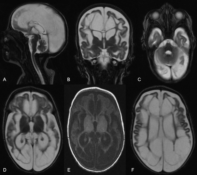
Neuroimaging findings in MMDS type 1 ( NFU1 -related disorder). ( A ) Sagittal and ( B ) coronal brain MR images disclosing corpus callosum dysgenesis, pontocerebellar atrophy, and marked diffuse cavitating lesions involving mainly the periventricular and profound supratentorial white matter in T2-weighted images. Axial brain MR images disclosing multiple cystic and cavitating lesions of the supratentorial white matter with relative sparing of the basal ganglia and anterior temporal lobe in T2 ( C,D,F ) and T1-weighted sequences ( E ). MR, magnetic resonance; MMDS, multiple mitochondrial dysfunction syndrome.
As a possible mitochondrial encephalomyopathy was initially suspected, Deltoid muscle biopsy was performed and disclosed only mild subsarcolemmal mitochondrial proliferation without typical ragged red fibers on modified Gömöri's trichrome stain, but with 10% of cytochrome oxidase (COX)-negative fibers. No specific analysis of activities of mitochondrial respiratory chain complexes was performed. Additionally, nerve conduction studies and needle electromyography were not performed. Whole-exome sequencing (WES) analysis revealed compound heterozygous mutations c.622G > T (pGly208Cys) and c.545G > A (p.Arg182Gln) in the NFU1 gene, both previously reported as pathogenic variants, 4 5 providing definite genetic diagnosis of MMDS type 1. After definite clinical and genetic diagnosis and introduction of L-carnitine (50 mg/kg per day, twice daily), coenzyme Q10 (ubiquinol, 5 mg/kg per day, twice daily), and riboflavin (100 mg per day), the patient remained clinically refractory with progressive neurological deterioration. After hospital admission for possible fever of undetermined origin, the patient required permanent use of nasogastric feeding tube, evolved with marked weight loss, and presented with refractory convulsive status epilepticus. The patient died at 8 months from aspiration pneumonia. Autopsy and postmortem studies were not performed in agreement with the parents.
Discussion
Cavitating genetic leukoencephalopathies represent a complex and heterogeneous group of inherited leukodystrophies with abnormal white matter areas, which develop cavitating lesions, such as those observed in IBA57 -related disorders (MMDS type 3), A8344G mutation in the MT-TK gene, mitochondrial complex I deficiency ( NDUFS1 gene mutation), mitochondrial complex III deficiency ( LYRM7 gene mutations), mitochondrial complex IV deficiency ( APOPT1 gene mutations), autosomal recessive progressive cavitating leukoencephalopathy, and MMDS type 1 ( NFU1 gene mutations). 1 2 6 As a rule, congenital cavitating lesions must be differentiated from congenital CMV infection of the central nervous system and other causes of cystic degeneration of the white matter also found in Alexander Disease, megalencephalic leukoencephalopathy with subcortical cysts ( MLC1 and HEPACAM gene mutations), leukoencephalopathy with anterior temporal cysts without megalencephaly ( RNASET2 gene mutations), incontinentia pigmenti, leukoencephalopathy with cerebral calcifications and cysts (Labrune syndrome), eIF2B-related disorders (leukoencephalopathy with vanishing white matter), combined oxidative phosphorylation deficiency type 11 ( RMND1 gene mutations), COL4A1- associated leukoencephalopathy, progressive vacuolating glycine leukoencephalopathy, L-2-hydroxygluatric aciduria, acute disseminated encephalomyelitis (ADEM), autosomal dominant acute necrotizing encephalopathy, hemophagocytic lymphohistiocytosis, and rarely in Canavan's disease, Wilson's disease, Aicardi–Goutières syndrome, and merosin-deficient congenital muscular dystrophy. 1 2 6 In most cases, clinical, laboratory, neurophysiological, and even other neuroimaging findings can provide differential diagnosis among each of the referred conditions.
NFU1 -related disorders represent an autosomal recessive expanding group of neurological syndromes, commonly referred to as MMDS type 1 (OMIM #605711), resulting from homozygous or compound heterozygous mutations in the NFU1 gene (2p13.3), encoding an iron-sulfur cluster protein involved in normal biosynthesis of lipoic acid and indirectly involved with the stability of mitochondrial respiratory chain complexes I and II. 3 7 8 Most cases generally have a neonatal onset of muscle weakness, early neuropsychomotor development delay, epileptic encephalopathy with severe compromise during intercurrent illnesses, and neuroimaging studies disclosing severe cystic and cavitating leukoencephalopathy resulting from white matter necrosis and spongiform neurodegeneration. Systemic features include failure to thrive, pulmonary hypertension, obstructive vasculopathy, and very early-onset acute respiratory failure. Metabolic evaluation commonly shows in most cases high plasma lactate levels, high urinary 2-hydroxybutyrate, and variable increases in glutaric acid and glycine in serum or cerebrospinal fluids. The absence of these metabolic findings does not rule out such diagnosis in a proper clinical suspicion context, but commonly represents important neurometabolic clues in differential diagnosis of patients with mitochondrial respiratory chain defects or very early-onset leukoencephalopathy. No specific curative therapeutic approaches are available, and treatment approaches are mainly based on symptomatic measures. 3 7 9
Conclusion
Clinicians must be aware about the wide group of inherited and acquired disorders, which can present with classical neuroimaging findings of cavitating leukoencephalopathies and MMDS, including NFU1 gene mutations. These must be included as a major differential diagnosis during evaluation and proper early introduction of specific therapeutic measures.
Funding Statement
Funding None.
Conflict of Interest None.
Note
Full consent was obtained from the patient's parents for the case report. This study was approved by the authors' ethics institution.
References
- 1.Schiffmann R, van der Knaap M S. Invited article: an MRI-based approach to the diagnosis of white matter disorders. Neurology. 2009;72(08):750–759. doi: 10.1212/01.wnl.0000343049.00540.c8. [DOI] [PMC free article] [PubMed] [Google Scholar]
- 2.Vanderver A, Prust M, Tonduti D et al. Case definition and classification of leukodystrophies and leukoencephalopathies. Mol Genet Metab. 2015;114(04):494–500. doi: 10.1016/j.ymgme.2015.01.006. [DOI] [PMC free article] [PubMed] [Google Scholar]
- 3.Invernizzi F, Ardissone A, Lamantea E et al. Cavitating leukoencephalopathy with multiple mitochondrial dysfunction syndrome and NFU1 mutations. Front Genet. 2014;5:412. doi: 10.3389/fgene.2014.00412. [DOI] [PMC free article] [PubMed] [Google Scholar]
- 4.Cameron J M, Janer A, Levandovskiy V et al. Mutations in iron-sulfur cluster scaffold genes NFU1 and BOLA3 cause a fatal deficiency of multiple respiratory chain and 2-oxoacid dehydrogenase enzymes. Am J Hum Genet. 2011;89(04):486–495. doi: 10.1016/j.ajhg.2011.08.011. [DOI] [PMC free article] [PubMed] [Google Scholar]
- 5.Navarro-Sastre A, Tort F, Stehling O et al. A fatal mitochondrial disease is associated with defective NFU1 function in the maturation of a subset of mitochondrial Fe-S proteins. Am J Hum Genet. 2011;89(05):656–667. doi: 10.1016/j.ajhg.2011.10.005. [DOI] [PMC free article] [PubMed] [Google Scholar]
- 6.Kohlschütter A, Eichler F. Childhood leukodystrophies: a clinical perspective. Expert Rev Neurother. 2011;11(10):1485–1496. doi: 10.1586/ern.11.135. [DOI] [PubMed] [Google Scholar]
- 7.Kurt Y G, Kurt B, Aydin I, Agilli M, Aydin F N. NFU1 gene mutation and mitochondrial disorders. Neurol India. 2016;64(04):630–632. doi: 10.4103/0028-3886.185402. [DOI] [PubMed] [Google Scholar]
- 8.Ferrer-Cortès X, Font A, Bujan N et al. Protein expression profiles in patients carrying NFU1 mutations. Contribution to the pathophysiology of the disease. J Inherit Metab Dis. 2013;36(05):841–847. doi: 10.1007/s10545-012-9565-z. [DOI] [PubMed] [Google Scholar]
- 9.Ahting U, Mayr J A, Vanlander A V et al. Clinical, biochemical, and genetic spectrum of seven patients with NFU1 deficiency. Front Genet. 2015;6:123. doi: 10.3389/fgene.2015.00123. [DOI] [PMC free article] [PubMed] [Google Scholar]