

Abstract
Background:
We intend to study the inhibitory effect of sulfur compound in Ramsar hot spring mineral on tumor-genesis ability of high natural background radiation.
Objective:
The radioprotective effect of sulfur compounds was previously shown on radiation-induced chromosomal aberration, micronuclei in mouse bone marrow cells and human peripheral lymphocyte. Ramsar is known for having the highest level of natural background radiation on Earth. This study was performed to show the radioprotective effect of sulfur-containing Ramsar mineral water on mouse bone marrow cells.
Method:
Mice were fed three types of water (drinking water, Ramsar radioactive water containing sulfur and Ramsar radioactive water whose sulfur was removed). Ten days after feeding, mice were irradiated by gamma rays (0, 2 and 4 Gy). 48 and 72 hours after irradiating, mice were killed and femurs were removed. Frequency of micronuclei was determined in bone marrow erythrocytes.
Results:
A significant reduction was shown in the rate of micronuclei polychromatic erythrocyte in sulfur-containing hot spring water compared to sulfur-free water in hot spring mineral water. Gamma irradiation induced significant increases in micronuclei polychromatic erythrocyte (MNPCE) and decreases in polychromatic erythrocyte/polychromatic erythrocyte + normochromatic erythrocyte ratio (PCEs/PCEs+NCEs) (P < 0.001) in sulfur-containing hot spring water compared to sulfur-free hot spring mineral water. Also, apparently there was a significant difference between drinking water and sulfur-containing hot spring water in micronuclei polychromatic erythrocyte and polychromatic erythrocyte/polychromatic erythrocyte+ normochromatic erythrocyte ratio.
Conclusion:
The results indicate that sulfur-containing mineral water could result in a significant reduction in radiation-induced micronuclei representing the radioprotective effect of sulfur compounds.
Keywords: High Natural Background Radiation (HNBR) , Sulfur , Gamma Rays , Bone Marrow Micronucleus Assay , Ramsar
Introduction
Ramsar is one of the most beautiful coastal cities in the North of Iran, which has great tourist attractions. It has been subject for high natural background radiation for many years [1]. This city has 9 main and over 50 small hot mineral spring water with different concentrations of Radium. High Natural Background Radiation (HNBR) in the “hot” areas of Ramsar is primarily due to the presence of very high amounts of 226Ra and its decay products, which were brought to the earth’s surface by hot springs [2,3]. Groundwater is heated by subsurface geologic activity and passes through relatively young and uraniferous igneous rock [4,5]. Radium is dissolved from the rocks by hot ground water. Uranium is not dissolved because the groundwater is anoxic and uranium is insoluble in anoxic waters [6,7]. A secondary cause of high local radiation levels is travertine deposits with a high thorium concentration [8]. The amount of radiation in Ramsar is considered far more than the maximum dose to radiographers (260 mSy y-1 compared to 20 mSy y-1), while the normal annual dose for people is less than 1 mSy y-1 [9,10]. Data on the effects of irradiation on blood component, as a result of exposure to low doses of ionizing radiation, is rare but the results of these studies describe no significant changes in hematologic parameters in HNBR residents [4,11]. The effects of high dose radiation on human health is known but the data on low and very low doses is controversial and contradictory which seems not harmful for the inhabitants [12-14]. Dose comparison in different regions of the world shows that radiation dose rate in Ramsar is about 90 times more than people from the contaminated areas of Chernobyl nuclear power. In general, data show that there is no significant increase in the number of death and abortion in high natural background radiation [15]. Researches did not find any positive correlation between indoor Radon levels and lung cancer rate in the inhabitants who lived in the dwellings with high levels of Radon for many generations in Ramsar [2,5]. Since the effect of sulfur as a radioprotector has been widely investigated [16-18], we conducted this study to assess the inhibitory effect of sulfur compound in Ramsar hot spring mineral on radiation-induced micronuclei.
Material and Methods
The in-vivo micronucleus test was performed as followed on the healthy young NMRI mice (7-12 weeks old, body weight 25-30 g, Pasture Institute, Karaj, Iran), which were randomly assigned to four groups (control and treatment groups: drinking water (DW); Ramsar hot spring water (RHSW) and the last group used Ramsar hot spring water whose sulfur was removed (RHSW-S).
In 2013, mice were caged in a group of five. Animal care and handling were performed according to guidelines of World Health Organization (Geneva, Switzerland). This study was approved by the animal ethical committee of Babol Medical University.
48 and 72 hours after exposing to Gamma irradiation (mice were whole body irradiated with 0, 2 and 4 Gy Gamma-rays generated from a cobalt-60 source (Teratron 780, Canada) at a dose rate of 1.02 Gy/min, with SSD = 80cm, field size: 20×20 in Shohada-e-Tajrish Hospital, Tehran, Iran), animals were killed by cervical dislocation and the femurs were excised. The bone marrow from both femurs was flushed into a tube using 2ml fetal bovine serum and centrifuged for 7min at 4°C. Finally, slides were prepared and stained.
Counting and Statistic Evaluation
For MNT cell and micronuclei counting, the slides prepared above were for blind analysis. A Nikon microscope with ×100 objective lens was used for scoring the cells. At least 1000 polychromatic erythrocytes per animal were scored for the presence of micronuclei (Figures 1 and 2). At the same time, the ratio of polychromatic to norm chromatic erythrocyte was determined by counting a total of 1000 erythrocytes (PCEs/PCEs+NCEs). PCEs and NCEs containing micronuclei were also counted and recorded. In order to study cytotoxic effect of gamma rays on the proliferation of bone marrow cells, the ratio of PCEs/PCEs+NCEs was calculated. In 2013, the data were statistically evaluated by the one-way ANOVA and Tukey’s test using SPSS18.
Figure1.
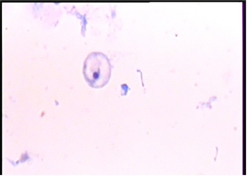
Microscopic Examination of MNPCE by Magnification of 1,000
Figure2.
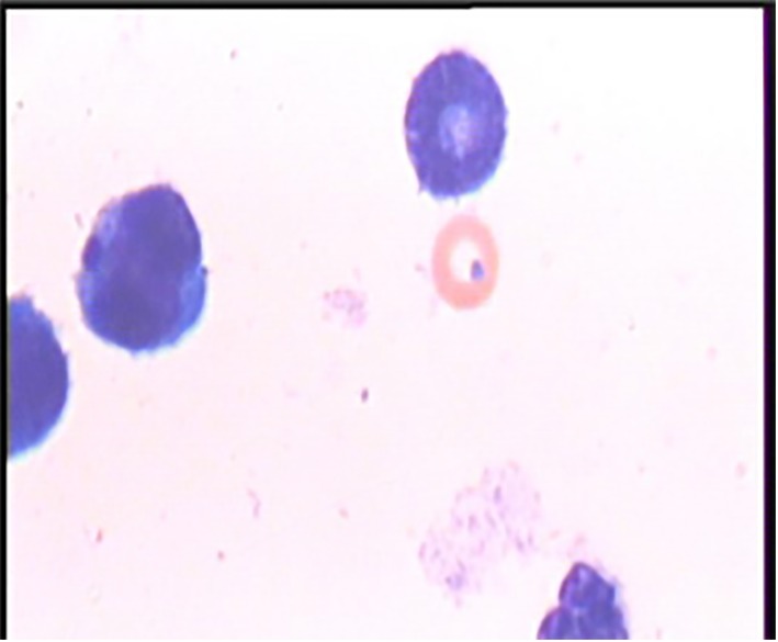
Microscopic Examination of MNNCE by Magnification of 1,000
Results
The frequency of MNPCE, MNNCE and PCE/PCE+NCE in control and irradiated groups (0, 2 and 4Gy, 48 and 72h) were shown in Figures 3 and 4 and Table 1. The results of statistical analyses showed no significant difference in MNNCE (p=1.0) and MNPCE (p=0.6) between control group and irradiated RHSW group (2Gy 48 hours), but there was a significant difference in PCE/PCE+NCE ratio (p<0.001) between these groups. MNNCE, MNPCE and PCE/PCE+NCE ratio between control group and irradiated 2Gy RHSW-S showed a significant difference in (p<0.001). The Dose Reduction Factor (DRF) is used to describe the effectiveness of a radioprotector and it was calculated as the rate of MNPCE in RHSW to RHSW-S group. The noticeable DRF for irradiated group after 48h was 8.45.
Figure3.
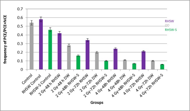
Frequency of PCE/PCE+NCE in Control, 0, 2 and 4 Gy DW, RHSW and RHSW-S Groups 48 and 72 Hours after Irradiation
Figure4.
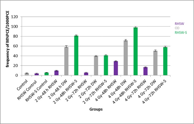
Frequency of MNPCE/1000PCE in Control, 0, 2 and 4 Gy DW, RHSW and RHSW-S Groups 48 and 72 Hours after Irradiation
Table 1.
Effect of Sulfur on Radiation-induced Micronuclei Induction in Mouse Bone Marrow.
| Group | MNPCE/1000PCE (mean ± SD) | MNNCE/1000NCE (mean ± SD) | PCE/PCE+NCE (mean ± SD) |
|---|---|---|---|
| Control | 4.60±0.51 | 3.34±0.69 | 0.54±0.003 |
| RHSW Control | 3.96±0.76 | 3.07±0.52 | 0.58±0.003 |
| RHSW-S Control | 5.78±0.23 | 3.86±0.71 | 0.46±0.002 |
| 2 Gy-48 h RHSW | 9.60±0.87 | 2.80±0.62 | 0.42±0.004 |
| 2 Gy-48 h DW | 58.00±2.63 | 11.64±0.53 | 0.28±0.003 |
| 2 Gy-48h RHSW-S | 81.20±1.68 | 8.83±1.23 | 0.16±0.001 |
| 2 Gy-72h RHSW | 5.60±0.51 | 4.06±0.30 | 0.34±0.002 |
| 2 Gy-72h DW | 39.80±0.86 | 12.79±0.26 | 0.20±0.001 |
| 2 Gy-72h RHSW-S | 41.00±1.58 | 8.77±0.19 | 0.10±0.001 |
| 4 Gy-48h RHSW | 29.00±1.00 | 2.34±0.11 | 0.24±0.001 |
| 4 Gy-48h DW | 71.60±1.96 | 5.96±0.18 | 0.11±0.001 |
| 4 Gy-48h RHSW-S | 98.00±2.34 | 7.66±0.14 | 0.07±0.001 |
| 4 Gy-72h RHSW | 16.80±0.80 | 5.02±0.62 | 0.21±0.001 |
| 4 Gy-72h DW | 50.40±1.99 | 7.91±0.21 | 0.10±0.001 |
| 4 Gy-72h RHSW-S | 58.00±2.09 | 7.29±0.29 | 0.06±0.001 |
RHSW: Ramsar Hot Spring Water, DW: Drinking Water, RHSW-S: Ramsar Hot Spring Water whose Sulfur was omitted.
Discussions
The results showed that radiation increases the number of micronuclei in bone marrow cells. The comparison between the effect of radioactivity in water with 0, 2 and 4 Gy external exposures showed significant differences in micronuclei. Radiation caused a significant decrease in the PCE/PCE+NCE (the ratio of cell proliferation). The frequency of MNPCE, MNNCE and PCE/PCE+NCE was a significant difference between 2 and 4Gy radiation groups. In radiation 4Gy group, MNPCE and MNNCE had increased and PCE/PCE+NCE had decreased compared to 2Gy group. These results were generally consistent with previous research. Also, the results showed that the type of drinking water could change the frequency of the bone marrow MN in mice. There were significant differences in MNPCE and PCE/PCE+NCE between DW and RHSW-S. Besides, there were significant differences in MNPCE and PCE/PCE+NCE between DW and RHSW. Amifostine with sulfur in its compound has been shown to specifically protect normal tissues from damage caused by radiation and chemotherapy [19] . Moreover, the radioprotective effect of famotidine which is recently introduced as a novel clinical radioprotector is attributed to sulfur atom in its structure [20-22]. Asefi [23] and Shanthi [24] also showed that the presence of Radium isotope in food, especially fruit and vegetables could cause internal exposure, which ultimately leads to a significant increase in cancer rates than other regions. Preliminary cytogenetic studies indicated no statistically significant difference in chromosomal abnormalities between residents of the High Natural Radiation Area (HBRAs) and people in control areas [25]. Also, there would be no public health advantage from relocating Ramsar inhabitants, and studies performed on the inhabitants of other HBRAs, like Yangjiang, China indicated that there is no harmful impact induced by natural radiation [26]. Monfared [27] indicated that both natural background radiation and occupational exposure could induce cytogenetic radioadaptive response and this process is more significant in high natural background radiation residents.
Finally, it seems that the radioprotective effect observed in our study could be related to sulfur atom in Ramsar mineral water.
Footnotes
Conflict of interests: None.
References
- 1.Borzoueisileh S, Monfared AS, Abediankenari S, Mostafazadeh A, Khosravifarsani M, Amiri M, et al. The comparison of CD4/CD8 ratio among high and ordinary background radiation areas in Ramsar, Iran. International Journal of Low Radiation. 2011;8:329–37. doi: 10.1504/IJLR.2011.046531. [DOI] [Google Scholar]
- 2.Monfared AS, Jalali F, Mozdarani H, Hajiahmadi M, Samavat H, editors . Living in high natural background radiation areas in Ramsar, Iran. Is it dangerous for health?. International Congress Series; 2005 . Amsterdam: Elsevier; 2005. pp. 438–9. [Google Scholar]
- 3.Borzoueisileh S, Monfared AS, Abediankenari S, Mostafazadeh A, editors The assessment of cytotoxic T cell and natural killer cells activity in residents of high and ordinary background radiation areas of Ramsar-Iran. J Med Phys. 2013;38:30–3. doi: 10.4103/0971-6203.106602. [ PMC Free Article] [DOI] [PMC free article] [PubMed] [Google Scholar]
- 4.Monfared AS, Mozdarani H, Hajiahmadi M. The inhabitants health status in high and low natural background radiation areas in Ramsar, north of Iran. Journal of Gorgan University of Medical Sciences. 2004;6:23–8. [Google Scholar]
- 5.Monfared AS, Jalali F, Sedaghat S, Mansoorizade E, Jarrahi A, Hajiahmadi M, et al. High natural background radiation areas in Ramsar, Iran: can inhabitants feel safe? International Journal of Low Radiation. 2006;3:171–7. doi: 10.1504/IJLR.2006.012016. [DOI] [Google Scholar]
- 6.Grandstaff D. A kinetic study of the dissolution of uraninite. Economic Geology. 1976;71:1493–506. [Google Scholar]
- 7.Langmuir D. Uranium solution-mineral equilibria at low temperatures with applications to sedimentary ore deposits. Geochimica et Cosmochimica Acta. 1978;42:547–69. doi: 10.1016/0016-7037(78)90001-7. [DOI] [Google Scholar]
- 8.Sohrabi M, editor . Recent radiological studies of high level natural radiation areas of Ramsar. Proceeding of International Conference on High Levels of Natural Radiations; 1993. [Google Scholar]
- 9.Borzoueisileh S, Monfared AS, Abediankenari S, Mostafazadeh A, Khosravifarsani M. The effects of residence duration in high background radiation areas on immune surveillance. J Nat Sci Biol Med. 2013;4:218–22. doi: 10.4103/0976-9668.107295. [ PMC Free Article] [DOI] [PMC free article] [PubMed] [Google Scholar]
- 10.Mortazavi S, Monfared A, Ghiassi-Nejad M, Mozdarani H. Radioadaptive responses induced in human lymphocytes of the inhabitants of high level natural radiation areas in Ramsar, Iran. Asian Journal of Experimental Science. 2005;19:19–31. [Google Scholar]
- 11.Borzoueisileh S, Shabestani Monfared A, Comby B, Khosravifarsani M, Roshan Shomal P, Saeid Ramezani M, et al. The highest background radiation school in the world and the health status of its students and their offspring. Isotopes Environ Health Stud. 2014;50:114–9. doi: 10.1080/10256016.2013.821986. [DOI] [PubMed] [Google Scholar]
- 12.Sobue T, Lee VS, Ye W, Tanooka H, Mifune M, Suyama A, et al. Residential randon exposure and lung cancer risk in Misasa, Japan: a case-control study. J Radiat Res. 2000;41:81–92. doi: 10.1269/jrr.41.81. [DOI] [PubMed] [Google Scholar]
- 13.Zou J, Sun Q, Akiba S, Yuan Y, Zha Y, Tao Z, et al. A case-control study of nasopharyngeal carcinoma in the high background radiation areas of Yangjiang, China. J Radiat Res. 2000;41:53–62. doi: 10.1269/jrr.41.S53. [DOI] [PubMed] [Google Scholar]
- 14.Tao Z, Zha Y, Akiba S, Sun Q, Zou J, Li J, et al. Cancer mortality in the high background radiation areas of Yangjiang, China during the period between 1979 and 1995. J Radiat Res. 2000;41:31–41. doi: 10.1269/jrr.41.S31. [DOI] [PubMed] [Google Scholar]
- 15.Mortazavi S, Ghaisinezhad M, Ikushima T, Assaie R, Heidary A, Varzegar R, et al. Are the inhabitants of high background radiation areas of Ramsar more radioresistant? Scope of the Problem and the Need for Future Studies. 2003;1:37–44. [Google Scholar]
- 16.Mazur L. Effects of sulphur-containing compounds and X-rays on the mouse erythropoietic system assayed by in-vivo peripheral blood micronucleus test. Strahlenther Onkol. 1996;172:25–9. [PubMed] [Google Scholar]
- 17.Dansette PM, Sassi A, Deschamps C, Mansuy D. Sulfur containing compounds as antioxidants . Antioxidants in therapy and preventive medicine: Springer; 1990. pp. 209–15. [DOI] [PubMed] [Google Scholar]
- 18.Chigareva NG, Strel’nikov Iu A, Alferova OF. Radio-protective effect of sulfur-containing methylfuran derivatives and the role of thiols in its realization. Radiobiologiia. 1983;23:816–9. [PubMed] [Google Scholar]
- 19.Kouvaris JR, Kouloulias VE, Vlahos LJ. Amifostine: the first selective-target and broad-spectrum radioprotector. Oncologist. 2007;12:738–47. doi: 10.1634/theoncologist.12-6-738. [DOI] [PubMed] [Google Scholar]
- 20.Razzaghdoust A, Mozdarani H, Mofid B, Aghamiri SM, Heidari AH. Reduction in radiation-induced lymphocytopenia by famotidine in patients undergoing radiotherapy for prostate cancer. Prostate. 2014;74:41–7. doi: 10.1002/pros.22725. [DOI] [PubMed] [Google Scholar]
- 21.Razzaghdoust A, Mozdarani H, Mofid B. Famotidine as a radioprotector for rectal mucosa in prostate cancer patients treated with radiotherapy: phase I/II randomized placebo-controlled trial. Strahlenther Onkol. 2014;190:739–44. doi: 10.1007/s00066-014-0602-8. [DOI] [PubMed] [Google Scholar]
- 22.Ching TL, de Jong J, Bast A. Structural characteristics of histamine H2 receptor antagonists that scavenge hypochlorous acid. Eur J Pharmacol. 1994;268:89–93. doi: 10.1016/0922-4106(94)90123-6. [DOI] [PubMed] [Google Scholar]
- 23.Asefi M, Fathivand A, Amidi J. Estimation of annual effective dose from 226Ra and 228Ra due to consumption of foodstuffs by inhabitants of Ramsar city, Iran. Iran Journal of Radiation Research. 2005;3:7–48. [Google Scholar]
- 24.Shanthi G, Maniyan C, Raj GAG, Kumaran JTT. Radioactivity in food crops from high-background radiation area in southwest India. Curr Sci. 2009;97:1331–5. [Google Scholar]
- 25.Ghiassi-nejad M, Mortazavi SM, Cameron JR, Niroomand-rad A, Karam PA. Very high background radiation areas of Ramsar, Iran: preliminary biological studies. Health Phys. 2002;82:87–93. doi: 10.1097/00004032-200201000-00011. [DOI] [PubMed] [Google Scholar]
- 26.Karam PA. The high background radiation area in Ramsar Iran: Geology, Norm, Biology, LNT, and possible regulatory fun. WM. 2002;2:24–8. [Google Scholar]
- 27.Monfared AS, Mozdarani H, Amiri M. Natural background radiation induces cytogenetic radioadaptive response more effectively than occupational exposure in human peripheral blood lymphocytes. Czechoslovak Journal of Physics. 2003;53:A791–A5. doi: 10.1007/s10582-003-0103-y. [DOI] [Google Scholar]