Abstract
Visceral leishmaniasis (VL) is one of the major public health problems in northwest Ethiopia, mainly in Libo-Kemkem and Metema districts, where Phlebotomus orientalis is the most probable vector of the disease. The aim of this study was to determine the physiological age, host preference and vectorial potential of P. orientalis in the highland and lowland foci of the region. Sand flies were collected using CDC light traps between May 2011 and April 2012 in Libo-Kemkem and October 2012 and September 2013 in Metema from household compounds, farm field and mixed forest. Females belonging to Phlebotomus were dissected for physiological age determination and Leishmania detection and isolation. Leishmania infections in sand flies were investigated using molecular methods. Freshly fed Phlebotomus females were tested to identify blood meal sources using PCR-RLB and ELISA. A total of 1149 (936 from Libo-Kemkem and 213 from Metema) blood unfed female P. orientalis were dissected for age determination. The parity rate was 45.6% and 66.2% in Libo-Kemkem and Metema, respectively. None of 798 female P. orientalis dissected (578 from Libo-Kemkem and 220 from Metema) was infected with Leishmania parasites. A total of 347 P. orientalis specimens collected from Libo-Kemkem were processed using PCR, of which 10 (2.8%) specimens were found with DNA of Leishmania spp. Of a total 491 freshly fed female P. orientalis analyzed for blood meal origins by RLB-PCR and ELISA, 57.6% (67.8% from Libo-Kemkem and 49.8% from Metema) were found to contain bovine blood while 4.9% (3.7% from Libo-Kemkem and 5.7% from Metema) were of human blood. In conclusion, the present study showed parity difference between the two populations of P. orientalis and that both populations have strong zoophilic behavior. Based on the presented evidences, the species is strongly implicated as a vector of kala-azar in both areas. Therefore, vector control should be a component of a strategy to manage visceral leishmaniasis in both study areas.
Introduction
Visceral leishmaniasis (VL) in the Old World is transmitted by female Phlebotomus species [1] which feed on blood of vertebrate hosts to obtain protein for the maturation of their eggs [2]. During blood feeding, a sand fly may ingest the etiological agent of VL along with the blood meal from an infected host, allowing biological development of the parasite in the gut and then transmitting to another host during a subsequent feeding [3].
Based on the circumstantial evidences, Phlebotomus orientalis has been implicated as a vector of Leishmania donovani in north, northwest and southwest of Ethiopia. Demonstration of natural infection in P. orientalis, which is one of the criteria of vector incrimination [4], has been difficult except one study by Hailu et al. [5] who found L. donovani promastigotes in one P. orientalis in the lower Omo plains. Gebre-Michael et al. [6] dissected 1219 females in the Awash Valley but none was infected with Leishmania spp. Similar unsuccessful attempts have been made with dissection of 607 and 618 P. orientalis from Libo-Kemkem and Metema-Humera lowlands [7,8].
Blood meal identification of sand flies provides information on the type of host they are fed and also to imply the potential reservoir host (s) [9]. A good reservoir host is a major blood meal source for sand flies [4]. In malaria epidemiology, it is always common to study the proportion of mosquitoes that have fed on humans (anthropophagic) and animals (zoophagic) in order to determine the human blood index (HBI) and bovine blood index (BBI) [10]; however, this has been rarely applied in sand flies.
Identification of the blood meal source of haematophagous insects has been performed using both serological and molecular techniques. The earliest and well known serological methods were precipitin test, hemagglutination inhibition assays, counter-current immune-electrophoresis (CCIE) and at the present enzyme linked immunosorbent assay (ELISA) is widely employed [11–15]. Although these methods yielded valuable information on the identity of the hosts of many blood feeding arthropods, they are not far from limitations, such as the need to produce species-specific antibodies for each potential host and the requirement for fresh blood [16]. However, such limitations are minimized after the molecular techniques [17]. A molecular method for the identification of blood meal origin involves the amplification of either mitochondrial or nuclear DNA by polymerase chain reaction (PCR) followed by species identification using restriction digestion (PCR-RFLP), terminal restriction length polymorphisms, heteroduplex mobility assays, and sequencing. All of these approaches require relatively large amounts of PCR product and do not detect multiple blood sources in a single insect. In order to overcome these setbacks Cytochrome b (cyt b) PCR- reverse line blotting (RLB) assay was developed and used for identifying blood meal sources [18].
Few studies have been undertaken on the blood meal sources of P. orientalis in Ethiopia. Mamo [19] analyzed some blood meal specimens of this species collected from the Awash Valley using CCIE and showed that domestic animals were predominately the source of blood meal. Similarly, Gebre-Michael et al. [8] from Metema-Humera lowlands and Gebresilassie et al. [20] from Tahtay Adiyabo district, northern Ethiopia reported similar results by analyzing blood meals using ELISA.
Understanding the physiological age (parity) of a sand fly population which is established as a vector is great importance in the epidemiology of leishmaniasis, as parous female could have acquired Leishmania infections by feeding a reservoir or an infected person and has potential to transmit parasites [21]. High parous rates in a sand fly population imply high proportion of longer lived individuals that are potentially infected and capable of transmitting disease.
In order to control sand flies and thereby to reduce leishmaniasis, the first important step is to identify the sand fly species, which has the ability to support full development of Leishmania parasites and transmit the parasites to human [22]. Identification or incrimination of vector species in Leishmania endemic focus should fulfill at least one of the five basic vector incrimination criteria set by WHO [4]. The aim of this study was to determine the physiological age, blood meal origins and detect natural infection of P. orientalis in two ecologically distinct foci of VL in northwestern Ethiopia (Libo-Kemkem and Metema districts) where the disease is endemic.
Materials and methods
Study areas
The study was conducted in two ecologically distinct areas of Amhara Regional State in northwestern Ethiopia, namely Libo-Kemkem and Metema districts. The former is situated in a highland with altitude of 2000 meter above sea level (masl) and the latter in a lowland area with altitude ranges 700-750masl. The Metema—Humera plains in the north-west, bordering east Sudan, are the long-recognized VL endemic areas in the country [23]. The two regions contribute more than 60% of the disease burden in the country [24]. In addition to the lowlands, kala-azar has disseminated to the highlands of Libo-Kemkem district and neighboring areas, wherein it claimed more than 200 people [25]. Detailed description of the study areas were presented elsewhere [26].
Sand fly collection and processing
Sand flies were collected using CDC light traps between May 2011 and April 2012 in Libo-Kemkem and October 2012 and September 2013 in Metema. For collection of flies, three villages from the highland district (namely Angot, Bura and Yifag) and three villages from the lowland district (Aftit, Bura and Mender-6) were selected. These villages were selected based on previous kala- azar cases report [25] and accessibility of the villages. In each village in both districts, three permanent sampling habitats were selected: household compound, farm field and mixed forest. The distance between the habitats ranged between 150 to 200 m. In each habitat, two light traps were deployed. During the sampling night, traps were hanged 30–50 cm above the ground from dusk to dawn. In the mornings, trapped sand flies were transported to a field laboratory where Phlebotomus females were categorized into different abdominal status (unfed, half-gravid, fully gravid, and freshly fed) under a dissecting microscope.
All freshly engorged Phlebotomus females were preserved for blood meal analysis. For this purpose, the head and tip of each fed female were severed from the rest of the body and slide-mounted separately for species identification. The rest of the body corresponding to the parts that were slide mounted was individually placed either in silica gel or absolute alcohol. The specimens preserved in silica gel stored at room temperature whereas those in absolute alcohol were stored at -20 °C until blood meal analysis carried on by either cyt b PCR-RLB or ELISA.
Blood meal analysis
Molecular method (cyt b PCR-RLB)
Blood meal analysis using cyt b PCR-RLB was conducted following the procedure of Abbasi et al. [18] in August 2012 in the Department of Microbiology and Molecular Genetics, Hadassah Medical School, Hebrew University, Jerusalem, Israel. Analysis of blood meal by this technique was carried out for some of the specimens collected from Libo-Kemkem (May 2011-April 2012). The rest blood meal samples from both Libo-Kemkem and Metema were analyzed by ELISA at Aklilu Lemma Institute of Pathobiology, Addis Ababa University in Ethiopia.
DNA extraction
For DNA extraction, each blood fed specimen (thorax and abdomen of the flies) was individually placed in eppendorf tube which contained a mixture of 200 μl lysis buffer (50mM NaCl, 10mM ethylene diamine teracacetic [EDTA], 50mM Tris-HCL 1% trition X-100) and 10 μl proteniase K and homogenized with pestle. This was followed by extraction of DNA by adding 180 μl phenol and 8 μl NaCl solutions and centrifuging at 1400 rpm for about two minutes. The DNA extract was transferred into another eppendorf tube, mixed with 400 μl of ethanol, preserved at -20 °C overnight and the DNA was precipitated, after which cold centrifugation at 1400 rpm for 10 min. The DNA was dried in oven for about 10 min and the DNA pellet was re-suspended in 50 μl double distilled water (ddH2O).
DNA amplification by PCR
A 25 μl solution of PCR was prepared by mixing 5 μl DNA with 20 μl master mix (containing forward and backward primers, Taq DNA polymerase, dNTPs, MgCl2, reaction buffers and ddH2O). The target DNA for amplification was 344 base pairs of the conserved region of cyt b gene. Amplification of this region was made using the following primer pairs: forward Cyto1: 5’-CCA TCA AAC ATA TCA GCA TGA TGA AA-3’ and reverse, Cyto 2: 5’-CCC CTC AGA ATG ATA TTT GTC CTC -3’. The thermo-cycling conditions consisted of 35 cycles at 94 °C for 30 sec, 55 °C for 30 sec, and an elongation step at 72 °C for 1 min.
Electrophoresis
The PCR products were loaded on agarose gel (1.5%) and electrophoresed at 120 V in 1x Tris-Acetate (TAE) buffer containing 10 μl ethidium. The gels were visualized under UV light for determination of the sizes of the amplicon. The PCR amplified products were used as probes in RLB hybridization reaction.
RLB hybridization reaction
This procedure was done in two steps (Immobilization of oligonucleotide to the membranes and hybridization and detection) following the method described in Abbasi et al. [18].
Immobilization of oligonucleotide to membranes
Biodyne C nylon membrane (5.5 × 15 cm, Gelman USA) was activated by washing three times using 0.1 M HCL for 10 min. Afterward, the membrane was rinsed in ddH2O three times for 6 min and soaked in 10% solution of 1-ethyl-3-[3’-dimethyl amino propyl] carbodiimide (EDC) for half an hour. Then, the membrane was rinsed in ddH2O and left to dry. Species specific 5’-end amino linked oligonucleotide probes for human, cow, sheep, goat, camel, donkey, dog, mouse, rat, chicken, and bird (avian) developed by Abbasi et al. [18] were diluted to 5 pmol/μL and added to the membrane. The probes were linked to nylon membrane through the formation of amide bonds between the carboxyl groups on the nylon membrane and the amino groups to the oligonucleotides using a manifold blotter apparatus (Immunetics, Cambridge, MA).
Hybridization and detection
The nylon membrane sheet with the oligonucleotide probes was cut into strips at 90 ° to the direction of the blot; hence each strip contained all the eleven probes. Strips were placed in an incubation tray, which had eight lanes and incubated in pre-hybridization solution (2 x sodium chloride and sodium citrate [SSC] with 0.1 Sodium dodecyl sulfate [SDS]) for 30 min at 46 °C with gentle shaking. Biotinylated PCR products were denatured in water bath at 95 °C for 10 min. Hybridization of the denatured PCR products took place at the same temperature of incubation of strips for an hour. Hybridized biotinylated DNA was detected by incubating the strips in streptavidin horseradish peroxidase (HRP) for 30 min at room temperature. After washing the strips three times using 2 xSSC, 0.1%SDS, freshly prepared TMB solution (0.1mg/ml of 3, 3’, 5, 5’ tetramethylbezidine, 0.003% H2O2 in 0.1M sodium citrate [pH 5.0]), was added for chromogenic detection. After a few minutes bands were observed.
Serological method (ELISA)
Blood meal analysis using ELISA was performed on the remaining specimens from Libo-Kemkem and all of the specimens from Metema following the procedure of Beier et al. [13] with some modifications. Briefly, each fed fly was triturated in 1.5 ml eppendrof tube with micro tissue grinder to which 50 μl of 0.01 M phosphate buffered saline (PBS), pH 7.4, was added. The triturated sample was kept at -20 °C until analysis. Then, 50 μl of the triturate sample was diluted in carbonate/bicarbonate coating buffer (CBB) (1:50) and 50 μl of the mixture was added to wells of polyvinyl chloride, U-shaped, 96-well microtiter plates (Dynatech Laboratories, Inc., Alexandria, Va), which were covered and incubated at 4 °C overnight. Each well was washed three times with washing solution (200 μl PBS containing Tween-20). Plates were subsequently blocked by adding bovine serum albumin (BSA) and incubated for one hour at 37 °C. Each well was washed three times with washing solution (PBS-Tw-20). This was followed by the addition of 50 μl host specific conjugate (antihost IgG, Human, bovine, donkey, goat, sheep and dog) diluted 1:2000 for human, 1:250 for bovine, 1:5000 for the rest of animals) in 0.5% boiled casein containing 0.025% Tween-20. The boiled casein was prepared by boiling 5 g casein in 100 ml 0.5 N NaOH and adding 900 ml PBS (pH 7.4), 0.1 g Thimerosal (sodium ethyl mercuri thio salicylate) and 0.02 gm phenol red. After 1 h, wells were washed three times with PBS-Tween-20, and 100 μl of ABTS peroxidase substrate were added to each well. Absorbance at 405 nm was determined with an ELISA reader 30 min after the addition of substrate. Each blood meal sample was considered positive if the absorbance value exceeded the mean plus three standard deviations of the mean of three negative controls and also by observing color change (green color). Negative controls were prepared using unfed insectary female P. orientalis from ALIPB. Positive controls were blood specimens of the six hosts.
The anti-immunoglobulin antisera were pre-screened to verify their antigentic specificity by reactions with blood meal samples of the six hosts. Cross- reaction was noted only between anti-goat and anti-sheep antisera and the result of these (goat and sheep) reported as one, i.e. goat/sheep.
Dissection of sand flies for determination of physiological age and detection of natural infection
Unfed females of Phlebotomus spp. were dissected for determination of parous state. Flies were first washed twice in 2% savlon in saline and once in sterile physiological saline. Then, each female fly was dissected in a drop of physiological saline on a glass slide under a dissecting microscope. The ovaries were pulled out along with the gut of the fly, and then covered with a small cover slip for examination under the microscope. Parous females were distinguished from nulliparous by the presence of granules in the accessory glands [21] as well as ovarian features described by Gebre-Michael et al. [27]. The gut of parous female was instantaneously examined with 10 x and 40 x objectives of a compound microscope for the presence of flagellated parasites. The guts of half and fully gravid females Phlebotomus were also examined. After microscopic examination, the guts of parous, half-gravid and gravid females in saline were transferred to 70% alcohol for further detection of parasites using molecular method if in case infection might have been overlooked.
Detection of Leishmania parasites using molecular method
Four procedures were followed to detect Leishmania parasites in the preserved guts of dissected and undissected sand flies. These were DNA extraction, DNA amplification, gel electrophoresis and sequencing of DNA samples to identify the species of Leishmania [28]. DNA extraction and gel electrophoresis steps were similar to that of the steps used in blood meal analysis using molecular method. However, DNA amplification step was quite different as it targeted different DNA base pairs and used different primers to that of the blood meal analysis. The target DNA for amplification was 346 bp of the whole internal transcribed spacer (ITS) in the ribosomal operon. The DNA was amplified using the primers LITSV (5’-ACACTCAGGTCTGTAAC-3’) and LITSR (5’-CTGGATCATTT-TCCGATG-3’). The positive controls were L. donovani, L. major and L. aethiopica whereas the negative control was distilled water.
Data analysis
The data were entered in SPSS sheet and statistical analysis was made with SPSS IBM version 20.0 (Armonk, NY, IBM Corpn.). Monthly variation in parity rate of P. orientalis was determined by Chi-square test. The human blood index (HBI) and bovine blood index (BBI) were calculated as the proportion of the sand flies fed on either human or bovine blood meals out of the total blood meals determined [10].
Results
Abdominal status of Phlebotomus spp. in both areas
A total of 1314 female P. orientalis were collected from Libo-Kemkem, and the majority, 958 (72.9%) were unfed, followed by 205 (15.6%) freshly fed and 151 (11.5%) gravid or half-gravid. In Metema, of a total 484 P. orientalis females examined, 213 (44.0%) were unfed, 192 (39.7%) freshly fed and the rest 79 (16.3%) gravid or half-gravid (Table 1).
Table 1. Abdominal status of female Phlebotomus species sampled from Libo-Kemkem and Metema.
| Species | No. collected | No. unfed (%) | No. gravid (%) | No. fresh fed (%) | ||||
|---|---|---|---|---|---|---|---|---|
| Libo-Kemkem | Metema | Libo-Kemkem | Metema | Libo-Kemkem | Metema | Libo-Kemkem | Metema | |
| P. orientalis | 1314 | 484 | 958(72.9) | 213(44) | 151(11.5) | 79(16.3) | 205(15.6) | 192(39.7) |
| P. rodhaini | 1 | 46 | 1 (100) | 39(84.8) | 0 | 7(15.2) | 0 | 0 |
| P. papatasi | 0 | 3 | 0 | 1(33.3) | 0 | 2(66.7) | 0 | 0 |
| P. duboscqi | 0 | 2 | 0 | 1(50) | 0 | 1(50) | 0 | 0 |
| P. bergeroti | 0 | 1 | 0 | 0 | 0 | 1(100) | 0 | 0 |
| Total | 1315 | 536 | 959 (72.9) | 254 (47.4) | 151 (11.5) | 90 (16.8) | 205 (15.6) | 192(35.8) |
Parity rates
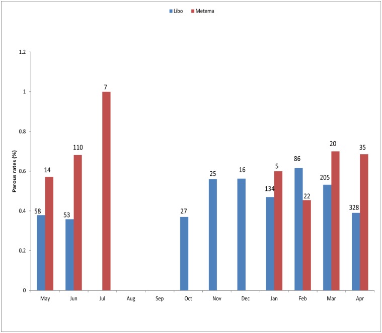
In Libo-Kemkem, a total of 936 unfed P. orientalis were dissected and the parity rate was 45.6% (n = 427), ranging from 37.0 in October to 61.6% in the middle of the dry season (February) with obvious seasonal trend with statistical significance was observed among the months (χ2 = 23.8, df = 9, P<0.05). In Metema, out of a total of 213 unfed P. orientalis dissected the mean parous rate was 66.2% (n = 141), ranging from 0 during the early dry season (October-December) to 100% at the beginning of the rainy season (July), thus showing clear seasonal pattern (Fig 1), however no statistical significance was observed among the months (P>0.05). From the small number of P. rodhaini dissected (n = 39) in Metema, 46.1% (n = 18) were parous.
Fig 1. Monthly parous rate of P. orientalis from Libo-Kemkem (May 2011-April 2012) and Metema (October 2012- September 2013) districts.
N. B. number above the bar indicates the total number of P. orientalis dissected per month.
Blood meal identification based on Cyt b PCR-RLB
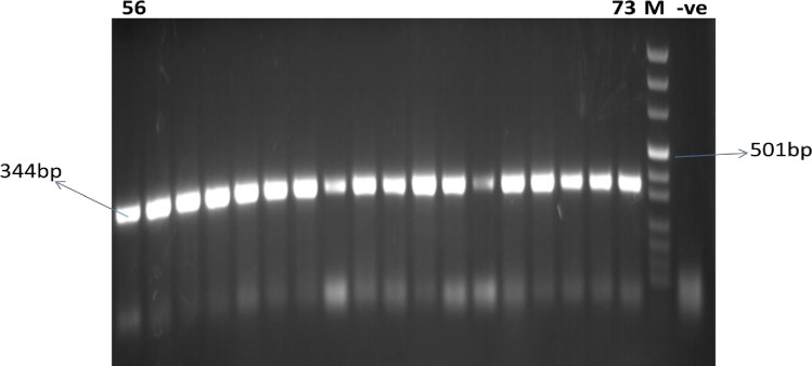
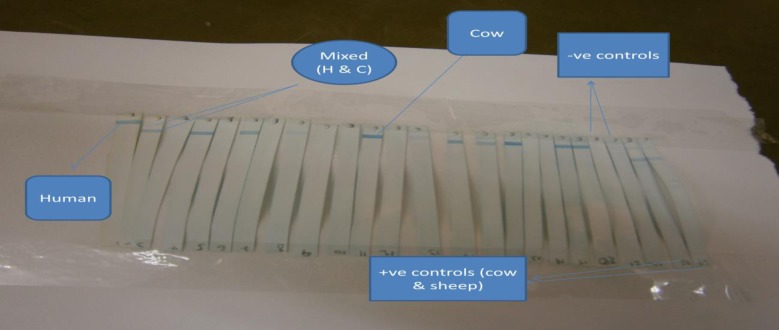
A total of 216 blood fed females of P. orientalis were collected from May 2011 to April 2012 in Libo-Kemkem. Out of these, 115 flies were analyzed using cyt b PCR-RLB to identify the blood meal sources. The remaining blood samples were analyzed by ELISA. Of the 115 P. orientalis, 113 (98.3%) were positive to cyt b PCR (Fig 2) and were used for blood meal identification using RLB and the results after immobilization, hybridization and chromogenic detection (RLB) revealed that 75 (66.4%) were of cow origin and 6 (5.3%) were from human origin. Mixed blood meal for human-cow was detected in 18 (15.9%) and the rest 14 (12.4%) were unidentified (Table 2 and Fig 3).
Fig 2. Gel image of cyt b PCR targeting DNA extracted from wild caught blood fed P. orientalis.
Lanes 56 to 73 are PCR products of blood fed sand fly amplified at the region of cyt b. M is DNA marker.–ve, negative control.
Table 2. Sources of blood meals of P. orientalis sampled from Libo-Kemkem and Metema districts and identified by cyt b PCR-RLB and ELISA.
| Blood meal sources | District | Total (%) 491 |
|||
|---|---|---|---|---|---|
| Libo-Kemkem | Metema | ||||
| RLB = 113 | ELISA = 101 | Combined = 214 | ELISA = 277 | ||
| Bovine | 75 (66.4) | 70 (69.3) | 145 (67.8) | 138 (49.8) | 283 (57.6) |
| Human | 6 (5.3) | 2 (1.9) | 8 (3.7) | 16 (5.7) | 24 (4.9) |
| Donkey | 0 | 0 | 0 | 15 (5.4) | 15 (3.1) |
| Dog | 0 | 0 | 0 | 4 (1.4) | 4 (0.8) |
| Sheep/Goat* | 0 | 1 (0.99) | 1 (0.5) | 4 (1.4) | 5 (1.0) |
| Human-Bovine | 18 (15.9) | 11 (10.9) | 29 (13.6) | 5 (1.8) | 34 (6.9) |
| Human-Donkey | 0 | 1 (0.99) | 1 (0.5) | 9 (3.2) | 10 (2.0) |
| Human-Dog | 0 | 0 | 0 | 3 (1.1) | 3 (0.6) |
| Bovine-Donkey | 0 | 1 (0.99) | 1 (0.5) | 6 (2.2) | 7 (1.4) |
| Bovine-Dog | 0 | 1 (0.99) | 1 (0.5) | 9 (3.2) | 10 (2.0) |
| Bovine-Sheep/Goat | 0 | 0 | 0 | 2 (0.7) | 2 (0.4) |
| Bovine-Donkey-Dog | 0 | 0 | 0 | 1 (0.4) | 1 (0.2) |
| Unidentified | 14(12.4) | 14 (13.9) | 28 (14.0) | 65 (23.5) | 93 (18.9) |
* Since there was cross-reaction between the two antisera the results are presented as goat/sheep
Fig 3. Reverse line blotting results of cyt b PCR products from wild caught blood fed P. orientalis.
H = human, C = cow.
Blood meal identification based on ELISA
In Libo-Kemkem, of the remaining 101 P. orientalis tested by ELISA, blood of five animals was identified. These were bovine blood alone 69.3% (n = 70), human alone 1.9% (n = 2) and goat/sheep alone 0.99% (n = 1). Mixed blood meals were also identified that included; 10.9% (n = 11) for human-bovine blood, 0.99% (n = 1) for human-donkey blood, 0.99% (n = 1) for bovine-donkey blood and 0.99% (n = 1) for bovine-dog blood. The remaining 13.9% (n = 14) blood meal samples were unidentified (Table 2).
The HBI (including mixed feeding) of P. orientalis in Libo-Kemkem based on both methods of analysis (cyt b PCR-RLB and ELISA) was a little over 0.17 whereas the BBI (including mixed feedings) was 0.82, showing its higher predilection for cattle.
In Metema, a total of 277 blood fed females P. orientalis were collected during the course of the study period and were all analyzed by ELISA. The pattern for single hosts was: 138 (49.8%) for bovine blood, 16 (5.7%) for human blood, 15 (5.4%) for donkey blood, 4 (1.4) for dog blood, 4 (1.4%) for sheep/goat blood. Mixed blood meals from either two hosts (six) or three hosts (one) were also identified that included: 9 (3.24%) of bovine-dog, 9 (3.24%) of human-donkey, 6 (2.2%) of bovine-donkey, 5 (1.8%) of bovine-human, 3 (1.1%) of human-dog, 2 (0.7%) of bovine-sheep/goat and including one tri-host (0.36%) from bovine-donkey-dog. Specimens for 65 (23.5%) blood meals remained unidentified (Table 2).
The HBI for P. orientalis (including the mixed blood feedings) in Metema was 0.12 while the bovine blood index (including the mixed feedings) was 0.58 showing a predilection for cattle, though much lower than in Libo-Kemkem.
Leishmania detection based on dissection and molecular method
In Libo-Kemkem, the guts of 578 females P. orientalis (427 parous and 151 gravid) were observed for detection of natural infection (Table 3) and none was positive. Similar results were also obtained from 251 Phlebotomus spp. (141 parous and 79 gravid P. orientalis, 18 parous and 7 gravid P. rodhaini, 1 parous and 2 gravid P. papatasi and 1 parous and 1 gravid P. duboscqi) in Metema.
Table 3. Guts dissection results in Libo-Kemkem and Metema districts.
| Species | Libo-Kemkem | Metema | Total dissected (% infection) | ||||
|---|---|---|---|---|---|---|---|
| No. parous Dissected | No. gravid dissected. | No. infection (%) | No. parous dissected | No. gravid dissected | No. infection (%) | ||
| P. orientalis | 427 | 151 | 0 | 141 | 79 | 0 | 798 (0) |
| P. rodhaini | 1 | 0 | 0 | 18 | 7 | 0 | 26 (0) |
| P. papatasi | 0 | 0 | 0 | 1 | 2 | 0 | 3 (0) |
| P. duboscqi | 0 | 0 | 0 | 1 | 1 | 0 | 2 (0) |
| P. bergeroti | 0 | 0 | 0 | 0 | 1 | 0 | 1 (0) |
| Total | 428 | 151 | 0 | 161 | 90 | 0 | 830 (0) |
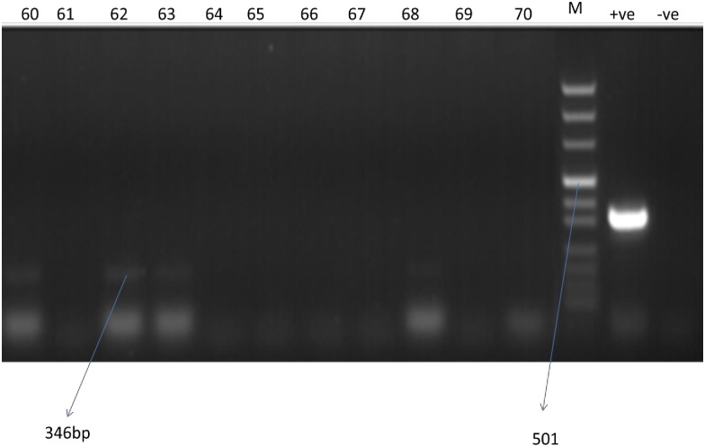
Of the PCR tested 347 P. orientalis from Libo-Kemkem, 10 (2.8%) were positive to Leishmania spp. (Fig 4), however, sequencing did not give positive result due to low concentration of DNA.
Fig 4. Agarose gel electrophoresis of PCR amplification of extracted from wild caught P. orientalis; Lanes 60 to 70 PCR products sand fly amplified for ITS region.
M is DNA marker. +ve- positive control, -ve- negative control.
Discussion
In this study the parity rate, blood meal sources and the status of Leishmania infection in P. orientalis and other Phlebotomus species in two geographically separate areas were investigated as part of an epidemiological study on the ecology of leishmaniasis in the northern and north west of Ethiopia. The findings are additional evidences for the role of P. orientalis in the transmission of leishmaniasis.
Recognition of parous females of sand flies has epidemiological significance as these flies must survive at least one oviposition cycle to be potentially infective with Leishmania parasites [29]. A focus that sustains a significant percentage of parous sand flies is expected to contain long lived individuals than a focus with low parity rate. In this study, the overall parous rate of P. orientalis was lower in Libo-Kemkem than Metema. This indicates that highland population of P. orientalis are short-lived, with most failing to survive long enough to take a second blood meal, presumably dying during or immediately after oviposition in comparison with the lowland population of the species. Similar observation was also noted by Anez et al. [30] in Venezuela. However, parous rates of the species in both populations were higher than that of previous observations reported from these areas [7, 8]. Such discrepancy might be due to seasonal differences as earlier investigations were done during a brief period of time in both study areas and relatively smaller numbers of females were dissected in Metema in the present study.
This is the first attempt to describe the natural blood meal sources of P. orientalis in Libo-Kemkem where five hosts (single and mixed) were identified as sources of blood meals for P. orientalis of which bovine was the most preferred host. The results indicated that humans were comparatively less attractive to this species and thus, the human blood index (0.17) was much lower than the bovine blood index (0.82), exhibiting the zoophily. A similar result was also obtained from Metema, however the BBI was lower. Such differences might be due to the lowland population of P. orientalis having more access to other hosts such as goat, sheep, and donkey as compared to the highland population, where most goats and sheep were kept inside human dwellings during the night. These data from both areas agree with a previous observation in Metema by Gebre-Michael et al. [8] who showed the zoophilic nature of P. orientalis, cattle being the major host (92%) and much higher than the present study. Study on host choice conducted recently in Sheraro, north Ethiopia, hascorroborated the present observation where cattle were found to attract the highest number of P. orientalis [20]. In contrast, a study on host attractiveness in a limited number of animal hosts (dog, mongoose, Nile rat and genet) to P. orientalis in eastern Sudan, showed that dog baited trap significantly attracted the highest number of P. orientalis, though no other domestic animals were used in the experiment [31]; there is yet no results available on blood meal analysis of P. orientalis from Sudan.
The driving force behind the tendency of P. orientalis for cattle to blood meals other hosts in the present study is not known; however from previous knowledge on host preferences of other haematophagous insects, different explanations can be given. One of these reasons could be the relative abundance of the hosts which might determine the host preference of vector species [32]. In both study areas large numbers of cattle with small number of goats, sheep, donkeys and dogs were present outside of human dwellings and these animals were guarded by two to three adult men during the night. The other reason could be difference in body size of the animals. This difference in size in turn results in variation in the amount of CO2 released by the animals [33]. Thus, larger animals like cattle could be more attractive than the rest, although some authors suggested that animal size had no any effect on host selection by sand flies in the cases of Lu. olmeca, Lu. panamensis and Lu. vespertilionis [34].
The role of cattle in the epidemiology of VL is controversial. In India, Barnett et al. [35] concluded that owning a large number of cattle is a risk factor for VL transmission. A similar result was also reported by Bucheton et al. [36]. These studies hypothesize the presence of plethora cattle in the areas might serve as blood meal source for the vector species and also the byproduct of these animals could be an important source of food for sand fly larvae and as a result of which the abundance of the vector species increases thereby increasing transmission of the disease. In contrast, Bern et al. [37] reported that increasing cattle density around house decreased kala-azar transmission by sand flies in Bangladesh. It has been suggested that the proximity of cattle to human dwellings diminish kala-azar transmission by enabling vector species to feed preferentially on animals, not susceptible to leishmaniasis (dead end host), thereby decreasing vector-human contact (zooprophylaxis effect) [38]. However, the role of cattle in both our study areas other than serving as main blood meal sources is not clear.
The detection of other blood meal hosts, though at lower proportion, might indicate the opportunistic behavior of P. orientalis, enabling it to feed on those readily available hosts. Such behavior is common in a number of species observed elsewhere [19, 39–42].
In both areas, particularly in the lowland region, a considerable proportion of P. orientalis analyzed had contact with more than one host (mixed feeding) during a single gonotrophic cycle. Of these, 13.7% had double meals and 0.4% contained triple meals, presumably on the same night. Such mixed blood meals are commonly observed in other sand flies [40, 41]. The exact cause of mixed feeding for this and other species of sand flies in nature is not apparent, however, it is believed to be a consequence of interrupted feedings due to difficulties faced while they engorge on a single host-due to host defensive behavior [42, 43], or when the fly encounters an unnatural host forcing it to divert to another nearby host before repletion from the first host, or the fly might be infected by Leishmania parasites promotes multiple feeding [44].
The numbers of unidentified blood meals in both areas were considerable: 28 (14.0%) in Libo-Kemkem and 65 (23.5%) in Metema. The failure to determine the blood meal sources of P. orientalis may have occurred as a result of either not using probes/antisera of both large (e.g. warthog) and small (e.g. murines and canines) wild mammals which were seen frequently in the study areas especially in Metema. It could also be due to the poor quality of the blood meal resulting from enzymatic degradation of the blood.
Owing to the populations of P. orientalis in both localities having a strong predilection for cattle, treating of these animals by topical application of insecticides [45] or systematic insecticides such as ivermectin [46] may help in alleviating the burden of the disease in the study areas. However, proper evaluations on these potentialities are required.
Identification of vectors and determination of natural infection rates with Leishmania spp. in wild flies are important for definition of risk factors and control of leishmaniasis [47]. In the current study, no natural infection was detected in dissected P. orientalis and other Phlebotomus. In Libo-Kemkem a few specimens were found with Leishmania; DNA sequencing, however, did not produce any results. Absence of promastigotes in the gut of dissected P. orientalis is not uncommon in Ethiopia. Ashford et al. [48] dissected a total of 1006 females of this species in Belessa and found no infection. Previous studies in Libo-Kemkem [7], Humera and Metema districts reported [8]. Natural infection rates in phlebotomine sand flies are usually low, and it is sometimes necessary to dissect thousands of sand flies to find an infected specimen [49]. This might be due to deficiencies in sampling, intensity of the disease or short life span of the vector. In previous studies by Gebre-Michael et al. [7, 8], the majority of dissected flies were nulliparous, while the parous rates were only 30–34% in both the highland (Libo-Kemkem) and lowlands (Humera and Metema). On the other hand, Hailu et al. [50] found natural L. donovani infection in one of 70 (1.4%) dissected female P. orientalis in southwest Ethiopia, while Ashford et al. [51] detected four infected among 48 P. orientalis (8.3%) during an epidemic of the disease in south Sudan.
Although natural infection was not detected in either methods in the present observations, P. orientalis appears to be the principal vector of L. donovani in northwestern Ethiopia mainly by the following reasons; firstly, P. orientalis is the only species in the genus Phlebotomus which is found in large numbers in both areas [23]; second, in a recent study on susceptibility of P. orientalis (colony of Libo-Kemkem population) to L. donovani showed that this species supports full development of L. donovani promastigotes in the gut and the parasite colonized in the anterior parts of the midgut and the stomodeal value of the flies [52] and lastly, P. orientalis is a proven vector in neighboring Sudan [51, 53].
In conclusion, the present study shows that the difference in the parity rates between the highland and lowland populations of P. orientalis being higher in the lowland population. It also indicates that both populations of P. orientalis have strong zoophilic behavior. Furthermore, in both areas the species is strongly implicated as a vector of kala-azar. Therefore, in the future vector control should be one of the approaches for the management of leishmaniasis in the two foci.
Acknowledgments
Authors are grateful to the people of Libo-Kemkem and Metema districts for their support during the study. We also thank our field and laboratory assistants, particularly Mr. Gashaw Mihiret for his helps in carrying out this study. The Bill and Melinda Gates Foundation Global Health Program (grant number OPPGH5336) provided financial support.
Data Availability
All relevant data are within the paper.
Funding Statement
The authors received funding from The Bill and Melinda Gates Foundation Global Health Program (grant number OPPGH5336).
References
- 1.Maroli M, Feliciangeli MD, Bichaud L., Charrel RN, Gradoni L. Phlebotomine sand flies and the spreading of leishmaniasis and other diseases of public concern. Med Vet Entomol. 2013; 27:123–47. doi: 10.1111/j.1365-2915.2012.01034.x [DOI] [PubMed] [Google Scholar]
- 2.Lane RP. Sand flies (Phlebotomine) In: Lane RP, Crosskey RW, editors. Medical Insects and Arachnids. Chapman and Hall; 1993. pp. 78–119. [Google Scholar]
- 3.Garlapati RB, Abbasi I, Warburg A, Poche D, Poche R. Identification of blood meals in wild caught blood fed Phlebotomus argentipes (Diptera: Psychodidae) using cytochrome b PCR and reverse line blotting in Bihar, India. J Med Entomol. 2012; 49:515–521. [DOI] [PubMed] [Google Scholar]
- 4.WHO. Control of the Leishmaniasis. WHO Technical Report Series. 2010; 949:1–186. [PubMed]
- 5.Hailu A, Balkew M, Berhe N, Meredith S, Gemetchu T. Is Phlebotomus (Larroussius) orientalis a vector of visceral leishmaniasis in South–west Ethiopia? Acta Trop. 1995; 60:15–20. [DOI] [PubMed] [Google Scholar]
- 6.Gebre-Michael T, Balkew M, Ali A, Ludovisi A, Gramiccia M. The isolation of Leishmania tropica and L. aethiopica from Phlebotomus (Paraphlebotomus) species (Diptera: Psychodidae) in the Awash Valley, northeastern Ethiopia. Trans Roy Soc Trop Med Hyg. 2004; 98:64–70. [DOI] [PubMed] [Google Scholar]
- 7.Gebre-Michael T, Balkew M, Alamirew T, Reta M. Preliminary entomological observations in a highland area of Amhara region, northern Ethiopia, with epidemic visceral leishmaniasis. Ann Trop Med Parasitol. 2007; 101: 367–370. doi: 10.1179/136485907X176382 [DOI] [PubMed] [Google Scholar]
- 8.Gebre-Michael T, Balkew M, Berhe N, Hailu A, Mekonnen Y. Further studies on the phlebotomine sand flies of the kala-azar endemic lowlands of Humera-Metema (north-west Ethiopia) with observations on their natural blood meal sources. Parasit Vec. 2010; 3:6. [DOI] [PMC free article] [PubMed] [Google Scholar]
- 9.Jaouadi K, Haouas N, Chaara D, Boudabous R, Gorcii M, Kidar A, et al. Phlebotomine (Diptera: Psychodidae) blood meals sources in Tunisian cutaneous leishmaniasis foci: could Sergentomyia minuta, which is not an exclusive Herpetophilic species, be implicated in transmission of pathogens? Ann. Entomol. Soc. Am. 2013; 106:79–85. [Google Scholar]
- 10.Garrett-Johns C. The human blood index of malaria vectors in relation to epidemiological assessment. Bull. World Health Organ. 1964; 30:241–61. [PMC free article] [PubMed] [Google Scholar]
- 11.Boreham PF. Some applications of blood meal identifications in relation to the epidemiology of vector-borne tropical diseases. Am J Trop Med Hyg. 1975; 78:83–91. [PubMed] [Google Scholar]
- 12.Dhanda J, Gill G. Double blood meals by Phlebotomus argentipes and P. papatasi in two villages of Mahrashtra. Indian J. Med. Res. 1982; 76:840–842. [PubMed] [Google Scholar]
- 13.Beier J, Perkins P, Writz R. Blood meal identification by direct enzyme linked immunosorbent assay (ELISA), tested on Anopheles (Diptera: Culicidae) in Kenya. J Med Entomol.1988; 25:9–16. [DOI] [PubMed] [Google Scholar]
- 14.Ogusuku E, Perez J, Nieto L. Identification of blood meal sources of Lutzomyia spp. in Peru. Ann Trop Med Parasitol. 1994; 88:329–335. [DOI] [PubMed] [Google Scholar]
- 15.Svobodova M, Sadlova J, Chang KP, Volf P. Distribution and feeding preferences of the sand flies Phlebotomus sergenti and P. papatasi in a cutaneous leishmaniasis focus in Sanliurfa, Turkey. Am J Trop Med Hyg. 2003; 68:6–9. [PubMed] [Google Scholar]
- 16.Blackwell A, Mordue A, Mordue W. Identification of blood meals of the Scottish biting midge, Culicoides impunctatus, by indirect enzyme-linked immunosorbent assays (ELISA). Med Vet Entomol. 1994; 8:20–24. [DOI] [PubMed] [Google Scholar]
- 17.Mukabana W, Takken W, Knols B. Analysis of arthropod blood meals using molecular genetic markers. Trends Parasitol. 2002; 18:505–509. [DOI] [PubMed] [Google Scholar]
- 18.Abbasi I, Cunio R, Warburg A. Identification of blood meals imbibed by phlebotomine sand flies using cytochrome b PCR and reverse line blotting. Vector Borne Zoonotic Dis. 2008; 9:79–86. doi: 10.1089/vbz.2008.0064 [DOI] [PubMed] [Google Scholar]
- 19.Mamo H. Production of specific antisera against selected mammals and identification of blood meals of phlebotomine sand flies transmitting visceral leishmaniasis in Ethiopia. MSc thesis. Addis Ababa University. 1999.
- 20.Gebresilassie A, Abbasi I, Aklilu E, Yared S, Kirstein O, Moncaz A, et al. Host-feeding preference of Phlebotomus orientalis (Diptera: Psychodidae) in an endemic focus of visceral leishmaniasis in northern Ethiopia. Parasit Vec. 2015; 8:270. [DOI] [PMC free article] [PubMed] [Google Scholar]
- 21.Lewis DJ, Lainson R, Shaw JJ. Determination of parous rates in phlebotomine sand flies with special reference to Amazonian species. Bull Entomol Res. 1970; 60:209–219. doi: 10.1017/S0007485300040736 [DOI] [PubMed] [Google Scholar]
- 22.Killick-Kendrick R. Recent advances and outstanding problems in the biology of phlebotomine sand flies. Acta Trop. 1978; 35:297–313. [PubMed] [Google Scholar]
- 23.Mengesha B, Abuhoy M. Kala-azar among labour migrants in Metema-Humera region of Ethiopia. Trop Geogr Med. 1978; 30:199–206. [PubMed] [Google Scholar]
- 24.Gadisa E, Tsegaw T, Abera A, Elnaiem D, Boer M, et al. Eco-epidemiology of visceral leishmaniasis in Ethiopia. Parasit Vec. 2015; 8:381. [DOI] [PMC free article] [PubMed] [Google Scholar]
- 25.Alvar J, Bashaye S, Argaw D, Cruz I, Aparicio P, Kassa A, et al. Kala-Azar Outbreak in Libo Kemkem, Ethiopia: Epidemiologic and Parasitologic Assessment. Am J Trop Med Hyg. 2007; 77: 275–282. [PubMed] [Google Scholar]
- 26.Aklilu E, Gebresilassie A, Yared S, Kindu M, Tekie H, Balkew M, et al. Studies on sand fly fauna and ecological analysis of Phlebotomus orientalis in the highland and lowland foci of kala-azar in northwestern Ethiopia. PloS One. 2017; 12. [DOI] [PMC free article] [PubMed] [Google Scholar]
- 27.Gebre-Michael T., Pratlong F. and Lane R. P. (1993). Phlebotomus (Phlebotomus) duboscqi (Diptera: Phlebotomine), naturally infected with Leishmania major in southern Ethiopia. Trans Roy Soc Trop Med Hyg. 1993; 87:10–11. [DOI] [PubMed] [Google Scholar]
- 28.Abbasi I, Aramin S, Hailu A, Shiferaw W, Kassahun A, Belay S, et al. Evaluation of PCR procedures for detecting and quantifying Leishmania donovani DNA in large numbers of dried human blood samples from visceral leishmaniasis focus in northern Ethiopia. BMC Infect Dis. 2013; 13:153 doi: 10.1186/1471-2334-13-153 [DOI] [PMC free article] [PubMed] [Google Scholar]
- 29.Lewis DJ. Phlebotomid sand flies. Bull World Health Organ. 1971; 77:535–551. [PMC free article] [PubMed] [Google Scholar]
- 30.Anez N, Nieves E, Cazorla D, Oviedo M, Yarbuh Y, Valera M. Epidemiology of cutaneous leishmaniasis in Merida, Venezuela. III. Altitudinal distribution, age structure, natural infection and feeding behavior of sand flies and their relation to the risk of transmission. Ann Trop Med Parasitol. 1994; 88:279–287. [DOI] [PubMed] [Google Scholar]
- 31.Hassan MM, Osman O, El-Raba’a F, Schallig H, Elnaiem D. Role of the domestic dog as a reservoir host of Leishmania donovani in eastern Sudan. Parasit Vec. 2009; 2:26. [DOI] [PMC free article] [PubMed] [Google Scholar]
- 32.Takken W, Verhulst O. Host preferences of blood feeding mosquitoes. Annu. Rev. Entomol. 2013; 58:433–453. doi: 10.1146/annurev-ento-120811-153618 [DOI] [PubMed] [Google Scholar]
- 33.Quinnell R, Dye C, Shaw J. Host preferences of the phlebotomine sand fly Lutzomyia longipalpis in Amazonian Brazil. Med Vet Entomol. 1992; 6:195–200. [DOI] [PubMed] [Google Scholar]
- 34.Christensen H, Herrer A. Panamanian Lutzomyia (Diptera: Psychodidae) host attraction profiles. J Med Entomol. 1980; 17:522–528. [DOI] [PubMed] [Google Scholar]
- 35.Barnett P, Singh S, Bern C, Hightower A, Sundar S. Virgin soil: the spread of visceral leishmaniasis into Uttar Pradesh, India. Am J Trop Med Hyg. 2005; 73:720–725. [PubMed] [Google Scholar]
- 36.Bucheton B, Kheir M, El-Safi S, Hammad A, Mergani A, Mary C, et al. The interplay between environmental and host factors during an outbreak of visceral leishmaniasis in eastern Sudan. Microb Infect. 2002; 4:1449–1457. [DOI] [PubMed] [Google Scholar]
- 37.Bern C, Courtenay O, Alvar J. Of cattle, sand flies and men: a systematic review of risk factor analyses for South Asian visceral leishmaniasis and implications for elimination. PLoS Negl. Trop. Dis. 2010; 4:599. [DOI] [PMC free article] [PubMed] [Google Scholar]
- 38.Mahande A., Mosha F., Mahande J. and Kweka E. Feeding and resting behaviour of malaria vector, Anopheles arabiensis with reference to zooprophylaxis. Malar J. 2007; 6:100 doi: 10.1186/1475-2875-6-100 [DOI] [PMC free article] [PubMed] [Google Scholar]
- 39.Foster W. Boreham P, Tempelis H. Studies on leishmaniasis in Ethiopia. IV. Feeding behavior of Phlebotomus longipes (Diptera: Psychodidae). Ann Trop Med Parasitol. 1972; 66:433–43. [PubMed] [Google Scholar]
- 40.Guy MW, Killick-Kendrick R, Gill GS, Rioux JA, Bray RS. Ecology of leishmaniasis in the south of France. 19. Determination of the hosts of Phlebotomus ariasi Tonnoir, 1921 in the Cevennes by blood meal analysis. Ann Parasitol Hum Comp. 1984; 59: 449–458. [PubMed] [Google Scholar]
- 41.Ngumbi P, Lawyer P, Johnson R, Kiilu G, Asiago C. Identification of phlebotomine sand fly blood meals from Baringo district, by direct enzyme-linked immunosorbent assay (ELISA). Med Vet Entomol. 1992; 6:385–388. [DOI] [PubMed] [Google Scholar]
- 42.Bongiorno G, Habluetzel A, Khoury C, Maroli M. Host preferences of phlebotomine sand flies at a hypoendemic focus of canine leishmaniasis in central Italy. Acta Trop. 2003; 88:109–116. [DOI] [PubMed] [Google Scholar]
- 43.Coleman R, Edman J. The influence of host behavior of sand fly (Lutzomyia longipalis) feeding success on laboratory mice. Bull Soc Vec Ecol. 1987; 12:539–540. [Google Scholar]
- 44.Rogers M, Bates P. Leishmania manipulation of sand fly feeding behavior results in enhanced transmission. PLoS Pathog. 2007; 3:91. [DOI] [PMC free article] [PubMed] [Google Scholar]
- 45.Courtenay O, Kovacic V, Gomes P, Garcez M, Quinnell R. A long-lasting topical deltamethrin treatment to protect dogs against visceral leishmaniasis. Med Vet Entomol. 2009; 23:245–256. doi: 10.1111/j.1365-2915.2009.00815.x [DOI] [PubMed] [Google Scholar]
- 46.Mascari TM, Mitchell MA, Rowton ED, Foil LD. Ivermectin as a rodent feed-through control of immature sand flies (Diptera: Psychodidae). J Am Mosq Control Assoc. 2008; 24:323–326. doi: 10.2987/5678.1 [DOI] [PubMed] [Google Scholar]
- 47.Oshaghi M, Maleki R, Javadian E, Mohebali M, Hajjaran H, Zare Z, et al. Vector incrimination of sand flies in the most important visceral leishmaniasis focus in Iran. Am J Trop Med Hyg. 2009; 81:572–577. doi: 10.4269/ajtmh.2009.08-0469 [DOI] [PubMed] [Google Scholar]
- 48.Ashford R, Hutchinson MP, Bray RS. Kala-azar in Ethiopia: epidemiological studies in a highland valley. Ethiop Med J. 1973; 11:259–264. [PubMed] [Google Scholar]
- 49.Killick-Kendrick R. Phlebotomine vectors of the leishmaniasis: a review. Med Vet Entomol. 1990; 4:1–24. [DOI] [PubMed] [Google Scholar]
- 50.Hailu A, Balkew M, Berhe N, Meredith S, Gemetchu T. Is Phlebotomus (Larroussius) orientalis a vector of visceral leishmaniasis in South–west Ethiopia? Acta Trop. 1995; 60:15–20. [DOI] [PubMed] [Google Scholar]
- 51.Ashford R, Seaman J, Schorscher J, Pratlong F. Epidemic visceral leishmaniasis in southern Sudan: identity and systematic position of the parasites from patients and vectors. Trans R Soc Trop Med Hyg. 1992; 86:379–380. [DOI] [PubMed] [Google Scholar]
- 52.Seblova V, Volfova V, Dvorak V, Pruzinova K, Votypka J, Kassahun A, et al. Phlebotomus orientalis sand flies from two geographically distant Ethiopian localities: biology, genetic analyses and susceptibility to Leishmania donovani. PLoS Negl. Trop. Dis. 2013; 4: 2187. [DOI] [PMC free article] [PubMed] [Google Scholar]
- 53.Elnaiem AD, Ward RD, Hassan HK, Miles MA, Frame IA. Infections rates of Leishmania donovani in Phlebotomus orientalis from focus of visceral leishmaniasis in eastern Sudan. Ann Trop Med Parasitol. 1998; 92:229–232. [DOI] [PubMed] [Google Scholar]
Associated Data
This section collects any data citations, data availability statements, or supplementary materials included in this article.
Data Availability Statement
All relevant data are within the paper.


