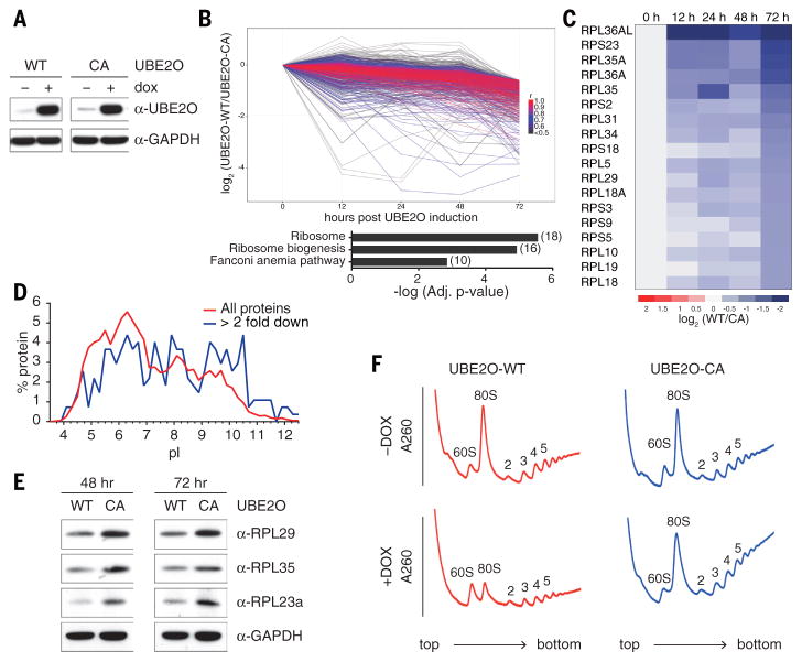
Fig. 3. UBE2O is sufficient to drive the elimination of ribosomes in HEK293 cells.
(A) UBE2O induction by doxycycline treatment in Flp-In T-REx 293 cells was assessed by immunoblotting, using antibodies to UBE2O. Full length human UBE2O-WT and UBE2O-CA genes were genomically integrated in an untagged form, and induced with doxycycline for 24 hr. 20 μg of cell lysate was loaded per lane. GAPDH is a loading control.
(B, C) Mass spectrometry (TMT) quantification of 7,808 proteins after UBE2O (WT or CA) induction for 0, 12, 24, 48 and 72 hours. All 685 proteins that were downregulated more than 50% after 72h of UBE2O-WT induction, in comparison to UBE2O-CA, are shown in (B). Coloring reflects Pearson correlation (r) to the median pattern from high (red) to low (black). The bar graph represents results from KEGG pathway enrichment analysis, which indicated highly significant enrichment (adj. p-value=2.77 x 10⁻⁶) of ribosomes, with 18 proteins downregulated (shown in C).
(D) Isoelectric point (pI) comparison of all quantified proteins (red), and all downregulated proteins (blue), based on data from panel B. pI values were obtained from the proteome pI database (46).
(E) After induction of UBE2O (WT or CA) for the indicated times (48 and 72 hours), ribosomal protein levels were analyzed by immunoblotting. GAPDH is a loading control. 20 μg of cell lysate was loaded per lane, and all samples were from the same experiment.
(F) Sucrose gradient analysis of cells overexpressing UBE2O-WT showed a reduced 80S monosome peak after 72 hours of doxycycline treatment.

