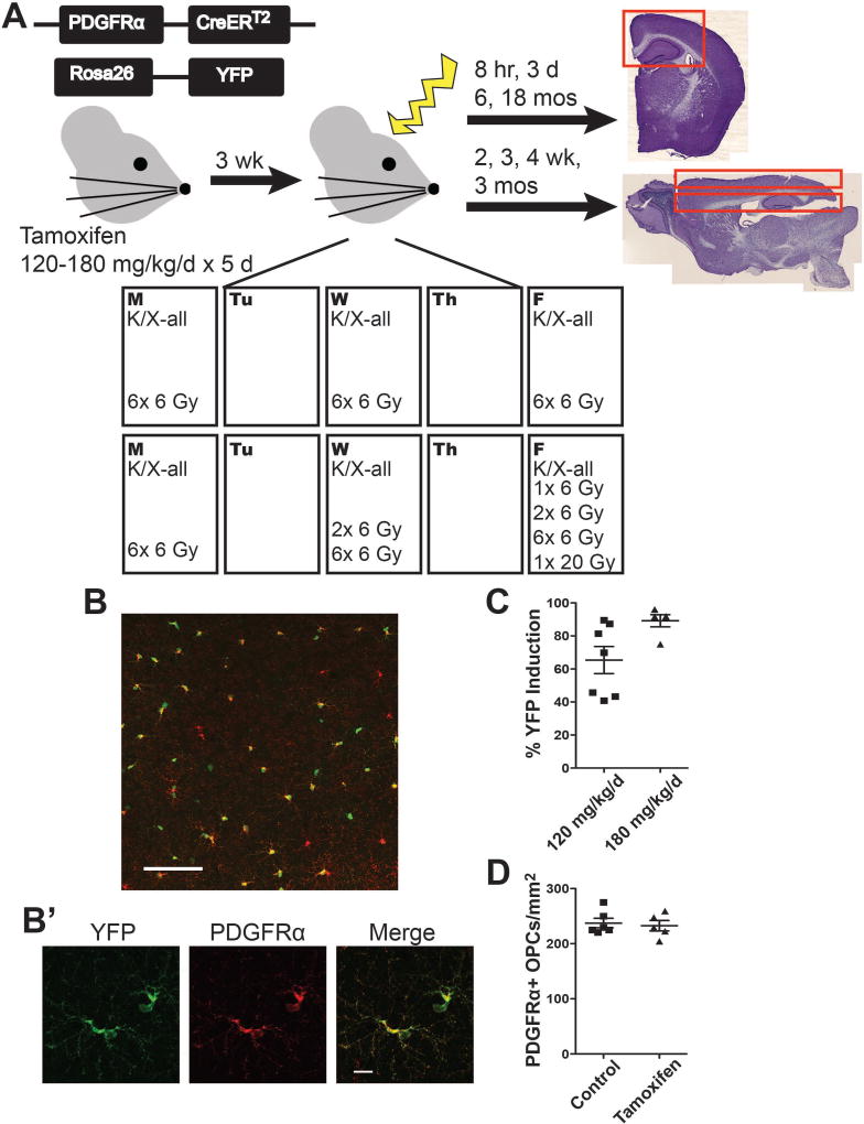
Figure 1. Experimental design.
A. Mice received intraperitoneal injections of 120–180 mg/kg/d tamoxifen for 5 consecutive days at 6–8 weeks of age. Three weeks was allowed for clearance of tamoxifen. Mice were anesthetized and exposed to 0 Gy, 1× 6 Gy, 2× 6 Gy, 6× 6 Gy, or 1× 20 Gy cranial irradiation, with exposures occurring Monday, Wednesday, and Friday for two consecutive weeks or on the final exposure days in the case of animals not exposed to the 6× 6 Gy paradigm. Animals were sacrificed at time points ranging from 8 hours to 18 months after exposure. Tissue was sectioned coronally for 8 hour, 3 day, 6 month, and 18 month time points and sagittally for 2, 3, 4 week and 3 month time points. B. Representative image shows YFP and PDGFRα expression in Pdgfrα-CreERT2:Rosa26R-YFP mice 3 weeks after intraperitoneal (i.p.) injection of 120 mg/kg/d tamoxifen for 5 days. Image scale bar = 100 µm. B’. Inset scale bar = 10 µm. C. YFP induction varied between 65.4% and 89.2% in the following conditions, respectively: 5 weeks following 5 days of 120 mg/kg/d tamoxifen and 5 weeks following 5 days of 180 mg/kg/d tamoxifen. n = 5–7 animals/group. D. Tamoxifen treatment did not alter the number of PDGFRα+ oligodendrocyte progenitor cells (OPCs) at 3 weeks post-injection of 120 mg/kg/d tamoxifen. n = 5–6 animals/group, Student’s t-test (p = 0.7189). Graphs represent mean ± SEM.

