Abstract
Statement of problem
Trueness and precision are used to evaluate the accuracy of intraoral optical impressions. Although the in vivo precision of intraoral optical impressions has been reported, in vivo trueness has not been evaluated because of limitations in the available protocols.
Purpose
The purpose of this clinical study was to compare the accuracy (trueness and precision) of optical and conventional impressions by using a novel study design.
Material and methods
Five study participants consented and were enrolled. For each participant, optical and conventional (vinylsiloxanether) impressions of a custom-made intraoral Co-Cr alloy reference appliance fitted to the mandibular arch were obtained by 1 operator. Three-dimensional (3D) digital models were created for stone casts obtained from the conventional impression group and for the reference appliances by using a validated high-accuracy reference scanner. For the optical impression group, 3D digital models were obtained directly from the intraoral scans. The total mean trueness of each impression system was calculated by averaging the mean absolute deviations of the impression replicates from their 3D reference model for each participant, followed by averaging the obtained values across all participants. The total mean precision for each impression system was calculated by averaging the mean absolute deviations between all the impression replicas for each participant (10 pairs), followed by averaging the obtained values across all participants. Data were analyzed using repeated measures ANOVA (α=.05), first to assess whether a systematic difference in trueness or precision of replicate impressions could be found among participants and second to assess whether the mean trueness and precision values differed between the 2 impression systems.
Results
Statistically significant differences were found between the 2 impression systems for both mean trueness (P=.010) and mean precision (P=.007). Conventional impressions had higher accuracy with a mean trueness of 17.0 ±6.6 µm and mean precision of 16.9 ±5.8 µm than optical impressions with a mean trueness of 46.2 ±11.4 µm and mean precision of 61.1 ±4.9 µm.
Conclusions
Complete arch (first molar-to-first molar) optical impressions were less accurate than conventional impressions but may be adequate for quadrant impressions.
Optical impressions can offer many advantages over conventional impressions because they do not undergo dimensional change and are not susceptible to damage from handling; need less armamentarium (no need for trays, adhesives, and dispensers); provide immediate on-screen feedback; they increase the scope of planning and diagnosis; permit easier communication with the dental laboratory with no cross infection risk; and allow easier duplication, storage, and retrievability. Furthermore, optical impressions allow chairside fabrication of definitive restorations in a single appointment.1 However, optical impressions have some limitations, such as the size of the scanning wand, which can create problems for patients with limited mouth opening, the procedure learning curve, and the difficulty in scanning subgingival margins.1 Although optical impressions have advantages, their adoption in daily dental practice and the shift toward a complete digital workflow are contingent upon the ability of complete arch optical impressions to replicate oral and dental structures at a level of accuracy at least comparable with that of conventional impressions.
The accuracy of optical impressions can be evaluated by in vitro assessment on a sturdy dental model, often fabricated from metal. This model is digitized with a reference (gold standard) scanner that has a validated accuracy to create a reference 3-dimensional (3D) model. An equivalent number of conventional and digital impressions are made from the dental model. Conventional impressions are digitized, either directly from the impressions or indirectly after they are poured in stone, using the same reference scanner. Deviations of the 3D models of each impression system from the reference are calculated after superimposing (overlaying) each model with the reference, using available software designed for that purpose.2–5 This protocol allows the measurement of 2 variables that are used to define the accuracy of any measurement device according to ISO standard no. 5725-1:1994, trueness and precision. Trueness represents mean deviation (error) of a group of measurements from an original structure or a reference.5–7 Precision represents mean deviation between repeated measurement.4,8 Higher trueness and precision are represented by lower values of those variables. For example, a measurement device that gives a mean deviation (error) of 10 µm from a reference value has less trueness than another device that gives a mean deviation of 5 µm from the same reference. Likewise, a device that has a mean deviation of 10 µm between its repeated measurements is less precise than another device that has a mean deviation of 5 µm.
The second method of evaluating the accuracy of optical impressions is by comparing the marginal and internal fit of restorations fabricated using optical impressions with restorations obtained by conventional impressions.9–19 This methodology evaluates the accuracy of the entire fabrication chain and not just the impression system. This fabrication chain includes many error prone steps beginning with optical scanning and the associated 3D model creation processes through all the stages of restoration fabrication (milling, 3D printing, casting) and postfabrication procedures (sintering, finishing, and polishing). Therefore, the importance of the first protocol for evaluating the impression systems cannot be overemphasized, especially in the early stages of the development of new intraoral scanners or in upgrading the existing ones.
Most studies that have evaluated the accuracy of optical impression systems were performed in vitro.2–6,20–22 The present authors are only aware of in vivo studies evaluating precision because the dental arches of research participants cannot be scanned using a reference scanner.23–25 However, a protocol is clearly needed that evaluates both the trueness and the precision of complete arch optical impressions in vivo, considering the trend toward digital dentistry. The purpose of this study was to compare the accuracy (trueness and precision) of optical impressions with that of conventional impressions in vivo by using a novel study design. The null hypothesis was that no differences would be found in the mean trueness and precision of complete arch conventional and optical impressions in vivo.
MATERIAL AND METHODS
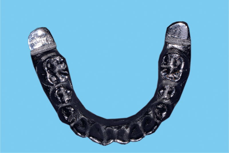
A convenience sample of 5 volunteers with complete dentition consented and were enrolled after Institutional Board approval from the University of North Carolina at Chapel Hill. The participants had no temporomandibular disorders, restricted mouth opening, or xerostomia, nor did they report the use of medications that might have caused xerostomia. A reference appliance was fabricated from cobalt-chromium (Co-Cr) alloy (Cobalt Base PB; IdentAlloy) for each participant. The appliance was designed to fit over the occlusal surface of the participant’s mandibular teeth, extending from first molar to first molar. Each appliance was first fabricated from acrylic resin material (Triad VLC; Dentsply Sirona) using mandibular impressions as a template to duplicate the mandibular teeth of the participant. Each reference appliance was then converted into removable partial denture Co-Cr alloy using 3D printing technology (3DRPD USA). Each appliance was designed with a slot placed after the distal molar to help orient the custom trays during the impression procedure and with a distal extension to aid in retrieving the appliance from the conventional impression with minimum distortion (Fig. 1). The purpose of this appliance was to facilitate the intraoral comparison of impression accuracy by acting as a removable device that could be impressed intraorally, using both impression systems and scanned extraorally by using a reference scanner in a way similar to the protocols described in the introduction.
Figure 1.
Reference appliance showing distal extension and slots designed to help in tray orientation and retrieval.
Five conventional and 5 optical impressions were made in a single visit for each participant by the same operator (M.A.). Body temperature was measured for each participant, using an infrared ear thermometer (ThermoScan 3; Braun) to exclude fever. The intaglio surface of each reference appliance was relined intraorally with rigid interocclusal record material (Futar D; Kettenbach) to enhance stability during the impression procedure. The temperature of each appliance was kept within 1°C of the average oral temperature with water bath until use. The appliance was then placed intraorally over the mandibular arch of each participant and impressed using vinylsiloxanether (VSXE) impression material (Identium; Kettenbach) in a custom tray. All conventional impressions for each participant were allowed to set according to the manufacturer’s instructions (≥5 minutes and 30 seconds total working time). After removal, each impression was evaluated for correct seating by looking for the uniform spread of the material around the appliance. The appliance was then separated from the impression and cleaned with rubbing alcohol (91% alcohol; Equate) to prepare for the next impression. All impressions were disinfected for 5 minutes (CaviCide; Metrex) and stored at room temperature.
Optical impressions were obtained using CEREC Omnicam (with CEREC SW 4.3 software). Although Omnicam is a powderless intraoral scanner, a powder (CEREC Optispray; Dentsply Sirona) was lightly used to help in scanning the reference appliance effectively because of the high reflectivity of its surface.
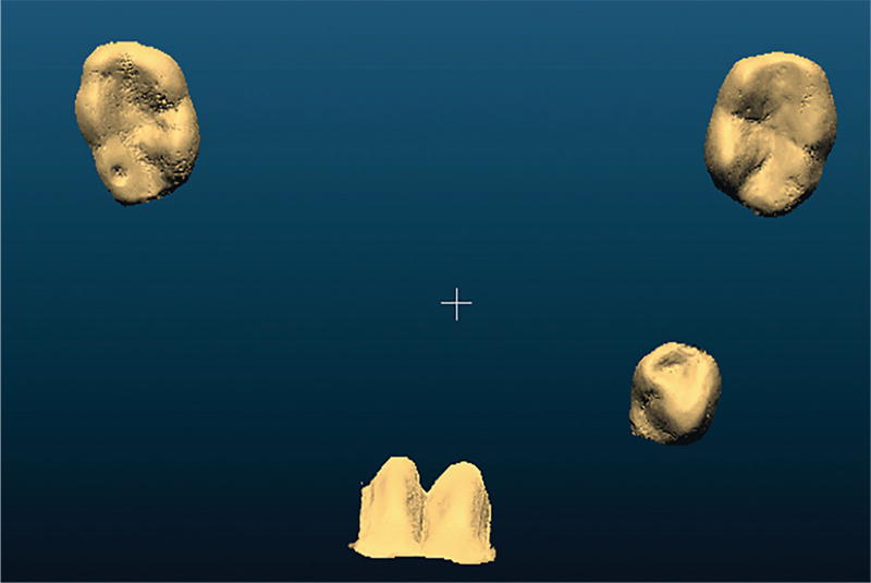
All reference appliances were lightly powdered with CEREC Optispray and scanned using a validated reference scanner (Infinite focus standard; Alicona Corp) with trueness of 5.3 ±1.1 µm and precision of 1.6 ±0.6 µm.3 A reference 3D model for each reference appliance was generated from the scan. Because of the time-consuming procedure and the limited availability of the reference scanner, 4 areas were selected from each reference appliance for comparison. These areas were the occlusal third of the right first molar, the lingual surface of the mandibular anterior teeth, the occlusal surface of the left first premolar, and the occlusal third of the left first molar (Fig. 2). The conventional impressions were poured in Type IV die stone (Elite Rock; Zhermack) after 2 days of storage at room temperature. All resultant stone casts were scanned using the same reference scanner, and 3D models were generated. The same areas of comparison were selected. The 3D models were generated from optical impressions by using CEREC inlab (SW 4.2.5).
Figure 2.
3D model showing areas selected for comparison.
Three-dimensional models of the conventional and optical groups were aligned to their respective reference models (IF-Measure Suite 5.1; Alicona Corp), and any areas outside the selected field of comparison were cropped (CloudCompare v2.6.2, 2016). Subsequently, automatic fine alignment was performed, and any remaining nonoverlapped areas after the second alignment were cropped again. Deviations between each impression and its respective reference model were then calculated using difference measurement module in the “nearest” mode, which calculated the mean absolute deviation between all the nearest signed neighbor points (trueness). This was done for all the impression replicates for each participant, resulting in 5 trueness values for each participant. These values were then averaged for each participant and then averaged across all participants to give the total mean trueness for each impression system.
Precision for both groups was measured by calculating mean absolute deviations between all the possible pairs of impression replicates for each participant. The mean absolute deviations were calculated from 10 precision values for each participant. These values were averaged for each participant and then averaged across all participants to give the total mean precision for each impression system. The numerical outputs of all comparisons were saved in a spreadsheet (Excel 15.24 for Macintosh; Microsoft Corp). In addition, the deviation maps were saved for the visual analysis of patterns of deviations (Fig. 3).
Figure 3.
Deviation maps of representative impressions. A, Conventional. B, Optical.
Statistical analysis was performed using SAS v9.3 software (SAS Institute Inc). Separately by impression system, repeated measures ANOVA was used to assess whether a systematic difference could be found in the trueness or precision of the replicate impressions among the participants. With the averaged values for each participant, we used repeated measures ANOVA with impression system as the explanatory variable to assess whether the mean trueness and precision values differed between the 2 impression systems (α=.05).
RESULTS
The trueness and precision values for the conventional impression group were consistently lower than those for the optical impression group across all participants. No statistically significant differences were found in the mean trueness and precision values among the participants for each impression system alone (Table 1), indicating consistency in the differences between the reference and impression among the participants within each impression system.
Table 1.
Mean trueness and precision for conventional and optical impression groups
| Mean ±SD Trueness (µm) | Mean ±SD Precision (µm) | |||
|---|---|---|---|---|
|
|
|
|||
| ID | Conventional | Optical | Conventional | Optical |
|
| ||||
| 1 | 21.1 ±8.9 | 36 ±13.4 | 22.4 ±7.5 | 55.2 ±24.2 |
|
| ||||
| 2 | 17.8 ±4.6 | 38.6 ±13.7 | 18.9 ±3.0 | 43.6 ±20.7 |
|
| ||||
| 3 | 10.5 ±1.5 | 63.2 ±19.2 | 10.9 ±2.3 | 83.0 ±35.0 |
|
| ||||
| 4 | 25.3 ±7.3 | 52.9 ±8.9 | 21.7 ±3.8 | 56.5 ±16.1 |
|
| ||||
| 5 | 10.4 ±0.9 | 39.5 ±10.7 | 10.7 ±1.2 | 67.4 ±32.7 |
|
| ||||
| P | .181 | .740 | .519 | .459 |
The total mean trueness for the conventional impression group was 17.0 ±6.6 µm and 46.2 ±11.4 µm for the optical impression group (Table 2). A statistically significant differences were found in the mean trueness between the conventional and optical impression groups (P=.010).
Table 2.
Overall trueness and precision
| Impression System | Mean ±SD Trueness | Mean ±SD Precision |
|---|---|---|
| Conventional | 17.0 ±6.6 | 16.9 ±5.8 |
| Optical | 46.2 ±11.4 | 61.1 ±14.9 |
| P | .010 | .007 |
The total mean precision for the conventional impression group was 16.9 ±5.8 µm and 61.1 ±14.9 µm for the optical impression group (Table 2). Statistically significant differences were found in the mean precision between the conventional and optical impression groups (P=.007).
Regarding the pattern of deviation from the reference models, the conventional impression group showed a uniform distribution of deviation across the area of comparison in 84% of the impressions. However, a band of higher deviation ranging between 80 and 140 µm appeared lingual to the central incisors in 16% of the impressions. The optical impression group, in contrast, showed clusters of larger deviations at one or both molars in 88% of the impressions. The deviation was more prominent at either the left or right molar in 44% of the impressions. In these cases, the deviation was ≥140 µm in 60% of the impressions and >250 µm in 28% of the impressions.
DISCUSSION
This study introduced a protocol for evaluating the accuracy (trueness and precision) of optical impressions in vivo. As far as the present authors are aware, this was the first study to evaluate both trueness and precision intraorally. Authors of in vitro studies have predicted that the in vivo accuracy of optical impression will be less than that reported in vitro because of the challenging nature of the intraoral environment with moisture, restricted space, and patient movement.2,3,6,21 Moreover, 2 in vivo studies that evaluated precision confirmed the negative effect of the oral environment on the precision of optical impressions.23,25
In this study, conventional impressions showed significantly better trueness and precision than optical impressions, and the null hypothesis was rejected. This finding was in agreement with other in vitro studies evaluating the accuracy (trueness and precision) of complete arch optical impressions compared with VSXE impression material.3,5
The findings of this study are comparable with those of 2 other in vivo studies. Flugge et al23 reported a significant effect of the intraoral environment when they compared the precision of direct intraoral digitization with the indirect extraoral digitization of stone casts, using the same intraoral scanner. Ender et al24 reported that all of the 7 popular optical impression systems they investigated showed decreased precision when they were used in vivo. They further explained that single image systems are more sensitive to patient, tongue, and camera movement (inadequate support) than high-frame-rate-capturing systems. They mentioned that, to some extent, software can detect artifacts that result from such movements and require the scan to be repeated. They reported different precision results for 2 scanners from the same manufacturer but with different software versions.
The deviations of the conventional impressions of the reference models in this study were mostly consistent across the area of comparison. However, the deviations of the optical impressions were mostly clustered at the distal molars with one of the sides being significantly more prominent than the other in almost half the impressions. These findings are comparable with the findings of 2 in vitro studies and 1 in vivo study.5,20,24 Moreover, in a recent in vivo study by Ender et al,25 quadrant optical impressions obtained by 3 different intraoral scanners showed better precision than previously reported complete arch optical impressions and comparable precision to quadrant conventional impressions.
No agreement has yet been reached on how much accuracy is needed for each clinical procedure. Acceptable marginal discrepancy is another poorly defined quantity, ranging from 40 µm to 150 µm.26–30 An in vitro study showed that cement dissolution in crowns with 150 µm marginal discrepancies was significantly more than crowns with 25- to 75-µm discrepancies.30 Moreover, although marginal discrepancies of less than 50 µm can be achieved for gold castings and metal-ceramic crowns, this might be harder for milled crowns.26,28 Therefore, the fabrication technique should be considered too. Anadioti et al12 reported in an in vitro study that the best marginal fit was obtained by combining the conventional polyvinyl siloxane impressions and pressed ceramic techniques. However, they also reported that crowns obtained by a combination of optical impressions and either IPS e.max press or IPS e.max CAD showed clinically acceptable mean marginal accuracy ranging between 74 and 76 µm.
Although there is disagreement in regard to the acceptable marginal discrepancy, the clinical success of single-tooth restorations obtained by optical impressions has been reported.10,31–33 A recent in vivo study compared the fit of zirconia crowns obtained by intraoral digitization using 3 intraoral scanners with the extraoral digitization of stone casts using a laboratory scanner. The study found that the marginal discrepancy of crowns obtained by optical impressions, with the exception of Omnicam, were comparable with the conventional ones.13 Also, several in vitro studies and reviews reported that the marginal and internal fit of 1 to 4 unit restorations obtained by optical impressions were comparable with those obtained by conventional impressions.11,14–19
Digital technology is constantly improving. We are witnessing rapid hardware and software improvements that are being applied in the field of optical impressions. A recent study reported that most of the newer intraoral scanners showed better precision than the older ones.15 Optical impression systems will continue to improve, resulting in a more reliable complete arch impression for different treatment indications.
This study has several limitations. Although Omni-cam is a powderless intraoral scanner, a powder was necessary to overcome the high reflectivity of the polished metal. The appliances could have been airborne-particle abraded to lower their surface reflectively, but it was decided to keep their surface smooth to prevent material residues from adhering that may have affected accuracy measurements. Also, had the surface been airborne-particle abraded, this could have introduced errors into the surface reproduction of the conventional impressions. Nevertheless, the powder application was as light as possible to allow scanning.
Another limitation is the choice of a metal appliance that is liable to thermal expansion and contraction. An effort to decrease the temperature fluctuation at the time of impression procedure was done by controlling the temperature of the appliance to within 1°C of average oral temperature before it was inserted intraorally. Additionally, the reference scanner was in a room with controlled temperature during the time of scanning. Therefore, the effects of dimensional change were standardized across patients and across the impression systems.
The selection of only 4 areas to represent the whole arch prevented an evaluation of the pattern of deviation in the remaining areas of the arch. However, the smaller datasets that resulted from this selection may decrease the number of possible errors that might result from superimposing large datasets as explained by Guth et al.22
The size of the appliance was limited to first molar-to-first molar. An appliance extending over the second molars was not possible using the presented design because of the limitation in the amount of comfortable mouth opening.
CONCLUSIONS
Within the limitations of this study, the following conclusions were drawn:
The protocol for evaluating both trueness and precision in vivo appeared applicable.
Complete arch (first molar to first molar) optical impressions obtained by CEREC Omnicam were less accurate than conventional VSXE impressions,
The CEREC Omnicam impressions may be sufficiently accurate for quadrant impressions.
Clinical Implications.
Complete arch optical impressions using Cerec Omnicam were less accurate than conventional impressions. However, they may be sufficiently accurate for quadrant impressions.
Acknowledgments
The authors thank the research participants for volunteering for this project; and Dr Ceib Philips for help with statistical analysis.
Supported by 2014 Ralph Phillips Student Research Award from the Academy of Operative Dentistry (to M.A.A.). Cerec Omnicam provided by Dentsply Sirona USA. Materials provided by Kettenbach USA and Dentsply Sirona.
References
- 1.Reich S, Vollborn T, Mehl A, Zimmermann M. Intraoral optical impression systems–an overview. Int J Comput Dent. 2013;16:143–62. [PubMed] [Google Scholar]
- 2.Ender A, Mehl A. Full arch scans: conventional versus digital impressions–an in-vitro study. Int J Comput Dent. 2011;14:11–21. [PubMed] [Google Scholar]
- 3.Ender A, Mehl A. Accuracy of complete arch dental impressions: a new method of measuring trueness and precision. J Prosthet Dent. 2013;109:121–8. doi: 10.1016/S0022-3913(13)60028-1. [DOI] [PubMed] [Google Scholar]
- 4.Ender A, Mehl A. Influence of scanning strategies on the accuracy of digital intraoral scanning systems. Int J Comput Dent. 2013;16:11–21. [PubMed] [Google Scholar]
- 5.Ender A, Mehl A. In-vitro evaluation of the accuracy of conventional and digital methods of obtaining full-arch dental impressions. Quintessence Int. 2015;46:9–17. doi: 10.3290/j.qi.a32244. [DOI] [PubMed] [Google Scholar]
- 6.Guth JF, Keul C, Stimmelmayr M, Beuer F, Edelhoff D. Accuracy of digital models obtained by direct and indirect data capturing. Clin Oral Investig. 2013;17:1201–8. doi: 10.1007/s00784-012-0795-0. [DOI] [PubMed] [Google Scholar]
- 7.Normung DDIfr. Accuracy (trueness and precision) of measurement methods and results. Part 1. [Accessed November 11, 2014];General principles and definitions. (ISO 5725-1:1994). Available at: www.iso.org.
- 8.Ziegler M. Digital impression taking with reproducibly high precision. Int J Comput Dent. 2009;12:159–63. [PubMed] [Google Scholar]
- 9.Almeida ESJS, Erdelt K, Edelhoff D, Araujo E, Stimmelmayr M, Vieira LC, et al. Marginal and internal fit of four-unit zirconia fixed dental prostheses based on digital and conventional impression techniques. Clin Oral Investig. 2014;18:515–23. doi: 10.1007/s00784-013-0987-2. [DOI] [PubMed] [Google Scholar]
- 10.Syrek A, Reich G, Ranftl D, Klein C, Cerny B, Brodesser J. Clinical evaluation of all-ceramic crowns fabricated from intraoral digital impressions based on the principle of active wavefront sampling. J Dent. 2010;38:553–9. doi: 10.1016/j.jdent.2010.03.015. [DOI] [PubMed] [Google Scholar]
- 11.Tidehag P, Ottosson K, Sjogren G. Accuracy of ceramic restorations made using an in-office optical scanning technique: an in vitro study. Oper Dent. 2014;39:308–16. doi: 10.2341/12-309-L. [DOI] [PubMed] [Google Scholar]
- 12.Anadioti E, Aquilino SA, Gratton DG, Holloway JA, Denry I, Thomas GW, et al. 3D and 2D marginal fit of pressed and CAD/CAM lithium disilicate crowns made from digital and conventional impressions. J Prosthodont. 2014;23:1–8. doi: 10.1111/jopr.12180. [DOI] [PubMed] [Google Scholar]
- 13.Boeddinghaus M, Breloer ES, Rehmann P, Wöstmann B. Accuracy of single-tooth restorations based on intraoral digital and conventional impressions in patients. Clin Oral Investig. 2015;19:2027–34. doi: 10.1007/s00784-015-1430-7. [DOI] [PubMed] [Google Scholar]
- 14.Seelbach P, Brueckel C, Wostmann B. Accuracy of digital and conventional impression techniques and workflow. Clin Oral Investig. 2013;17:1759–64. doi: 10.1007/s00784-012-0864-4. [DOI] [PubMed] [Google Scholar]
- 15.Svanborg P, Skjerven H, Carlsson P, Eliasson A, Karlsson S, Ortorp A. Marginal and internal fit of cobalt-chromium fixed dental prostheses generated from digital and conventional impressions. Int J Dent. 2014;2014:534382. doi: 10.1155/2014/534382. [DOI] [PMC free article] [PubMed] [Google Scholar]
- 16.Tsirogiannis P, Reissmann DR, Heydecke G. Evaluation of the marginal fit of single-unit, complete-coverage ceramic restorations fabricated after digital and conventional impressions: a systematic review and meta-analysis. J Prosthet Dent. 2016;116:328–35. doi: 10.1016/j.prosdent.2016.01.028. [DOI] [PubMed] [Google Scholar]
- 17.Kim JH, Jeong JH, Lee JH, Cho HW. Fit of lithium disilicate crowns fabricated from conventional and digital impressions assessed with micro-CT. J Prosthet Dent. 2016;116:551–7. doi: 10.1016/j.prosdent.2016.03.028. [DOI] [PubMed] [Google Scholar]
- 18.Chochlidakis KM, Papaspyridakos P, Geminiani A, Chen CJ, Feng IJ, Ercoli C. Digital versus conventional impressions for fixed prosthodontics: a systematic review and meta-analysis. J Prosthet Dent. 2016;116:184–90. doi: 10.1016/j.prosdent.2015.12.017. [DOI] [PubMed] [Google Scholar]
- 19.Ahlholm P, Sipilä K, Vallittu P, Jakonen M, Kotiranta U. Digital versus conventional impressions in fixed prosthodontics: a review. J Prosthodont. 2016 Aug 2; doi: 10.1111/jopr.12527. [Epub ahead of print.] [DOI] [PubMed] [Google Scholar]
- 20.Luthardt RG, Loos R, Quaas S. Accuracy of intraoral data acquisition in comparison to the conventional impression. Int J Comput Dent. 2005;8:283–94. [PubMed] [Google Scholar]
- 21.Patzelt SB, Emmanouilidi A, Stampf S, Strub JR, Att W. Accuracy of full-arch scans using intraoral scanners. Clin Oral Investig. 2014;18:1687–94. doi: 10.1007/s00784-013-1132-y. [DOI] [PubMed] [Google Scholar]
- 22.Guth JF, Edelhoff D, Schweiger J, Keul C. A new method for the evaluation of the accuracy of full-arch digital impressions in vitro. Clin Oral Investig. 2016;20:1487–94. doi: 10.1007/s00784-015-1626-x. [DOI] [PubMed] [Google Scholar]
- 23.Flugge TV, Schlager S, Nelson K, Nahles S, Metzger MC. Precision of intraoral digital dental impressions with iTero and extraoral digitization with the iTero and a model scanner. Am J Orthod Dentofacial Orthop. 2013;144:471–8. doi: 10.1016/j.ajodo.2013.04.017. [DOI] [PubMed] [Google Scholar]
- 24.Ender A, Attin T, Mehl A. In vivo precision of conventional and digital methods of obtaining complete arch dental impressions. J Prosthet Dent. 2016;115:313–20. doi: 10.1016/j.prosdent.2015.09.011. [DOI] [PubMed] [Google Scholar]
- 25.Ender A, Zimmermann M, Attin T, Mehl A. In vivo precision of conventional and digital methods for obtaining quadrant dental impressions. Clin Oral Investig. 2016;20:1495–504. doi: 10.1007/s00784-015-1641-y. [DOI] [PubMed] [Google Scholar]
- 26.Christensen GJ. Marginal fit of gold inlay castings. Oper Dent. 1966;16:297–305. doi: 10.1016/0022-3913(66)90082-5. [DOI] [PubMed] [Google Scholar]
- 27.McLean JW, von Fraunhofer JA. The estimation of cement film thickness by an in vivo technique. Br Dent J. 1971;131:107–11. doi: 10.1038/sj.bdj.4802708. [DOI] [PubMed] [Google Scholar]
- 28.Belser UC, MacEntee MI, Richter WA. Fit of three porcelain-fused-to-metal marginal designs in vivo: a scanning electron microscope study. J Prosthet Dent. 1985;53:24–9. doi: 10.1016/0022-3913(85)90058-7. [DOI] [PubMed] [Google Scholar]
- 29.Fransson B, Oilo G, Gjeitanger R. The fit of metal-ceramic crowns, a clinical study. Dent Mater. 1985;1:197–9. doi: 10.1016/s0109-5641(85)80019-1. [DOI] [PubMed] [Google Scholar]
- 30.Jacobs MS, Windeler AS. An investigation of dental luting cement solubility as a function of the marginal gap. J Prosthet Dent. 1991;65:436–42. doi: 10.1016/0022-3913(91)90239-s. [DOI] [PubMed] [Google Scholar]
- 31.Reiss B. Clinical results of Cerec inlays in a dental practice over a period of 18 years. Int J Comput Dent. 2006;9:11–22. [PubMed] [Google Scholar]
- 32.Reiss B, Walther W. Clinical long-term results and 10-year Kaplan-Meier analysis of Cerec restorations. Int J Comput Dent. 2000;3:9–23. [PubMed] [Google Scholar]
- 33.Otto T, De Nisco S. Computer-aided direct ceramic restorations: a 10-year prospective clinical study of Cerec CAD/CAM inlays and onlays. Int J Prosthodont. 2002;15:122–8. [PubMed] [Google Scholar]


