Abstract
The incidence of esophagogastric cancer is rapidly rising but only a minority of patients derive durable benefit from current therapies. Chemotherapy as well as anti-HER2 and PD-1 antibodies are standard treatments. To identify predictive biomarkers of drug sensitivity and mechanisms of resistance, we implemented prospective tumor sequencing of metastatic esophagogastric cancer patients. There was no association between HRD defects and response to platinum-based chemotherapy. Patients with MSI-H tumors were intrinsically resistant to chemotherapy but more likely to achieve durable responses to immunotherapy. The single EBV+ patient achieved a durable, complete response to immunotherapy. The level of ERBB2 amplification as determined by sequencing was predictive of trastuzumab benefit. Selection for a tumor subclone lacking ERBB2 amplification, deletion of ERBB2 exon 16, and co-mutations in the receptor tyrosine kinase, RAS, PI3K pathways were associated with intrinsic and/or acquired trastuzumab resistance. Prospective genomic profiling can identify patients most likely to derive durable benefit to immunotherapy and trastuzumab, and guide strategies to overcome drug resistance.
INTRODUCTION
Esophagogastric cancer is the tumor type with the most rapidly increasing incidence in the US, particularly in young patients (1). These tumors have a high metastatic potential and frequently recur. Recent large-scale sequencing initiatives, including the retrospective studies performed by The Cancer Genome Atlas (TCGA), have revealed that most esophagogastric cancers are characterized by chromosomal instability with frequent amplifications of receptor tyrosine kinases (RTKs) (2–5). Additional molecularly defined esophagogastric cancer subsets that may be therapeutically relevant include those characterized by homologous recombination deficiency (HRD), Epstein-Barr virus (EBV)-related tumors, and tumors with hypermutation, in particular those with microsatellite instability (MSI) (2–5).
The combination of a fluoropyrimidine and a platinum is the standard first-line systemic therapy for patients with esophagogastric cancer (6). For patients with human epidermal growth factor receptor 2 (HER2/ERBB2) positive tumors, trastuzumab (a HER2-directed antibody) in combination with chemotherapy is standard of care (7). Pembrolizumab and nivolumab (anti-PD-1 antibodies) are approved for use in chemotherapy-refractory esophagogastric cancer patients. Pembrolizumab was recently approved in the U.S. for programmed death-ligand 1 (PD-L1) positive or MSI-high (MSI-H) esophagogastric cancer (8–10). However, PD-L1 status was not predictive of survival with nivolumab therapy in the ATTRACTION 2 study (11), and nivolumab is approved in Asia for treatment of irrespective of PD-L1 status.
Despite the recent increase in therapeutic options, responses to systemic therapy in patients with esophagogastric cancer are most often short lived, and less than 5% of patients with metastatic disease survive beyond 5 years (1). With the goal of identifying predictive biomarkers of response and molecular mechanisms of resistance to trastuzumab and immune checkpoint inhibitors, as a prelude to the development of rational combination strategies, we performed prospective targeted next generation sequencing (12) and clinicopathologic analysis of patients with recurrent or metastatic esophagogastric cancer.
RESULTS
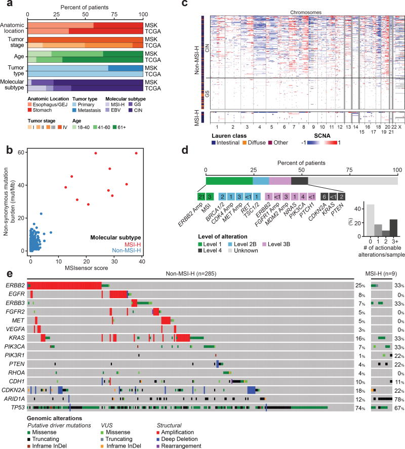
With the goal of identifying predictive biomarkers of drug response, we, in 2014, initiated an effort to perform prospective, targeted next generation sequencing analysis of paired tumor and normal samples from all patients with stage IV esophagogastric adenocarcinoma (Supplementary Table 1, 2) treated at our center. Tumors were analyzed using MSK-IMPACT, a capture based, next-generation sequencing platform that can detect mutations, copy-number alterations, and select rearrangements in 341 or more cancer genes (see Methods). Here, we report the results of the first 295 patients profiled along with accompanying prospectively captured detailed clinical annotation and treatment response data. The majority of samples were acquired from endoscopic biopsies of primary tumors. Unlike the TCGA dataset of early stage tumors, this Memorial Sloan Kettering (MSK)-cohort was comprised of specimens from predominantly younger patients with exclusively stage IV disease with detailed clinical annotation, including survival and therapeutic response data, available for all patients (Figure 1A).
Figure 1. Molecular characterization of esophagogastric tumors.
A, Comparison of clinical characteristics between the MSK and TCGA cohorts. B, Correlation between the MSIsensor score (x-axis) to non-synonymous mutation burden (y-axis). Samples are colored according to molecular subtype. C, DNA copy number changes categorized by molecular subtype. Chromosomes are presented from left to right, samples from top to bottom. Regions of losses appear in shades of blue while regions of gains are in shades of red. D, Highest level of clinical actionability across the cohort, as defined by OncoKB. Standard therapeutic implications include FDA–recognized or NCCN-guideline listed biomarkers that are predictive of response to an FDA-approved drug in a specific indication (Level 1). Investigational therapeutic implications include FDA-approved biomarkers predictive of response to an FDA-approved drug detected in an off-label indication (Level 2B), FDA- or non–FDA-recognized biomarkers that are predictive of response to novel targeted agents that have shown promising results in clinical trials (Level 3B), and non–FDA-recognized biomarkers that are predictive of response to novel targeted agents on the basis of compelling pre-clinical data (Level 4). E, Alterations of known drivers in esophagogastric cancer. Gene alteration types, patterns and overall frequencies are shown for non-MSI-H and MSI-H tumors separately. Tumors are shown from left top right. Mutations are color-coded by type and by presumed oncogenicity, as defined by prior knowledge and recurrence (cancerhotspots.org).
We achieved a mean sequencing coverage of 744X and identified an average of 5 non-synonymous mutations per tumor sample (range 1–63) (Supplementary Table 3). MSI status was inferred from the sequencing data using a clinically validated algorithm (13), with MSI-H defined as an MSIsensor score >=10 (Figure 1B). MSI-H tumors possessed a high ratio of small insertions and deletions to substitutions, a mutational signature consistent with mismatch repair deficiency. Notably, only nine samples in the MSK-cohort were MSI-H (3%), which is significantly lower than the fraction in the TCGA cohort (16%, P=8e-10, Fisher’s exact test, Figure 1B). This difference is likely attributable to the higher prevalence of metastatic cancers in the MSK-cohort, as MSI-H was a favorable prognostic marker in the TCGA cohort. EBV positivity could not be established from the targeted sequencing data and thus was not known for all patients. To assess for EBV in select patients receiving immunotherapy, Epstein-Barr encoding region in situ hybridization (EBER-ISH) was performed in 26 patients who received immunotherapy with only a single patient testing positive.
The MSK cohort was comprised predominantly of tumors with a signature of chromosomal instability (CIN, 63%), characterized by a high degree of copy-number alterations and low mutational burden (Figure 1C). TP53 was the most frequently mutated gene (73%), followed by ARID1A (15%) and CDKN2A (12%). In total, 53% of patients had at least one potentially actionable alteration as defined by OncoKB (14), a precision oncology knowledgebase that annotates the functional consequence and therapeutic implications of cancer mutations (Figure 1D, E). Focal amplifications and mutations in receptor tyrosine kinases and members of the RAS and PI3-kinase pathways were common in the CIN subset, with frequent oncogenic or likely oncogenic alterations in ERBB2 (25%), KRAS (16%), EGFR (8%), ERBB3 (7%), PIK3CA (7%), FGFR2 (5%), and MET (5%). Genomically stable (GS) tumors (34%), conversely, were more frequently of diffuse histology (32% vs. 9%, P=3e-5, Fisher’s test) and CDH1-mutant (20% vs. 7%, P=0.01, Fisher’s test). A comparison of the non-MSI-H tumors to those in the TCGA cohort found few statistically significant differences (Fisher’s test, 15% FDR): TP53 mutations were enriched in the MSK cohort (73% vs 62%, q=0.11), whereas KMT2C (2% vs 9%, q=0.06), GRIN2A (1% vs 6%, q=0.10), PTPRD (4% vs 11%, q=0.10) and CTNNB1 (1% vs 6%,q=0.10) were less frequently mutated (Supplementary Figure 1A). Notably, there were no significant differences in the alteration frequencies of any genes between primary and metastatic samples (Supplementary Figure 1B).
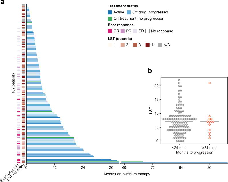
To identify potential biomarkers of response to systemic chemotherapy in an unbiased manner, we correlated the genomic findings with treatment response and patient outcomes in the 187 patients with HER2-negative disease treated with first-line fluoropyrimidine/platinum. In this setting, the median PFS was similar to the published literature (6.9 vs 5.3 months), with favorable OS (26.3 months vs 10.17 months) (15). In this analysis, no single mutant allele or gene, including those with a role in DNA repair pathways, such as BRCA1/2, were significantly associated with treatment response (Figure 2A).
Figure 2. Genomic determinants of response to cytotoxic chemotherapy.
A, Swimmer’s plot showing months on first-line platinum-based therapy for 185 patients with metastatic, HER2-negative esophageal cancer. The annotation tracks on the left of the y-axis indicate the patient's best response to platinum and the estimated LST score. The color of individual bars indicate the current status of the patient on this line of treatment. B, Distribution of LST scores in patients that progressed on platinum treatment before 24 months compared to patients with prolonged response (>24 months). Horizontal bars represent the median by group.
As an association between defects in homologous recombination deficiency (HRD) and response to platinum-based chemotherapy has been identified in other cancer types, we inferred a surrogate marker for HRD from the copy-number data (large-scale state transitions, LST (16,17)) and correlated the results with overall response and duration of treatment with chemotherapy. LST score was not predictive of progression free survival (HR=0.99, p=0.947, log-rank test) and was not higher in patients with responses to first line therapy lasting over 24 months (P=0.6, two-tailed t-test; Figure 2B). Notably, the majority of patients with prolonged response to platinum-based combination chemotherapy, including the two patients with the longest outlier responses (68 and 104 months, respectively), harbored no somatic alterations in known HR genes.
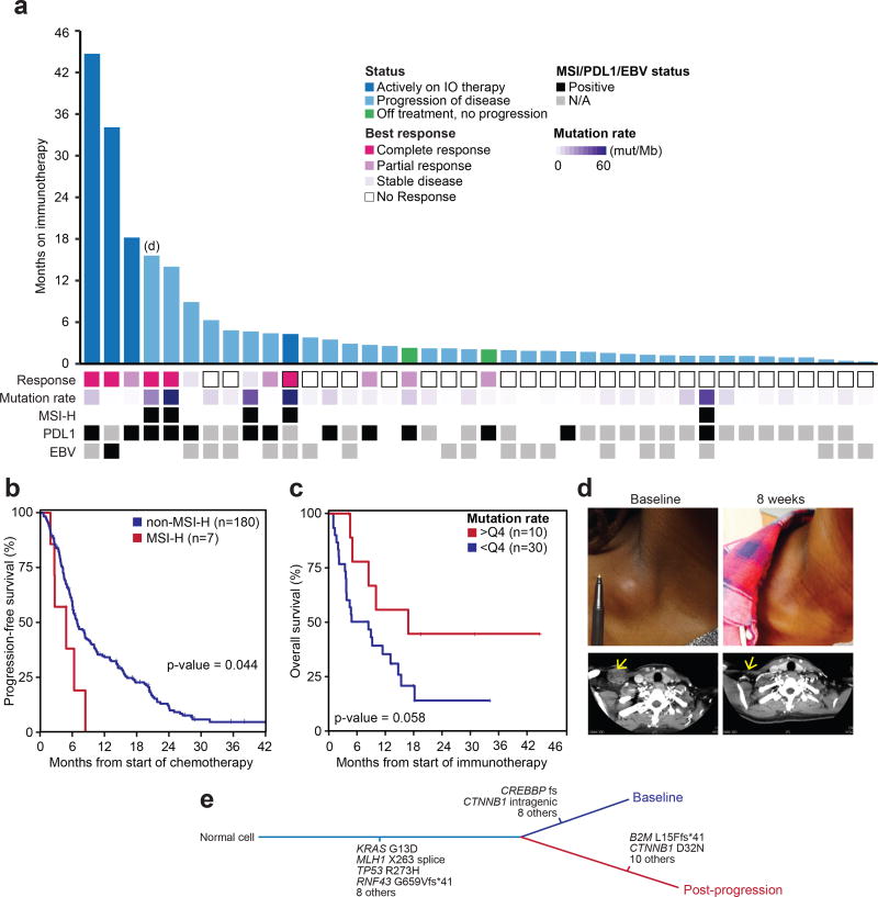
As outlined above, only 9 patients (3%) in the cohort had MSI-H tumors. Patients with MSI-H tumors suffered rapid disease progression on standard cytotoxic therapy, with a significantly shorter PFS on first-line chemotherapy when compared with non-MSI-H tumors (median PFS 4.8 vs 6.9 months for non-MSI-H patients, HR=0.4; P=0.027, log-rank test, Figure 3B), results consistent with those recently reported by the Adjuvant Gastric Chemotherapy MAGIC trial (18). On the basis of the sequencing results and prior data suggesting that MSI-H tumors arising in other disease sites may respond to immune checkpoint inhibitors, MSI-H patients who had progressed on standard cytotoxic chemotherapy were preferentially directed to immunotherapy. These patients received anti-PD1 antibodies (durvalumab, pembrolizumab, nivolumab), alone or in combination with anti-CTLA4 antibodies (ipilimumab, tremelimumab), on clinical trials or as part of compassionate use programs. In total, five patients with MSI-H tumors and 35 patients with non-MSI-H tumors received immune checkpoint inhibitors, with time on therapy ranging from 0.3 to 44.7 months (Figure 3A). Although 28% of patients had radiographic tumor regression with immunotherapy, responses were often transient, and the duration of response to immune checkpoint blockade was less than 6 months in all but five (12.5%) patients. However, all five of these patients remain alive 19.5 to 44.7 months following initiation of immunotherapy despite prior rapid progression on standard fluoropyrimidine, platinum, taxane and irinotecan based therapies. Of these five patients with durable responses to immunotherapy, four tumors tested positive for PDL-L1, whereas one patient had insufficient tumor material for PD-L1 testing. Three of the five patients with durable immunotherapy responses had tumors with high mutational burden (59.4 mut/Mb, 28.3 mut/Mb, and 14.2 mut/Mb), and 2 of these 3 were MSI-H. Overall, higher tumor mutational burden was associated with a better outcome on immunotherapy (Figure 3C), with patients in the top quartile of tumor mutational burden (>9.7 mut/Mb) having the best outcomes (median OS 16.8 compared to 6.62 months for patients with lower mutational burdens; 2 year OS: 44% vs 14%; HR=0.40, log-rank test P=0.058). As two of the five patients with durable outlier responses (>12 months) to immunotherapy had tumors with low mutational burden (1.9 and 3.3 mut/Mb), we explored these tumors in greater detail by performing EBV in situ hybridization. Notably, the outlier responder with the second longest duration on immunotherapy (>30 months and still on therapy) was EBV-positive, the only EBV+ tumor (of the 26 tested) in the cohort.
Figure 3. Genomic determinants of response to immune checkpoint inhibitors.
A, Months on immune checkpoint inhibitors for 40 patients with metastatic, chemotherapy-refractory esophagogastric cancer. The annotation tracks below x-axis indicate EBV and MSI status, mutational burden, and best response to immunotherapy (see legend). B, Kaplan-Meier progression free survival on first-line platinum-based therapy for patients with MSI-H vs non-MSI-H tumors, demonstrating shorter PFS and chemotherapy-resistance in MSI-H esophagogastric cancers. C, Kaplan-Meier overall survival curve of patients receiving immunotherapy demonstrating favorable OS for those in the top quartile of tumor non-synonymous mutational burden (those with >9.7 mut/Mb). D, Photograph and corresponding CT image showing complete response in a biopsy-proven lymph node metastases of a patient with Stage IV MSI-H chemotherapy-refractory esophagogastric cancer treated with anti-PD-1 monotherapy in 4th line setting. E, Genomic comparison of matched pre- and post-progression primary tumor sample from patient in (D):12 mutations were private to the post-treatment sample, including a loss-of-function mutation in exon 1 of the B2M gene, which encodes β2-microglobulin.
Of the five patients who achieved durable responses to immunotherapy lasting 12 month or longer, two have developed acquired resistance. One of these patients with acquired resistance to immune checkpoint blockade is highlighted in Figure 3D. This patient had a PD-L1+, MSI-H, chemotherapy-refractory tumor and achieved a complete response to anti-PD-1 monotherapy followed by disease progression in the distal esophageal tumor at 14 months. Genomic analysis of baseline and post-progression samples identified 33 somatic mutations, including 12 that were private to the post-treatment sample (Figure 3E). Most notably, the post-treatment sample harbored a loss-of-function mutation in exon 1 of the B2M gene, which encodes for β2-microglobulin, loss of which was confirmed by immunohistochemistry. Mutations in B2M have been associated with acquired resistance to immune checkpoint blockade in melanoma (19). In the MSK-cohort, 44% (4 of 9) of MSI-H tumors had likely deleterious alterations in B2M. While the B2M mutation was acquired post-treatment in the patient highlighted in Figure 3D, B2M mutations were present prior to therapy in other patients, and when present did not preclude response to checkpoint blockade.
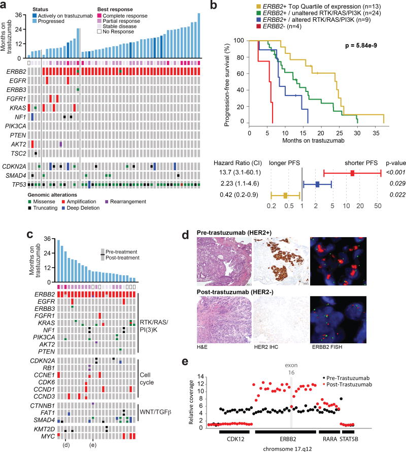
The addition of trastuzumab to cytotoxic chemotherapy is standard-of-care in esophagogastric cancer patients whose tumors overexpress HER2 protein. Of the 295 patients in the MSK-cohort, 68 were HER2+ based upon standard clinical criteria (immunohistochemistry or fluorescence in situ analysis; 44 had samples collected only pre-treatment, 14 only post-treatment, 10 both pre- and post-treatment with trastuzumab). There was a strong correlation between ERBB2 copy number as determined by sequencing and HER2 IHC/FISH (Supplementary Figure 2). Overall, we observed a concordance rate of 93.7% between IHC/FISH and NGS, with a positive predictive value (PPV) of 90% and a negative predictive value (NPV) of 96.9%. A total of 50 patients with HER2+ tumors collected before treatment received first-line trastuzumab. Of these, 92% (46/50) were ERBB2-amplified by NGS (20) (Supplementary Table 4). Detailed analysis of the four discordant patients indicated that the discordance was attributed to either tumor heterogeneity for ERBB2 amplification or equivocal IHC/FISH positivity. Additionally, the four patients with discordant cases exhibited significantly shorter PFS on first-line trastuzumab/chemotherapy compared to patients with ERBB2 amplified tumors by NGS (median PFS 5.8 vs 14.0 months; P=1e-6, log-rank test, Figure 4A, B).
Figure 4. Intrinsic and acquired trastuzumab resistance.
A, Duration, best response, and pre-treatment genomic alterations for 50 patients with HER2+ metastatic esophagogastric cancer treated with first-line trastuzumab/chemotherapy. The first four tumors had no ERBB2 amplification detected by sequencing, the next set of samples had co-alterations in the RTK/RAS/PI3K pathways, and the third set had no co-occurring alterations in these pathways. B, Kaplan-Meier progression free survival curves (top panel) and multivariate analysis (bottom panel) showing favorable outcome in patients with ERBB2-amplified and RTK/RAS/PI3K-wildtype tumors. Patients with tumors that were ERBB2-negative or ERBB2-amplified and RTK/RAS/PI3K pathway-activated had significantly shorter time to progression on first-line trastuzumab therapy, and patients in the highest quartile of ERBB2 levels as determined by sequencing had the best outcome. C, Analysis of somatic alterations in 23 pairs of matched pre- and post-trastuzumab samples. The oncoprint illustrates several oncogenic alterations, grouped by pathway, that are shared between or private to the paired pre- or post-treatment samples. The cells of the oncoprint are split, with the alteration status in the pre- and post-treatment samples shown in the top and bottom, respectively. D, A representative case illustrating loss of ERBB2 amplification and HER2 protein expression in the post-treatment sample, confirmed by FISH and IHC, respectively. E, The structure of the acquired ERBB2 exon 16 deletion in a post-trastuzumab specimen. The relative DNA-sequencing coverage is shown for each exon of ERBB2 and the adjoining genes on chromosome 17, as well as select intragenic regions. The post-trastuzumab sample had a distinct, more focal, amplification that did not include exon 16 of ERBB2.
The level of ERBB2 amplification by FISH has been shown to predict for sensitivity and prolonged survival with trastuzumab therapy in metastatic gastric cancer (21). Here, we also observed a strong correlation between the level of ERBB2 amplification as quantitated by NGS and PFS on first-line trastuzumab, with patients in the top quartile of ERBB2 amplification levels having a significantly longer progression-free survival on trastuzumab (median PFS 24.3, Figure 4B). Beyond ERBB2 itself, we observed significant heterogeneity in the pattern of co-mutational events in the HER2+ cohort. Patients with co-alterations in receptor tyrosine kinase (RTK)-RAS-PI3K/AKT pathway genes had significantly shorter PFS (median PFS 8.4) (Figure 4A, B), suggesting that activation of this pathway may contribute to intrinsic trastuzumab-resistance. In a multivariate analysis, ERBB2 levels and co-alterations in the PI3K pathway independently contributed to the differences in progression-free survival (Figure 4C). Alterations in cell cycle related genes, which were previously reported to be associated with less clinical benefit from trastuzumab based therapy in an Asian population (22,23), were not associated with response differences in the MSK cohort (median time on treatment for patients without a cell cycle alteration was 12.2 months (n = 23), compared to 14.0 months for patients with a cell cycle alteration (n = 27, p = 0.11, log-rank test).
To identify mechanisms of acquired resistance, we analyzed matched tumors collected from individual patients both pre- and post-trastuzumab treatment. Given the small number of post-trastuzumab progression and paired samples in the prospective series, we augmented this cohort with a retrospective analysis of additional paired samples from 20 patients, assembling in total 23 matched pre- and post-trastuzumab tumor pairs. The site of clinical and radiographic disease progression determined the location of the second biopsy, with 11 biopsies obtained from the same anatomical site. Overall, the concordance between genomic alterations found in pre and post-treatment samples was high, and most discordances were attributable to mutations found only in the post-treatment samples (Supplementary Figure 3). In this paired sample analysis, we identified two patients with loss of ERBB2 amplification and one patient with a focal ERBB2 exon 16 deletion exclusively in the sample collected following disease progression on trastuzumab (Figure 4C). In sum, in the 44 post-trastuzumab samples from patients that were HER2+ by clinical IHC/FISH testing prior to treatment with trastuzumab, 7 (16%) were ERBB2-negative by targeted sequencing. HER2 IHC analysis of the post-progression samples used for NGS further confirmed that HER2 expression was either lost or significantly lower at the time of disease progression in all 7 of these patients as compared to their corresponding pretreatment samples. In one representative patient shown in Figure 4D, the pre-trastuzumab tumor was IHC 3+/FISH>20 and ERBB2-amplified by NGS, whereas the sample collected following disease progression on trastuzumab did not express HER2 protein (IHC 0) and had no evidence of ERBB2 amplification by either FISH (FISH 1.1) or sequencing analysis (Figure 4D); additionally, the sample had a newly acquired PIK3CA E545K mutation. Together, these results suggest that selection for a ERBB2 non-amplified clone is a recurrent mechanism of trastuzumab resistance in esophagogastric cancer.
Among the notable somatic alterations in post-trastuzumab tumors not present in pretreatment tumors was a genomic rearrangement resulting in deletion of ERBB2 exon 16 (Figure 4E). This somatic alteration results in expression of the delta-16 HER2 variant, which is constitutively hyperphosphorylated compared to the wild-type isoform, and which has been shown to be resistant to anti-HER2 therapies in preclinical breast cancer models (24–26). Oncogenic co-mutations in KRAS were also identified in one tumor (1/50, 2%) collected prior to trastuzumab treatment and in three tumor samples (3/23, 13%) collected following disease progression. Similarly, known activating PIK3CA mutations were present in one (1/50, 2%) tumor collected prior to trastuzumab therapy and in two (2/23, 8.6%) tumors collected following disease progression. As the presence of a co-occurring alteration in the RTK/RAS/PI3K pathway in pre-treatment tumors was associated with shorter time to progression on trastuzumab therapy, these results suggest that, along with secondary alterations to ERBB2 and selection for a clone lacking ERBB2 amplification, co-mutations that induce RAS or PI3-kinase pathway activation may be mechanisms of both intrinsic and acquired resistance to trastuzumab in patients with esophagogastric cancer.
DISCUSSION
We report the largest experience with prospective next-generation sequencing using a comprehensive cancer gene panel to guide therapy and identify predictive biomarkers of drug response in patients with esophagogastric cancer. We demonstrated that multiplex sequencing of tumor and matched blood samples from esophagogastric cancer patients is efficient and permits interpretation and utilization of results in clinical practice. We generated an extensive dataset of manually reviewed mutations, copy-number alterations, and genomic rearrangements from 318 tumors from 295 patients with mature clinical annotation of treatment response and survival analyses on first-line platinum chemotherapy, trastuzumab and immune checkpoint inhibitor therapy. All genomic and clinical data are publically available through the cBioPortal for Cancer Genomics (27,28) (http://www.cbioportal.org/study?id=egc_msk_2017) to facilitate integration of this dataset with those generated by other institutions. Within the context of the AACR Project GENIE consortium (29), we have also committed to making all future data as part of our clinical sequencing of esophagogastric cancer patients publically available promptly upon data generation.
Based on FDA-approval of trastuzumab and pembrolizumab, reflex ERBB2 and MSI testing with the goal of guiding treatment selection in patients with esophagogastric cancer should now be standard practice. Among Level 2 alterations identified in the MSK cohort, BRCA1/2 alterations may have a role in identifying patients likely to respond to poly (ADP-ribose) polymerase (PARP) inhibitors or platinum chemotherapies. Notably, among the potentially targetable kinase targets identified (ERBB2, EGFR, MET, CDK4, FGFR1), many were found to be concurrent in individual patients, suggesting that the clinical actionability of these mutations will likely depend on developing effective combination strategies.
NGS analysis identified patients with ERBB2-amplified or MSI-H tumors with high concordance with standard assays. Within the clinical HER2+ cohort (as defined by IHC/FISH), patients with ERBB2 amplified, RAS/PI3K wild-type tumors derived the greatest benefit from trastuzumab-based therapy, with clinical benefit greatest in those patients with the highest levels of ERBB2 amplification. Notably, 30% of clinical HER2+ patients lacked ERBB2 amplification by sequencing or had co-mutations in the RTK-RAS-PI3K pathway, and such patients had rapid disease progression and minimal benefit from trastuzumab based therapy. ERBB2 amplification as defined by NGS may thus be a more robust biomarker of clinically meaningful response to trastuzumab than current IHC/FISH testing. In several of the patients with HER2 discordance between FISH/IHC and NGS, discordance could be attributed to tumor heterogeneity in regards to ERBB2 amplification. These results and the loss of ERBB2 amplification in tumors collected post-progression on trastuzumab suggest that selection for a non-ERBB2 amplified clone is a common mechanism of acquired resistance to trastuzumab-based therapy in patients with esophagogastric cancer.
Patients with MSI-H tumors could be robustly identified by next generation sequencing. Our data indicate that in the metastatic setting MSI-H esophagogastric tumors are rare, but that such patients may represent a chemotherapy refractory subset. Pembrolizumab was recently FDA-approved for MSI-H tumors, irrespective of site-of-origin, and the data presented here suggest that immunotherapy should be considered in MSI-H esophagogastric patients early in their disease course as such patients are unlikely to respond to cytotoxic chemotherapy. We also observed a complete and durable response (32 months and ongoing) to immune checkpoint blockade in the only patient with an EBV+ tumor. This dramatic outlier response is consistent with the activity of immunotherapy in other virus-associated tumors, such as Merkel cell carcinoma (30), and suggests that routine EBV testing may aid in the prospective identification of esophagogastric cancer patients most likely to benefit from immunotherapy.
A limitation of the current study was that the targeted capture approach employed could not by definition detect alterations in genes not included in the assay design, epigenetic mechanisms of gene suppression such as promoter methylation of the BRCA1/2 genes, or viral EBV DNA sequences. To address the latter, probes designed to capture viral DNA sequences will be included in future iterations of our clinical NGS platform. This study also highlights that tumor heterogeneity and acquisition of additional mutational events under the selective pressure of therapy is common in esophagogastric cancer. Sampling a single site of disease can never fully assess clonal complexity and tumor heterogeneity in patients with multi-site metastatic disease. Therefore, circulating cell free DNA methods capable of detecting genomic alterations present in genomically heterogeneous metastatic sites should be pursued in future studies of this disease.
While the favorable overall survival we observed in the MSK cohort compared to the published literature may have been due in part to access to novel therapies such as immune checkpoint inhibitors, it also likely reflects the high proportion of patients with ECOG 0–1 functional status (90% of patients) that were thus sufficiently fit to receive 2nd and 3rd line therapies. Additional clinical factors such as a multidisciplinary approach, specialized nursing care, frequent symptom reporting and aggressive early intervention in a highly specialized practice likely further contributed to the favorable outcomes of esophagogastric cancer patients in the MSK cohort compared to published data and warrant future study.
In sum, the results reported here indicate that targeted sequencing methods can robustly identify established and investigational biomarkers of treatment response and drug resistance, including MSI status, ERBB2 amplification, and others, and can potentially guide choice of therapy. Given the limited material available for genomic profiling and the high degree of genomic heterogeneity present in esophagogastric tumors, a multiplexed approach to the detection of multiple known biomarkers of response, possibly using tumor derived cell free DNA as input, will be needed to realize the promise of precision medicine in patients with this aggressive and often fatal disease.
METHODS
Patients with metastatic esophageal, gastric, and gastroesophageal junction adenocarcinoma receiving therapy at Memorial Sloan Kettering Cancer Center (MSK) were consented to an institutional review board approved protocol for prospective tumor genomic profiling between February 2014 and February 2017. The studies were conducted in accordance with the Declaration of Helsinki, International Ethical Guidelines for Biomedical Research Involving Human Subjects (CIOMS), Belmont Report, or U.S. Common Rule.
All tumors were prospectively reviewed to confirm histologic subtype, Lauren classification, and to estimate tumor content. Of 376 tumors submitted for sequencing, 318 samples were included in the final analysis (see CONSORT diagram in Supplementary Figure 4). We integrated genomic data with clinical characteristics, treatment history, response, and survival data (as of September 2017). Overall survival (OS) time was measured from the date of diagnosis of Stage IV disease until the date of death or last follow-up. Progression-free survival (PFS) and OS on first-line platinum therapy and first-line chemotherapy with trastuzumab and immune checkpoint inhibitors in chemotherapy-refractory patients was calculated from the date of start of treatment to the date of radiographic disease progression, death or last evaluation. Clinical HER2 status was based on HER2 protein expression by immunohistochemistry (IHC, positive defined as 3+) or ERBB2 gene amplification by FISH using College of American Pathologists (CAP)/American Society of Clinical Oncology (ASCO) criteria. IHC analysis of mismatch repair (MMR) proteins, and beta 2 microglobulin (B2M), and Epstein-Barr encoding region (EBER) in situ hybridization analysis was performed on a subset of tumors from patients treated with checkpoint inhibitors.
The MSK-IMPACT assay was performed in a CLIA-certified laboratory, initially using a 341 and more recently 410 and 468 gene panels (Supplementary Table 5), as previously described, with results reported in the electronic medical record (12,31). The assay is capable of detecting mutations, small insertions and deletions, copy number alterations and select structural rearrangements. In a previously published validation set, ERBB2 amplification calls on this NGS assay had an overall concordance of 98.4% with the combined IHC/FISH results (20). The PPV and NPV of our NGS assay with respect to HER2 IHC/FISH was calculated in this cohort.
Tumors were assigned to consensus TCGA molecular subtypes: CIN, GS, EBV, MSI-H (3). We assessed tumors for microsatellite instability (MSI) using the MSI-sensor method, and samples with a score >=10 were classified as MSI-high (MSI-H). Tumors were characterized as genomically stable (GS) if the fraction of the autosomal genome affected by DNA copy number alterations of any kind (FGA) was less than 5%. We classified tumors that were neither EBV-positive, MSI, or GS as CIN (chromosomal instability). The OncoKB Precision Oncology Knowledge Base was used (data from May 2017) (14) to infer the oncogenic effect and clinical actionability of individual somatic mutations. Recurrent mutational hotspots were annotated using cancerhotspots.org (32). We inferred allele-specific DNA copy number using FACETS (33) to determine the zygosity of key mutant tumor suppressors as well as to generate estimates of tumor purity. We also inferred large-scale state transition (LST) scores (34), based on the copy number data, from tumors with an analytically estimated tumor purity greater than 20%. Samples with <20% tumor content were excluded from the analysis.
Supplementary Material
SIGNIFICANCE.
Clinical application of multiplex sequencing can identify biomarkers of treatment response to contemporary systemic therapies in metastatic esophagogastric cancer. This large prospective analysis sheds light on the biologic complexity and the dynamic nature of therapeutic resistance in metastatic esophagogastric cancers.
Acknowledgments
Financial Support: 2013 Conquer Cancer Foundation ASCO Career Development Award (Y.Y.J), Cycle for Survival Award (Y.Y.J and N.S), the Department of Defense Congressionally Directed Medical Research Program CA 150646 (Y.Y.J), Marie-Josée and Henry R. Kravis Center for Molecular Oncology, National Cancer Institute Cancer Center Core Grant (P30-CA008748), Robertson Foundation (B.S.T. and N.S.), Center for Metastasis Research of the Sloan Kettering Institute (F.S.V.)
Disclosure of Potential Conflicts of Interest: Y.Y. Janjigian has received research funding from Boehringer Ingelheim, Bayer, Genentech/Roche, Bristol-Myers Squibb, Eli Lilly, Merck and serves on advisory boards for Merck Serono, Bristol-Myers Squibb, Eli Lilly, Pfizer, Merck.
References
- 1.Siegel RL, Miller KD, Jemal A. Cancer Statistics, 2017. CA: Cancer J Clin. 2017;67:7–30. doi: 10.3322/caac.21387. [DOI] [PubMed] [Google Scholar]
- 2.Cancer Genome Atlas Research Network, Analysis Working Group: Asan University, BC Cancer Agency, Brigham and Women’s Hospital, Broad Institute, Brown University et al. Integrated genomic characterization of oesophageal carcinoma. Nature. 2017;541:169–75. doi: 10.1038/nature20805. [DOI] [PMC free article] [PubMed] [Google Scholar]
- 3.Cancer Genome Atlas Research Network. Comprehensive molecular characterization of gastric adenocarcinoma. Nature. 2014;513:202–9. doi: 10.1038/nature13480. [DOI] [PMC free article] [PubMed] [Google Scholar]
- 4.Cristescu R, Lee J, Nebozhyn M, Kim K-M, Ting JC, Wong SS, et al. Molecular analysis of gastric cancer identifies subtypes associated with distinct clinical outcomes. Nat Med. 2015;21:449–56. doi: 10.1038/nm.3850. [DOI] [PubMed] [Google Scholar]
- 5.Secrier M, Li X, de Silva N, Eldridge MD, Contino G, Bornschein J, et al. Mutational signatures in esophageal adenocarcinoma define etiologically distinct subgroups with therapeutic relevance. Nat Genet. 2016;48:1131–41. doi: 10.1038/ng.3659. [DOI] [PMC free article] [PubMed] [Google Scholar]
- 6.Elimova E, Janjigian YY, Mulcahy M, Catenacci DV, Blum MA, Almhanna K, et al. It Is Time to Stop Using Epirubicin to Treat Any Patient With Gastroesophageal Adenocarcinoma. J Clin Oncol Off J Am Soc Clin Oncol. 2017;35:475–7. doi: 10.1200/JCO.2016.69.7276. [DOI] [PubMed] [Google Scholar]
- 7.Bang Y-J, Van Cutsem E, Feyereislova A, Chung HC, Shen L, Sawaki A, et al. Trastuzumab in combination with chemotherapy versus chemotherapy alone for treatment of HER2-positive advanced gastric or gastro-oesophageal junction cancer (ToGA): a phase 3, open-label, randomised controlled trial. Lancet. 2010;376:687–97. doi: 10.1016/S0140-6736(10)61121-X. [DOI] [PubMed] [Google Scholar]
- 8.Muro K, Chung HC, Shankaran V, Geva R, Catenacci D, Gupta S, et al. Pembrolizumab for patients with PD-L1-positive advanced gastric cancer (KEYNOTE-012): a multicentre, open-label, phase 1b trial. Lancet Oncol. 2016;17:717–26. doi: 10.1016/S1470-2045(16)00175-3. [DOI] [PubMed] [Google Scholar]
- 9.Le DT, Uram JN, Wang H, Bartlett BR, Kemberling H, Eyring AD, et al. PD-1 Blockade in Tumors with Mismatch-Repair Deficiency. New Engl J Med. 2015;372:2509–20. doi: 10.1056/NEJMoa1500596. [DOI] [PMC free article] [PubMed] [Google Scholar]
- 10.Fuchs CS, Doi T, Jang RW-J, Muro K, Satoh T, Machado M, et al. KEYNOTE-059 cohort 1: Efficacy and safety of pembrolizumab (pembro) monotherapy in patients with previously treated advanced gastric cancer. J Clin Oncol. American Society of Clinical Oncology. 2017;35:4003–4003. [Google Scholar]
- 11.Kang Y-K, Boku N, Satoh T, Ryu M-H, Chao Y, Kato K, et al. Nivolumab in patients with advanced gastric or gastro-oesophageal junction cancer refractory to, or intolerant of, at least two previous chemotherapy regimens (ONO-4538-12, ATTRACTION-2): a randomised, double-blind, placebo-controlled, phase 3 trial. Lancet. 2017 doi: 10.1016/S0140-6736(17)31827-5. [DOI] [PubMed] [Google Scholar]
- 12.Zehir A, Benayed R, Shah RH, Syed A, Middha S, Kim HR, et al. Mutational landscape of metastatic cancer revealed from prospective clinical sequencing of 10,000 patients. Nat Med. 2017;23:703–13. doi: 10.1038/nm.4333. [DOI] [PMC free article] [PubMed] [Google Scholar]
- 13.Niu B, Ye K, Zhang Q, Lu C, Xie M, McLellan MD, et al. MSIsensor: microsatellite instability detection using paired tumor-normal sequence data. Bioinforma. 2014;30:1015–6. doi: 10.1093/bioinformatics/btt755. [DOI] [PMC free article] [PubMed] [Google Scholar]
- 14.Chakravarty D, Gao J, Phillips SM, Kundra R, et al. OncoKB: A Precision Oncology Knowledge Base. JCO Precis Oncol. 2017 Jul. doi: 10.1200/PO.17.00011. [DOI] [PMC free article] [PubMed] [Google Scholar]
- 15.Ohtsu A, Shah MA, Van Cutsem E, Rha SY, Sawaki A, Park SR, et al. Bevacizumab in combination with chemotherapy as first-line therapy in advanced gastric cancer: a randomized, double-blind, placebo-controlled phase III study. J Clin Oncol Off J Am Soc Clin Oncol. 2011;29:3968–76. doi: 10.1200/JCO.2011.36.2236. [DOI] [PubMed] [Google Scholar]
- 16.Mirza MR, Monk BJ, Herrstedt J, Oza AM, Mahner S, Redondo A, et al. Niraparib Maintenance Therapy in Platinum-Sensitive, Recurrent Ovarian Cancer. New Engl J Med. 2016;375:2154–64. doi: 10.1056/NEJMoa1611310. [DOI] [PubMed] [Google Scholar]
- 17.Telli ML, Timms KM, Reid J, Hennessy B, Mills GB, Jensen KC, et al. Homologous Recombination Deficiency (HRD) Score Predicts Response to Platinum-Containing Neoadjuvant Chemotherapy in Patients with Triple-Negative Breast Cancer. Clinical cancer research: an official journal of the American Association for Cancer Research. 2016;22:3764–73. doi: 10.1158/1078-0432.CCR-15-2477. [DOI] [PMC free article] [PubMed] [Google Scholar]
- 18.Smyth EC, Wotherspoon A, Peckitt C, Gonzalez D, Hulkki-Wilson S, Eltahir Z, et al. Mismatch Repair Deficiency, Microsatellite Instability, and Survival: An Exploratory Analysis of the Medical Research Council Adjuvant Gastric Infusional Chemotherapy (MAGIC) Trial. JAMA oncology. 2017;3:1197–203. doi: 10.1001/jamaoncol.2016.6762. [DOI] [PMC free article] [PubMed] [Google Scholar]
- 19.Zaretsky JM, Garcia-Diaz A, Shin DS, Escuin-Ordinas H, Hugo W, Hu-Lieskovan S, et al. Mutations Associated with Acquired Resistance to PD-1 Blockade in Melanoma. New Engl J Med. 2016;375:819–29. doi: 10.1056/NEJMoa1604958. [DOI] [PMC free article] [PubMed] [Google Scholar]
- 20.Ross DS, Zehir A, Cheng DT, Benayed R, Nafa K, Hechtman JF, et al. Next-Generation Assessment of Human Epidermal Growth Factor Receptor 2 (ERBB2) Amplification Status: Clinical Validation in the Context of a Hybrid Capture-Based, Comprehensive Solid Tumor Genomic Profiling Assay. J Mol Diagn JMD. 2017;19:244–54. doi: 10.1016/j.jmoldx.2016.09.010. [DOI] [PMC free article] [PubMed] [Google Scholar]
- 21.Gomez-Martin C, Plaza JC, Pazo-Cid R, Salud A, Pons F, Fonseca P, et al. Level of HER2 gene amplification predicts response and overall survival in HER2-positive advanced gastric cancer treated with trastuzumab. J Clin Oncol Off J Am Soc Clin Oncol. 2013;31:4445–52. doi: 10.1200/JCO.2013.48.9070. [DOI] [PubMed] [Google Scholar]
- 22.Lee JY, Hong M, Kim ST, Park SH, Kang WK, Kim K-M, et al. The impact of concomitant genomic alterations on treatment outcome for trastuzumab therapy in HER2-positive gastric cancer. Scientific reports. 2015;5:9289. doi: 10.1038/srep09289. [DOI] [PMC free article] [PubMed] [Google Scholar]
- 23.Kim J, Fox C, Peng S, Pusung M, Pectasides E, Matthee E, et al. Preexisting oncogenic events impact trastuzumab sensitivity in ERBB2-amplified gastroesophageal adenocarcinoma. The Journal of clinical investigation. 2014;124:5145–58. doi: 10.1172/JCI75200. [DOI] [PMC free article] [PubMed] [Google Scholar]
- 24.Castiglioni F, Tagliabue E, Campiglio M, Pupa SM, Balsari A, Ménard S. Role of exon-16-deleted HER2 in breast carcinomas. Endocrine-related Cancer. 2006;13:221–32. doi: 10.1677/erc.1.01047. [DOI] [PubMed] [Google Scholar]
- 25.Turpin J, Ling C, Crosby EJ, Hartman ZC, Simond AM, Chodosh LA, et al. The ErbB2ΔEx16 splice variant is a major oncogenic driver in breast cancer that promotes a pro-metastatic tumor microenvironment. Oncogene. 2016;35:6053–64. doi: 10.1038/onc.2016.129. [DOI] [PMC free article] [PubMed] [Google Scholar]
- 26.Tagliabue E, Campiglio M, Pupa SM, Balsari A, Ménard S. The HER2 World: Better Treatment Selection for Better Outcome. J Natl Cancer Institute Monogr. 2009;2011:82–5. doi: 10.1093/jncimonographs/lgr041. [DOI] [PubMed] [Google Scholar]
- 27.Cerami E, Gao J, Dogrusoz U, Gross BE, Sumer SO, Aksoy BA, et al. The cBio cancer genomics portal: an open platform for exploring multidimensional cancer genomics data. Cancer Discov. 2012;2:401–4. doi: 10.1158/2159-8290.CD-12-0095. [DOI] [PMC free article] [PubMed] [Google Scholar]
- 28.Gao J, Aksoy BA, Dogrusoz U, Dresdner G, Gross B, Sumer SO, et al. Integrative analysis of complex cancer genomics and clinical profiles using the cBioPortal. Sci Signal. 2013;6:pl1. doi: 10.1126/scisignal.2004088. [DOI] [PMC free article] [PubMed] [Google Scholar]
- 29.AACR Project GENIE Consortium. AACR Project GENIE: Powering Precision Medicine through an International Consortium. Cancer Discov. 2017 doi: 10.1158/2159-8290.CD-17-0151. [DOI] [PMC free article] [PubMed] [Google Scholar]
- 30.Gulley JL, Rajan A, Spigel DR, Iannotti N, Chandler J, Wong DJL, et al. Avelumab for patients with previously treated metastatic or recurrent non-small-cell lung cancer (JAVELIN Solid Tumor): dose-expansion cohort of a multicentre, open-label, phase 1b trial. Lancet Oncol. 2017;18:599–610. doi: 10.1016/S1470-2045(17)30240-1. [DOI] [PMC free article] [PubMed] [Google Scholar]
- 31.Cheng DT, Mitchell TN, Zehir A, Shah RH, Benayed R, Syed A, et al. Memorial Sloan Kettering-Integrated Mutation Profiling of Actionable Cancer Targets (MSK-IMPACT): A Hybridization Capture-Based Next-Generation Sequencing Clinical Assay for Solid Tumor Molecular Oncology. J Mol Diagn JMD. 2015;17:251–64. doi: 10.1016/j.jmoldx.2014.12.006. [DOI] [PMC free article] [PubMed] [Google Scholar]
- 32.Chang MT, Asthana S, Gao SP, Lee BH, Chapman JS, Kandoth C, et al. Identifying recurrent mutations in cancer reveals widespread lineage diversity and mutational specificity. Nat Biotechnol. 2016;34:155–63. doi: 10.1038/nbt.3391. [DOI] [PMC free article] [PubMed] [Google Scholar]
- 33.Shen R, Seshan VE. FACETS: allele-specific copy number and clonal heterogeneity analysis tool for high-throughput DNA sequencing. Nucleic acids Res. 2016;44:e131. doi: 10.1093/nar/gkw520. [DOI] [PMC free article] [PubMed] [Google Scholar]
- 34.Marquard AM, Eklund AC, Joshi T, Krzystanek M, Favero F, Wang ZC, et al. Pan-cancer analysis of genomic scar signatures associated with homologous recombination deficiency suggests novel indications for existing cancer drugs. Biomark Res. 2015;3:9. doi: 10.1186/s40364-015-0033-4. [DOI] [PMC free article] [PubMed] [Google Scholar]
Associated Data
This section collects any data citations, data availability statements, or supplementary materials included in this article.


