Abstract
Euglena gracilis Z (Euglena) is a unicellular, photosynthesizing, microscopic green alga. It contains several nutrients such as vitamins, minerals, and unsaturated fatty acids. In this study, to verify the potential role of Euglena consumption on human health and obesity, we evaluated the effect of Euglena on human adipose-derived stem cells. We prepared a Euglena extract and evaluated its effect on cell growth and lipid accumulation, and found that cell growth was promoted by the addition of the Euglena extract. Interestingly, intracellular lipid accumulation was inhibited in a concentration-dependent manner. Quantitative real-time PCR analysis and western blotting analysis indicated that the Euglena extract suppressed adipocyte differentiation by inhibiting the gene expression of the master regulators peroxisome proliferator-activated receptor-γ (PPARγ) and one of three CCAAT-enhancer-binding proteins (C/EBPα). Further Oil Red O staining experiments indicated that the Euglena extract inhibited the early stage of adipocyte-differentiation. Consistent with these results, we observed that down-regulation of gene expression was involved in the early stage of adipogenesis represented by the sterol regulatory element binding protein 1 c (SREBP1c), two of three CCAAT-enhancer-binding proteins (C/EBPβ, C/EBPδ), and the cAMP regulatory element-binding protein (CREB). Taken together, these data suggest that Euglena extract is a promising candidate for the development of a new therapeutic treatment for obesity.
Introduction
Obesity is an abnormal health condition in which body fat is excessively accumulated [1, 2]. Various recent studies have reported that obesity is associated with several chronic diseases, such as type 2 diabetes mellitus (T2DM), asthma, and cardiovascular disease [3–5]. Strikingly, it was reported that there was a positive correlation between body mass index and mortality caused by those diseases [6, 7]. Contrary to these aspects, adipocytes have crucial functions that contribute to the maintenance of lipid homeostasis [8] and intracellular energy balance by storing/releasing triglycerides or fatty acids, depending on the extracellular environment [9, 10]. Thus, to understand the physiology of adipocytes, many studies involving adipocyte-differentiation and its regulatory system have been carried out in the murine 3T3-L1 cells and in vivo.
The peroxisome proliferator-activated receptor-γ (PPARγ) and the CCAAT-enhancer-binding protein family (C/EBPα, C/EBPβ, C/EBPδ), particularly C/EBPα, have been identified as master regulators that control adipocyte-differentiation, and thus, their physiological roles have been widely analyzed [11]. When cells are exposed to isobutylmethylxanthine (IBMX) or dexamethasone (DEX), C/EBPβ and C/EBPδ are expressed immediately, resulting in the upregulation of PPARγ [12, 13]. Activated PPARγ induces the expression of C/EBPα, which turns on several genes involved in adipocyte-differentiation to mature adipocyte [14].
In addition to C/EBPβ and C/EBPδ, several studies have revealed the physiological role of the sterol regulatory element binding protein 1 (SREBP1), which is associated with adipogenesis, insulin sensitivity, and fatty acid metabolism [15]. SREBP1 exists as two isozymes (SREBP1a and SREBP1c) derived from alternative splicing of the first exon [16]. SREBP1a is predominantly expressed in the spleen, whereas SREBP1c is predominantly expressed in the adipose tissue, liver, adrenal glands, and skeletal muscle in response to insulin [17]. In vitro experiments suggested that SREBP1c contributes to adipocyte-differentiation through the activation of PPARγ as well as C/EBPβ and C/EBPδ, either through the induction of enzymes responsible for endogenous ligand generation and/or by enhancing gene expression [18, 19]. Not only C/EBPβ and C/EBPδ, but also the cAMP regulatory element-binding protein, CREB, which also participates in the early stage of adipocyte-differentiation. Klemm and Lane reported that CREB is activated early during adipocyte-differentiation in response to isobutylmethylxanthine, which increases cellular cAMP in 3T3-L1 cells, resulting in the activation of C/EBPβ [20, 21].
PPARγ is a functional receptor for insulin-sensitizing drugs known as thiazolidinediones, which are used for the treatment of T2DM [22]. Anti-obesity drugs such as sibutramine and orlistat were developed after much pharmacological research to prevent and reduce obesity. However, it has been indicated that intake of these drugs might cause serious adverse reactions such as insomnia, constipation, headache, and cardiovascular stroke [23, 24]. Thus, many researchers have sought anti-obesity drugs that do not cause such adverse reactions. For instance, it has been reported that coffee or seaweed extract suppresses the gene expression of PPARγ and C/EBPα as well as lipid accumulation in the 3T3-L1 cell line, but reductions in cell viability were observed when these additives were used at high concentrations [25, 26]. However, studies on adipocyte -differentiation in human-derived stem cells have been limited. Thus, the development of anti-obesity drugs without adverse reactions against human-derived cells would be promising.
Euglena gracilis Z (Euglena) is a unicellular, photosynthesizing, microscopic green alga found in fresh water. Several studies revealed that the alga belongs to the phylum Euglenozoa, which is a member of Excavata, a root of eukaryote [27, 28]. Furthermore, it is also known that Euglena contains several nutrients such as vitamins, minerals, and unsaturated fatty acids. Moreover, it accumulates the reserve polysaccharide clystalline β-1,3-glucan, known as paramylon, which is considered a functional dietary fiber for health purposes [29, 30]. Therefore, the effect of consumption of Euglena and/or paramylon on human health has been assessed in various studies. For example, it was reported that Euglena consumption alleviated hyperglycemia in OLETF rats [31] and that paramylon reduced the development of atopic dermatitis-like skin lesions in NC/Nga mice [32]. However, to our knowledge, there has been no report on the effect of Euglena consumption on obesity in human-derived stem cells. In this study, as part of finding a new effect of Euglena on human cell, the effects of Euglena on adipocyte-differentiation and lipid accumulation in human adipose-derived stem cells (hASCs) were investigated.
Materials and methods
Preparation of Euglena extract
Euglena dry powder was obtained from euglena Co., Ltd. (Tokyo, Japan), and 0.5 g of powder was suspended in 20 mL of water and heated at 95°C for 2 h. For removal of the insoluble fraction and sterilization, the supernatant was collected after centrifugation and was filtered with a 0.45-μm filter purchased from Merck Millipore Co., Ltd. (Billerica, MA, USA). The sterilized supernatant (called Euglena extract) was used as 100% of concentration and diluted by any concentrations with medium before cultivation of cells. The Euglena extract was stored at 4°C until use.
Cell culture and differentiation
hASCs were purchased from Lonza Walkersville, Inc. (Walkersville, MD, USA); the cells were derived from a female donor (non-diabetic, BMI 26 kg/m2, 36 years old) (PT5006 lot 0000410257). The cells (passage 4) were pre-cultured as described by Yamada et al. [33]. Briefly, 2 × 10⁴ cells/mL of the cells were cultured in 50% Dulbecco’s modified Eagle’s medium (DMEM)/50% α minimum essential medium supplemented with 1% fetal bovine serum (FBS), 1 × ITS, 10 ng mL-1 bFGF (PeproTech Inc., NJ, USA), and 400 ng mL-1 of hydrocortisone on a 24-well plate for 3 days, until cells became confluent. To induce adipocyte-differentiation, post-confluent preadipocytes were stimulated in MDI differentiation medium (DMEM containing 10% FBS, 1 μM DEX, 0.5 mM IBMX, 0.2 mM indomethacin, 10 μg mL-1 insulin, and 33 μM biotin) for 7 days (Day 0–7). During cultivation, the culture was replaced with fresh MDI differentiation medium every 2 days with or without Euglena extract. Then, cells were maintained for a further 7 days (Day 7–14) in adipocyte nutrition medium (DMEM containing 10% FBS, 10 μg mL-1 insulin, and 33 μM biotin), which was replaced with fresh culture every 2 days. All cells were cultured at 37°C under a 5% CO2 atmosphere. All the above reagents were purchased from Sigma-Aldrich (St. Louis, MO, USA).
Determination of cell viability
The cytotoxicity of Euglena extract against hASCs was estimated by a modified MTT assay using the Cell Counting-kit 8 (Dojindo, Kumamoto, Japan); 100% confluent hASCs preadipocytes were cultured in D/α (-) medium (50% DMEM/50% α minimum essential medium (αMEM) supplemented with 1% FBS, 1 × ITS, and 400 ng mL-1 hydrocortisone) on a 96-well plate were treated with various concentrations of water or Euglena extract (1.25%, 2.5%, 5%, 10%, 20%, 40%) at 37°C for 48 h under a 5% CO2 atmosphere. 100 μL of the Cell Counting-Kit 8 solution was added to cells after aspirating the culture supernatant. After incubation for 30 min at 37°C under a 5% CO2 atmosphere, absorbance was measured at 450 nm with the microplate reader SH-1200Lab (Hitachi High-Tech Science Co., Ltd., Tokyo, Japan). Cell viability was determined by comparison of absorbance with the absorbance of the cells treated with no additives as control.
Estimation of lipid accumulation by staining with Oil Red O solution
After adipocyte-differentiation in MDI differentiation medium and adipocyte nutrition medium without or with various concentration of water (5%, 10% or 20%) or Euglena extract (5%, 10% or 20%) for 14 days (Day 0–14) or 7 days (Day 0–7 or Day 8–14) dependent on assay estimating the effect of the extract on lipid accumulation at early or late stage of adipocyte-differentiation. The cells fixed with 4% paraformaldehyde were washed with 500 μL phosphate-buffered saline (PBS), and 500 μL of 60% isopropanol was added to each well. After aspirating the culture supernatant, cells were treated with Oil Red O solution (0.3 g in 100 mL isopropanol; Sigma-Aldrich) for 15 min at room temperature, and the cells were photographed. To estimate adipogenesis, after removing the staining solution, the dye retained in the cells was eluted into 500 μL isopropanol, and absorbance was measured at 520 nm. Oil Red O content of cells was calculated from comparison with the absorbance of the cells treated with no additives as control for induction of adipocyte-differentiation.
RNA extraction and analysis of gene expression
Total RNA was extracted from cells by RNAiso plus (TaKaRa, Shiga, Japan) according to the manufacturer’s instructions. cDNA was synthesized from 200 ng of total RNA using PrimeScript Master Mix (TaKaRa) PCR primers are listed in Table 1 and S1 Table. Quantitative real-time PCR (RT-qPCR) was performed in StepOne Plus using the PowerUp SYBR Green Master Mix (Applied Biosystems Inc., Warrington, UK). The amount of target gene relative to the reference gene (GAPDH) was calculated based on the comparative threshold (Ct) method [34]. All data were normalized with the amount of GAPDH at induction of adipocyte-differentiation (Day 0).
Table 1. Primers used in quantitative RT-qPCR.
| Primers | Sequence (direction: 5´ to 3´) | |
|---|---|---|
| PPARγ | Forward | GAACGACCAAGTAACTCTCCTCAAAT |
| Reverse | TCTTTATTCATCAAGGAGGCCAGCATT | |
| C/EBPα | Forward | GGGTCTGAGACTCCCTTTCCTT |
| Reverse | CTCATTGGTCCCCCAGGAT | |
| C/EBPβ | Forward | GACAAGCACAGCGACGAGTA |
| Reverse | AGCTGCTCCACCTTCTTCTG | |
| C/EBPδ | Forward | TTCAGCGCCTACATCGACTC |
| Reverse | TTGAAGAGGTCGGCGAAGAG | |
| SREBP1c | Forward | TTGAGGACAGCAAGGCAAAG |
| Reverse | GGACAGGCAGAGGAAGACGA | |
| CREB | Forward | ACGAAAGCAGTGACGGAGGA |
| Reverse | TACGGTGGGAGCAGATGATG |
Western blotting analysis
Western blotting analysis was conducted to identify the gene expression of adipogenesis master regulator proteins, PPARγ and C/EBPα. Preparation of cell lysate and western blotting analysis were conducted as described by Aji et al. [35]. Briefly, cells were lysed in RIPA buffer (Wako) containing cOmplete EDTA-free Protease Inhibitor Cocktail (Roche, Mannheim, Germany) for 15 min on ice before sonication. The obtained cell lysate was centrifuged at 12,000 rpm for 15 min at 4°C, and the supernatant was sampled. The protein concentration was evaluated by using the BCA method (TaKaRa). Approximately 25 μg of protein samples were loaded on a 12% SDS-PAGE after denaturation at 95°C for 5 min. After electrophoresis, the separated proteins were transferred to a polyvinylidene difluoride membrane by using WSE-4115 PoweredBlot Ace (ATTO Co. Ltd., Tokyo, Japan), and the membrane was blocked by incubation in TBS-(T) (TBS containing 0.1% Tween 20 (Sigma-Aldrich)) and 5% skimmed milk for 1 h. The membrane was probed with antibodies against PPARγ (1:1000, Cell Signaling TECHNOLOGY, MA, USA), C/EBPα (1:1000, Abcam, Cambridge, UK), or GAPDH (1:2500, Abcam) as an internal loading control overnight at 4°C. The membrane was washed three times with TBS-(T) before treatment with antibody against rabbit IgG conjugated with horseradish peroxidase (1:5000, Abcam) for 1 h at room temperature. Signals were developed using an Amersham ECL western blotting analysis system (GE Healthcare, Little Chalfont, UK) by using MyECL imager (Thermo Fisher Scientific, MA, USA).
Statistical analysis
All data are represented as mean ± SEM from 3 independent experiments. Statistical significance was analyzed using Student’s t-test, and P < 0.05 was considered significant.
Results
Euglena extract inhibits lipid accumulation in adipocyte-differentiation
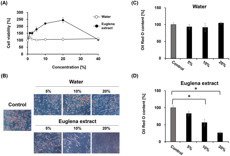
To verify the effects of Euglena on cytotoxicity and lipid accumulation in hASCs, it was considered that dissolving Euglena dry powder into the culture would have been convenient for estimating its effects, but the dry powder could not be dissolved into the cultures. Thus, we prepared a Euglena extract by eluting the powder with water at 95°C. First, to evaluate the effect of the extract on cytotoxicity, hASCs (passage 4, 2 × 10⁴ cells/mL) were cultivated to confluence on DMEM for 3 days, and DMEM was replaced with D/α (-) medium, which maintains the cells. Simultaneously, various concentrations of Euglena extract (1.25%, 2.5%, 5%, 10%, 20%, 40%), water (1.25%, 2.5%, 5%, 10%, 20%, 40%), or without these materials as a control were added to the medium. After the cells were cultured for 48 h, a WST-8 assay was conducted to estimate viable cells and compared to control. Cytotoxicity was not observed by the addition of water or Euglena extract into the culture. Interestingly, treatment with Euglena extract increased cell viability in a concentration-dependent manner, even when cells were at confluence, which raised to approximately 250% compared to control at 20% of concentration (Fig 1(A)). It was reported that some adipose tissue-derived stem cells exhibited apparent lack of contact inhibition and piled up in extended culture [36]. Notably, the hASCs used in this study also exhibited this characteristic in the presence or absence of Euglena extract in D/α (-) medium. This result indicated that Euglena extract exhibited no cytotoxicity against hASCs.
Fig 1. Euglena extract reduces lipid accumulation in hASCs.
(A) Effect of Euglena extract on cell viability. The confluent hASCs were cultured in D/α (-) medium with water (white circles) or Euglena extract (black circles) for 2 days at 37°C under 5% CO2 atmosphere before WST-8 assay. (B) Microscopic images of hASCs stained with Oil Red O. (C) and (D) Relative abundance of accumulated Oil Red O in hASCs treated with water (C) or Euglena extract (D) compared with control (no additive, dark grey bar in (C) and (D)). Data represent the mean ± SEM from 3 independent experiments. *P <0.05 (Student’s t-test).
Euglena extract may represent a source of nutrition for hASCs. To determine whether the Euglena extract inhibited lipid accumulation, confluent hASCs were stimulated with MDI differentiation medium to induce adipocyte-differentiation for 7 days (Day 0 to Day 7). Simultaneously, various concentrations of Euglena extract (5%, 10% or 20%), water (5%, 10% or 20%), or without these materials as control were added to the medium on Day 0. Stimulated cells at Day 14 were stained with Oil Red O and photographed (Fig 1(B)–1(D)). Compared with control, the cells treated with water contained Oil Red O as well as control, indicating that there was no effect on adipocyte differentiation (Fig 1(C)). Interestingly, the Oil Red O content was reduced to 83%, 56%, and 26% by the addition of Euglena extract at concentrations of 5%, 10%, and 20%, respectively (Fig 1(B) and 1(C)). In an attempt to identify the effective compound(s) in the Euglena extract, we prepared and tested another Euglena extract eluted with dimethyl sulfoxide (DMSO extract), and the DMSO extract was added into MDI differentiation medium at various concentrations (1.25%, 2.5%, 5%, or 20%). The cell viability significantly reduced at more than 2.5% of concentration of DMSO, but in the case of DMSO extract, cell piling was observed by 2.5% (S1(A) Fig). Intriguingly, DMSO extract had no effect on adipocyte-differentiation compared with only DMSO (S1(B) and S1(C) Fig). Furthermore, when using Euglena extracts eluted at various temperatures (25°C, 50°C, 75°C, or 120°C), the observed inhibition of adipocyte differentiation was reproduced in all extracts (S2 Fig).
Euglena extract suppresses gene expression of master regulators of adipocyte-differentiation PPARγ and C/EBPα
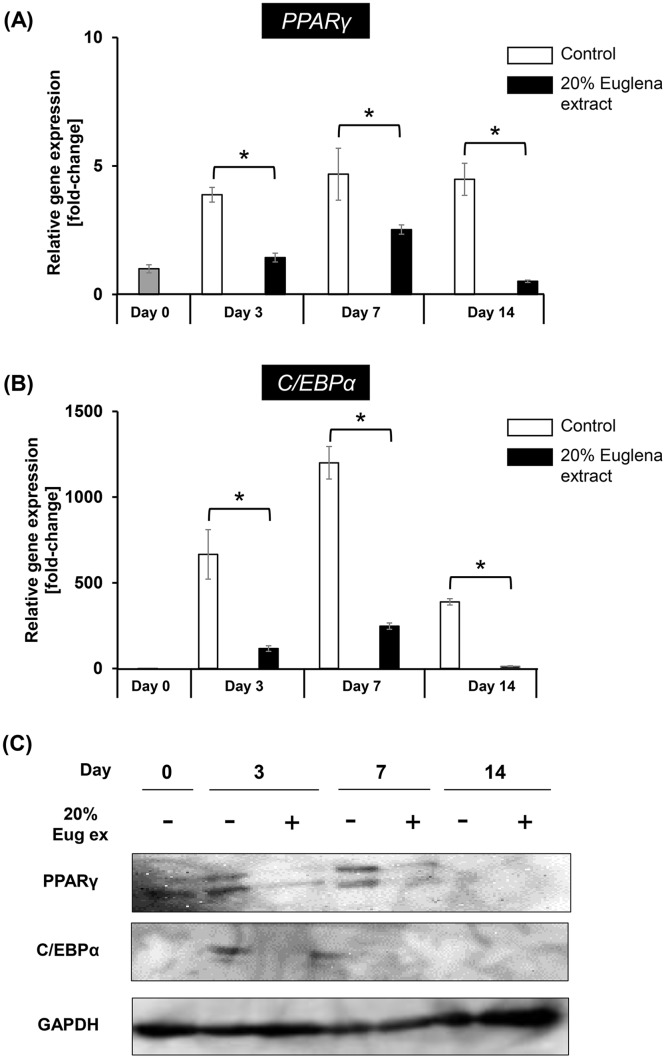
Hitherto, it was indicated that treatment of hASCs with Euglena extract inhibited lipid accumulation by arresting adipocyte-differentiation. Based on this observation, it was speculated that the gene expression of master regulators involved in adipocyte-differentiation, especially PPARγ and C/EBPα, would be inhibited by Euglena extract. To verify the hypothesis, the relative mRNA abundances was measured by RT-qPCR. Cells were cultured in MDI differentiation medium with or without 20% Euglena extract from Day 0 to 7, and then, the MDI differentiation medium was replaced with adipocyte nutrition medium with or without 20% Euglena extract on which cells were cultured for a further 7 days (Day 8–14). During cell cultivation, MDI differentiation medium and adipocyte nutrition medium were exchanged with fresh medium with or without 20% Euglena extract every 2 days. cDNA was synthesized from mRNA extracted on Day 0, Day 3, Day 7, or Day 14 and was used for RT-qPCR. As a result, in controls, the gene expression levels of PPARγ and C/EBPα were elevated during adipocyte-differentiation. Conversely, the gene expression levels of PPARγ and C/EBPα were repressed by 23% on average (Fig 2(A) and 2(B)). Consistent with this result, the protein amount of PPARγ and C/EBPα was significantly reduced by the addition of Euglena extract in adipocyte-differentiation (Fig 2(C)). It is reported that PPARγ and C/EBPα regulate the adipocyte-differentiation marker gene represented to aP2, adiponectin, and LPL, lipoprotein lipase, which promote lipid accumulation to form mature adipocytes [37]. Therefore, it was expected that the gene expression of aP2 and LPL was also reduced. Further RT-qPCR analysis supported this hypothesis, because treatment with Euglena extract on adipocyte-differentiation inhibited the gene expression of aP2 and LPL (S3 Fig). These results indicated that the inhibitory effect of Euglena extract on adipocyte-differentiation was occurring through suppression of master regulators.
Fig 2. Euglena extract represses the gene expression of master regulators of adipocyte-differentiation.
Relative gene abundances of (A) PPARγ and (B) C/EBPα in hASCs after treatment with no additive (control) or 20% Euglena extract for Day 3, Day 7, or Day 14 were monitored by RT-qPCR. Data represent mean ± SEM (n = 3). *P < 0.05 vs. control (Student’s t-test). (C) Western blotting analysis of PPARγ, C/EBPα, and GAPDH (as internal control). Cell lysate (25 μg) derived from soluble fraction was loaded in each lane. The data shown in (C) are representative from 3 independent experiments. Day 0 shown in (A)-(C) means before induction of adipocyte-differentiation.
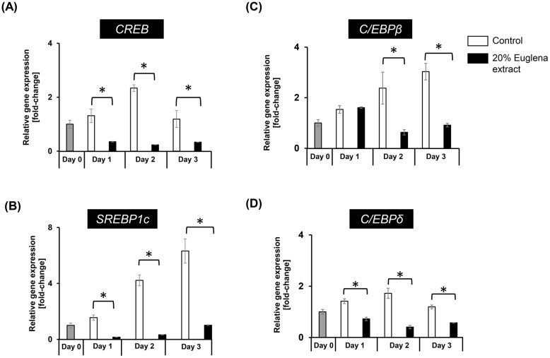
The expression of PPARγ is also activated by C/EBPβ and C/EBPδ at the early phase of adipocyte-differentiation [12, 13]. It is also known that SREBP1c and CREB are involved in the activation of PPARγ expression [18, 19]. To clarify the inhibitory effect of Euglena extract on early stage of adipocyte-differentiation, the abundance of mRNA of CREB, SREBP1c, C/EBPβ, and C/EBPδ was evaluated by using RT-qPCR, after cell cultivation for 1 day (Day 1), 2 days (Day 2), and 3 days (Day 3) with stimulation to differentiate adipogenesis. The gene expression level of CREB and SREBP1c was reduced by the addition of Euglena extract by 22% or 11% on average compared to control (Fig 3(A) and 3(B)). Not only CREB and SREBP1c were affected, but the gene expression level of C/EBPβ and C/EBPδ was also reduced by 48% and 50% on average, respectively. Taken together, these findings indicate that the inhibitory effect of Euglena extract on adipocyte-differentiation was caused by repressing the early stage of adipocyte-differentiation.
Fig 3. Expression of genes involved in the early stage of adipocyte-differentiation was suppressed by Euglena extract.
Relative gene abundances of (A) CREB, (B) SREBP1c, (C) C/EBPβ, and (D) C/EBPδ in hASCs after treatment with no additive (control) or 20% Euglena extract for Day 3, Day 7, or Day 14 were monitored by RT-qPCR. Day 0 indicates before induction of adipocyte-differentiation. Data represent the mean ± SEM (n = 3). *P < 0.05 vs. control (Student’s t-test).
Early stage of adipocyte-differentiation was inhibited by Euglena extract
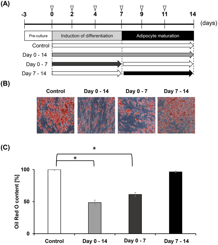
To verify the physiological role for the Euglena extract on the early stage of adipocyte-differentiation, adipogenesis was evaluated by eluting Oil Red O from cells treated with 20% of extract during adipocyte-differentiation and from those cells treated during adipocyte maturation (Fig 4(A)). It was speculated that if the extract had an inhibitory effect on adipocyte-differentiation, supplementation on Day 0–7 would be crucial for lipid accumulation, while if the extract exhibited the inhibition effect on adipogenesis, supplementation on Days 7–14 would be crucial. Compared with controls, constant supplementation (Day 0–14) with Euglena extract inhibited lipid accumulation by approximately 50%. Supplementation with extract on Day 0–7 also inhibited lipid accumulation by approximately 60%. Notably, approximately 96% of the accumulated lipids remained in the cells that were treated with the extract on Day 7–14 (Fig 4(B) and 4(C)). These results could support that Euglena extract suppresses adipocyte-differentiation at the early stage.
Fig 4. Euglena extract suppresses adipocyte-differentiation.
(A) Experimental scheme. Inverted triangles show medium exchange points with or without Euglena extract. White bar shows no additive as control. Euglena extract (20%) was added to hASCs during adipocyte differentiation and maturation (Day 0–14, grey bar), induction of differentiation only (Day 0–7, dark grey bar), or adipocyte maturation only (Day 7–14, black bar). (B) Microscopic images of hASCs stained with Oil Red O. (C) Relative Oil Red O content in hASCs treated with Euglena extract compared with controls (no additive). Data represent mean ± SEM (n = 3). *P < 0.05 vs. control (Student’s t-test).
Discussion
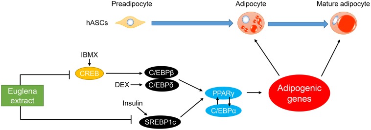
Algae have been increasingly recognized as resources of bioactive compounds for the improvement of human health [38]. Several recent studies have revealed that treatment with Euglena improves hyperglycemia and induces apoptosis of lung and breast cancer cells [31, 39]. However, to our knowledge, there has been no report showing the influence of Euglena on adipogenesis in hASCs. In the present study, the inhibitory effect of Euglena extract on adipocyte-differentiation in hASCs was evaluated, and the Euglena extract was capable of inhibiting adipocyte-differentiation without cytotoxicity (Fig 1). The regulatory mechanism of adipocyte-differentiation, in particular, the roles of the C/EBPα and PPARγ has been well documented [12, 40, 41]. RT-qPCR analysis and western blotting analysis for C/EBPα and PPARγ revealed that the Euglena extract inhibited these master regulators for adipocyte-differentiation at mRNA and protein levels, and further analysis indicated that other regulator genes of early stage of adipogenesis, C/EBPβ, C/EBPδ, and SREBP1c, were suppressed by the Euglena extract (Figs 2 and 3). Consistent with these results, it might be suggested that the Euglena extract inhibited lipid accumulation at the early stage of adipocyte-differentiation (Fig 4). Therefore, the above results allowed us to propose a working model for the effect of Euglena extract (Fig 5). However, the mechanisms underlying our observations remain unclear.
Fig 5. Proposed working model for the inhibitory effect of Euglena extract on adipocyte-differentiation.
In the early phase of adipogenesis, CREB activated by IBMX induces the expression of C/EBPβ, and C/EBPδ is induced by DEX [12, 13, 20]. Another transcription factor, SREBP1c, is induced by insulin [17]. These transcription factors induce PPARγ, which also induces C/EBPα. Cross-regulation exists between PPARγ and C/EBPα and is considered a key component of transcriptional control for adipogenic genes [14]. Our observations indicate that Euglena extract inhibits the gene expression of CREB and SREBP1c, resulting in the downregulation of PPARγ and C/EBPα and inhibition of adipocyte-differentiation (Figs 2 and 3).
In the search for natural products having an antiobesity effect, several researchers have reported that coffee, extract from red and brown algae, pomegranate seed, or brown seaweed exhibited an antiobesity effect [25, 26, 42, 43]. Although fucoxanthin derived from brown algae and xanthigen, the mixture of fucoxanthin and punicic acid, derived from pomegranate seed and brown seaweed, have been known to significantly suppress adipocyte-differentiation through an inhibition of gene expression of the PPARγ and C/EBPs family [44, 45], there is no report that Euglena is capable of synthesis of these compounds. To identify the antiobesity compound(s) in the Euglena extract, a metabolome analysis was conducted, but fucoxanthin and punicic acid were not detected, with the exception of basic metabolites such as carbohydrates, amino acids, vitamins, and nucleic acids.
One possibility for the mechanism underlying the downregulation of C/EBPβ/δ by the Euglena extract could be that some compounds inhibited the enzymatic activity or gene expression of CREB, which participates in the induction of C/EBPβ/δ [46, 47]. Recently, lanostane, a triterpene derived from the lanosterol found in the fruiting bodies of Ganoderma lucidum, was found to repress not only PPARγ, but also C/EBPα and SREBP1 expression in the 3T3-L1 cell line [48]. The lanosterol synthesis pathway, in which lanosterol is synthesized from squalene and converted into 24,25-dihydrolanosterol or water-soluble 24-methylene lanosterol, is conserved among mammals and fission yeasts [49]. An earlier study also reported that Euglena is capable of synthesizing up to 40% of the total sterols, such as squalene, triterpenes, and 4α-methylsterols, including water-soluble 24-methylene lanosterol [50]. According to these reports, it is speculated that Euglena can produce lanosterol, and thus one of the effective components in Euglena extract may be lanostane. However, the complete genome sequence of Euglena is not yet available, and whether it has the ability to convert lanosterol into lanostane remains unclear. Thus, the active component(s) in Euglena extract should be further investigated and identified to elucidate the underlying mechanism of the inhibitory effect of the extract on adipocyte-differentiation in hASCs.
Supporting information
(XLSX)
(PDF)
(A) Effect of Euglena extract by elution with DMSO (DMSO extract) on cell viability. The confluent hASCs were cultured in D/α (-) medium with DMSO (white squares) or DMSO extract (black squares) for 2 days at 37°C under 5% CO2 atmosphere before WST-8 assay. (B) and (C) Relative abundance of accumulated Oil Red O in hASCs treated with DMSO (B) or DMSO extract (C) compared with control (no additive, dark grey bar in (B) and (C)). Data represent mean ± SEM from 3 independent experiments. *P <0.05 (Student’s t-test).
(TIF)
hASCs were stimulated and cultured in MDI medium to induce adipocyte-differentiation from Day 0 to Day 14 with Euglena extract (grey; 5%, dark grey; 10% or black bar; 20%) or without (as control, shown as Ctr, white bar). At Day 14, cells were fixed and stained with Oil Red O solution to determine Oil Red O content compared with control. Data represent mean ± SEM (n = 3). * P < 0.05 (Student’s t-test).
(TIF)
Relative abundance of mRNA of (left panel) aP2 and (right panel) LPL in hASCs after treatment with no additive (white bar, as control) or 20% Euglena extract (black bar) for 1, 2, or 3 days compared with control. Day 0 means before induction of adipocyte-differentiation. Data represent mean ± SEM (n = 3). *P < 0.05 (Student’s t-test).
(TIF)
Acknowledgments
We would like to thank Editage (www.editage.jp) for English language editing.
Data Availability
Data are available from figshare: https://figshare.com/s/b5719689d4975233db78.
Funding Statement
This research did not receive any specific grant from funding agencies in the public, commercial, or not-for-profit sectors. But, we are employed by euglena Co., Ltd., and we apologize for inadequate description. To be exact, the funder provided support in the form of salaries for authors RS, NO, KA, YM, OI, AN, KS, but did not have any additional role in the study design, data collection and analysis, decision to publish, or preparation of the manuscript.
References
- 1.Kershaw EE, Flier JS. Adipose tissue as an endocrine organ. J Clin Endocrinol Metab. 2004;89: 2548–2556. doi: 10.1210/jc.2004-0395 [DOI] [PubMed] [Google Scholar]
- 2.Spiegelman BM, Flier JS. Adipogenesis and obesity: rounding out the big picture. Cell. 1996;87: 377–389. [DOI] [PubMed] [Google Scholar]
- 3.Thompson D, Edelsberg J, Coditz GA, Bird AP, Oster G. Lifetime health and economic consequences of obesity. Arch Intern Med. 1999;159(18): 2177–2183. [DOI] [PubMed] [Google Scholar]
- 4.Reilly JJ, Methven E, McDowell ZC, Hacking B, Alexander D, Stewart L, et al. Health Consequences of Obesity. Arch Dis Child. 2003;88:748–752. doi: 10.1136/adc.88.9.748 [DOI] [PMC free article] [PubMed] [Google Scholar]
- 5.Tran BX, Nair AV, Kuhle S, Ohinmaa A, Veugelers PJ. Cost analyses of obesity in Canada: scope, quality, and implications. Cost Eff Resour Alloc. 2013;11(1): 1–9. [DOI] [PMC free article] [PubMed] [Google Scholar]
- 6.Peeters A, Barendregt JJ, Willekens F, Mackenbach JP, AI MA, Bonneux L, et al. Obesity in adulthood and its consequences for life expectancy: a life-table analysis. Ann Intern Med. 2003: 138:24–32. [DOI] [PubMed] [Google Scholar]
- 7.Whitlock G, Lewington S, Sherliker P, Clarke R, Emberson J, Halsey J, et al. Body-mass index and cause-specific mortality in 900 000 adults: collaborative analyses of 57 prospective studies. Lancet. 2009;373: 1083–1096. doi: 10.1016/S0140-6736(09)60318-4 [DOI] [PMC free article] [PubMed] [Google Scholar]
- 8.Yu YH, Ginsberg HN. Adipocyte signaling and lipid homeostasis: sequelae of insulin-resistant adipose tissue. Circ Res. 2005;96(10): 1042–1052. doi: 10.1161/01.RES.0000165803.47776.38 [DOI] [PubMed] [Google Scholar]
- 9.Leibel RL. The role of leptin in the control of body weight. Nutr Rev. 2002;60: S15–19. [DOI] [PubMed] [Google Scholar]
- 10.Friedman JM. The function of leptin in nutrition, weight, and physiology. Nutr Rev. 2002;60(10): S1–14. [DOI] [PubMed] [Google Scholar]
- 11.Freytag SO, Paielli DL, Gilbert JD. Ectopic expression of the CCAAT enhancer-binding protein α promotes the adipogenic program in a variety of mouse fibroblastic cells. Genes Dev. 1994;8: 1654–1663. [DOI] [PubMed] [Google Scholar]
- 12.Wu Z, Xie Y, Bucher NL, Farmer SR. Conditional ectopic expression of C/EBP beta in NIH-3T3 cells induces PPARγ and stimulates adipogenesis. Genes Dev. 1995;9: 2350–2363. [DOI] [PubMed] [Google Scholar]
- 13.Yeh W, Cao Z, Classon M, McKnight SL. Cascade regulation of terminal adipocyte differentiation by three members of the C/EBP family of leucine zipper proteins. Genes Dev. 1995;15: 168–181. [DOI] [PubMed] [Google Scholar]
- 14.Wu Z, Rosen ED, Brun R, Hauser S, Adelmant G, Troy AE, et al. Cross-regulation of C/EBPα and PPARγ controls the transcriptional pathway of adipogenesis and insulin sensitivity. Mol Cell. 1999;3: 151–158. [DOI] [PubMed] [Google Scholar]
- 15.Spiegelman BM. PPAR-γ: adipogenic regulator and thiazolidinedione receptor. Diabetes. 1998; 47: 507–514. [DOI] [PubMed] [Google Scholar]
- 16.Tontonoz P, Kim J, Graves R., Spiegelman BM. ADD1: a novel helix-loop-helix transcription factor associated with adipocyte determination and differentiation. Mol Cell Biol. 1993;13: 4753–4759. [DOI] [PMC free article] [PubMed] [Google Scholar]
- 17.Shimomura I, Shimano H, Horton JD, Goldstein J, Brown MS. Differential expression of exons 1a and 1c in mRNAs for sterol regulatory element binding protein-1 in human and mouse organs and cultured cells. J Clin Invest. 1997;99: 838–845. doi: 10.1172/JCI119247 [DOI] [PMC free article] [PubMed] [Google Scholar]
- 18.Kim JB, Wright HM, Wright M, Spiegelman BM. ADD1/SREBP1 activates PPARγ through the production of endogenous ligand. Proc Natl Acad Sci USA. 1998;95: 4333–4337. [DOI] [PMC free article] [PubMed] [Google Scholar]
- 19.Fajas L, Schoonjans K, Gelman L, Kim JB, Najib J, Martin G, et al. Regulation of peroxisome proliferator-activated receptor gamma expression by adipopcyte differentiation and determination factor 1/sterol regulatory element binding protein 1: implications for adipocyte differentiation and metabolism. Mol Cell Biol. 1999;19: 5495–503. [DOI] [PMC free article] [PubMed] [Google Scholar]
- 20.Zhang JW, Klemm DJ, Vinson C, Lane MD. Role of CREB in trascriptional regulation of CCAAT/enhancer-binding protein beta gene during adipogenesis. J Biol Chem. 2004;279: 4471–4478. [DOI] [PubMed] [Google Scholar]
- 21.Student AK, Hsu RY, Lane MD. Induction of fatty acid synthethesis in differentiating 3T3-L1 preadipocytes. J Biol Chem. 1980;255:47450–4750. [PubMed] [Google Scholar]
- 22.Johnson MD, Campbell LK, Campbell RK. Troglitazone: Review and assessment of its role in the treatment of patients with impaired glucose tolerance and diabetes mellitus. Ann Pharmacother. 1998;32(3): 337–48. doi: 10.1345/aph.17046 [DOI] [PubMed] [Google Scholar]
- 23.Ho C, Kingree JB, Thompson MP. Associations between Juvenile delinquency and weight-related variables: Analysis from a national sample of high school students. Int J Eat Disord. 2006;39(6): 477–483. doi: 10.1002/eat.20271 [DOI] [PubMed] [Google Scholar]
- 24.Sung YY, Yoon T, Yang W kyung, Kim SJ, Kim HK. Inhibitory effects of Elsholtzia ciliata extract on fat accumulation in high-fat diet-induced obese mice. J Appl Biol Chem. 2011;54(3): 388–394. [Google Scholar]
- 25.Aoyagi R, Funakoshi-Tago M, Fujiwara Y, Tamura H. Coffee inhibits adipocyte differentiation via inactivation of PPARγ. Biol Pharm Bull 2014;37(11): 1820–1825. [DOI] [PubMed] [Google Scholar]
- 26.Kang M-C, Kang N, Ko S-C, Kim Y-B, Jeon Y-J. Anti-obesity effects of seaweeds of Jeju Island on the differentiation of 3T3-L1 preadipocytes and obese mice fed a high-fat diet. Food Chem Toxicol. 2016;90: 36–44. doi: 10.1016/j.fct.2016.01.023 [DOI] [PubMed] [Google Scholar]
- 27.Hample V, Hug L, Leigh JW, Dacks JB, Lang BF, Simpson AGB, et al. Phylogenomic analyses support the monophyly of Excavata and resolve relationships among eukaryotic “supergroups”. Proc Natl Acad Sci USA. 2009;106: 3859–3864. doi: 10.1073/pnas.0807880106 [DOI] [PMC free article] [PubMed] [Google Scholar]
- 28.Bruki F. The eukaryotic tree of life from a global phylogenomic perspective. Cold Spring Harb Perspect Med. 2014;6:a016147. [DOI] [PMC free article] [PubMed] [Google Scholar]
- 29.Sumida S, Ehara T, Osafune T, Hase E. Ammonia- and light-induced degeradation of paramylum in Euglena gracilis. Plant Cell Physiol. 1987;28: 1587–1592. [Google Scholar]
- 30.Briand J, Clavayrac R. Paramylon synthesis in heterotrophic and photoheterotrophic Euglena (euglenophyceae). J Phycol. 1980;16(2): 234–239. [Google Scholar]
- 31.Shimada R, Fujita M, Yuasa M, Sawamura H, Watanabe T, Nakashima A, et al. Oral administration of green algae, Euglena gracilis, inhibits hyperglycemia in OLETF rats, a model of spontaneous type 2 diabetes. Food Funct. 2016;7(11): 4655–4659. doi: 10.1039/c6fo00606j [DOI] [PubMed] [Google Scholar]
- 32.Sugiyama A, Hata S, Suzuki K, Yoshida E, Nakano R, Mitra S, et al. Oral administration of paramylon, a β-1,3-d-glucan isolated from Euglena gracilis Z inhibits development of atopic dermatitis-like skin lesions in NC/Nga mice. J Vet Med Sci. 2010;72(6): 755–763. [DOI] [PubMed] [Google Scholar]
- 33.Yamada T, Akamatsu H, Hasegawa S, Yamamoto N, Yoshimura T, Hasebe Y, et al. Age-related changes of p75 neurotrophin receptor-positive adipose-derived stem cells. J Dermatol Sci. 2010;58: 36–42. doi: 10.1016/j.jdermsci.2010.02.003 [DOI] [PubMed] [Google Scholar]
- 34.Livak KJ, Schmittgen TD. Analysis of relative gene expression data using real-time quantitative PCR and 2-DDCT method. Methods. 2001;25(4): 402–408. doi: 10.1006/meth.2001.1262 [DOI] [PubMed] [Google Scholar]
- 35.Aji K, Maimaijiang M, Aimaiti A, Rexiati M, Azhati B, Tusong H et al. Differentiation of human adipose derived stem cells into smooth muscle cells is modulated by CaMKIIγ. Stem Cells Int. 2016; doi: 10.1155/2016/1267480 [DOI] [PMC free article] [PubMed] [Google Scholar]
- 36.Ning H, Liu G, Garcia M, Li L-C, Lue TF, Lin C-S. Identification of an aberrant cell line among human adipose tissue-derived stem cell isolates. Differentiation. 2009;77:172–180. doi: 10.1016/j.diff.2008.09.019 [DOI] [PMC free article] [PubMed] [Google Scholar]
- 37.Moseti D, Regassa A, Kim W-K. Molecular regulation of adipogenesis and potential anti-adipogenic bioactive molecules. Int J Mol Sci. 2016;17: 1–24. [DOI] [PMC free article] [PubMed] [Google Scholar]
- 38.Pulz O, Gross W. Valuable producs from biotechnology of microaglae. Appl Microbiol Biotechnol. 2004;65: 635–648. doi: 10.1007/s00253-004-1647-x [DOI] [PubMed] [Google Scholar]
- 39.Panja S, Ghate NB, Mandal N. A microalga, Euglena tuba induces apoptosis and suppresses metastasis in human lung and breast carcinoma cells through ROS-mediated regulation of MAPKs. Cancer Cell Int. 2016;16(1):51. [DOI] [PMC free article] [PubMed] [Google Scholar]
- 40.Wu Z, Bucher NL, Farmer SR. Induction of peroxisome proliferator-activated receptor gamma during the conversion of 3T3 fibroblasts into adipocytes is mediated by C/EBPβ, C/EBPδ, and glucocorticoids. Mol Cell Biol. 1996;16(8): 4128–4136. [DOI] [PMC free article] [PubMed] [Google Scholar]
- 41.Lee J-E, Ge K. Transcriptional and epigenetic regulation of PPARγ expression during adipogenesis. Cell Biosci. 2014;4(1):29. [DOI] [PMC free article] [PubMed] [Google Scholar]
- 42.Maeda H, Hosokawa M, Sashima T, Murakami-Funayama K, Myyashita K. Anti-obesity and anti-diabetic effects of fucoxanthin on diet-induced obesity conditions in a murine model. Mol Med Rep. 2009;2:897–902. doi: 10.3892/mmr_00000189 [DOI] [PubMed] [Google Scholar]
- 43.Kim K-M, Kim S-M, Cho D-Y, Park S-J, Joo N-S. The effect of Xanthigen on the expression of brown adipose tissue assessed by 18F-FDG PET. Yonsei Med J. 2016;57:1038–1041. doi: 10.3349/ymj.2016.57.4.1038 [DOI] [PMC free article] [PubMed] [Google Scholar]
- 44.Kang S-I, Ko H-C, Shin H-S, Kim H-M, Hong Y-S, Lee N-H, et al. Fucoxanthin exerts differing effects on 3T3-L1 cells according to differentiation stage and inhibits glucose uptake in mature adipocytes. Biochem Biophys Res Commun. 2011;409:769–74. doi: 10.1016/j.bbrc.2011.05.086 [DOI] [PubMed] [Google Scholar]
- 45.Lai C-S, Tsai M-L, Badmaev V, Jimenez M, Ho C-T, Pan M-H. Xanthigen suppresses preadipocyte differentiation and adipogenesis through down-regulation of PPARγ and C/EBPs and modulation of SIRT-1, AMPK, and FoxO pathways. J Agric Food Chem. 2012;60: 1094–1101. doi: 10.1021/jf204862d [DOI] [PubMed] [Google Scholar]
- 46.Zhang JW, Klemm DJ, Vinson C, Lane MD. Role of CREB in Transcriptional regulation of CCAAT/enhancer-binding protein β gene during adipogenesis. J Biol Chem. 2004;279(6): 4471–4478. doi: 10.1074/jbc.M311327200 [DOI] [PubMed] [Google Scholar]
- 47.Cao Z, Ume R, McKnight S. Regulated expression of three C/EBP isoforms during adipose conversion of 3T3-L1 cells. Genes Dev. 1991;5: 1538–1552. [DOI] [PubMed] [Google Scholar]
- 48.Lee I, Seo J, Kim J, Kim H, Youn U, Lee J, et al. Lanostane triterpenes from the fruiting bodies of Ganoderma lucidum and their inhibitory effects on adipocyte differentiation in 3T3-L1 Cells. J Nat Prod. 2010;73(2): 172–176. doi: 10.1021/np900578h [DOI] [PubMed] [Google Scholar]
- 49.Espenshade PJ, Hughes AL. Regulation of sterol synthesis in eukaryotes. Annu Rev Genet. 2007;41(July): 401–427. [DOI] [PubMed] [Google Scholar]
- 50.Andinq C, Brandt RD, Ourisson G. Sterol Biosynthesis in Euglena gracilis Z. Eur J Biochem. 1971;24: 259–263. [DOI] [PubMed] [Google Scholar]
Associated Data
This section collects any data citations, data availability statements, or supplementary materials included in this article.
Supplementary Materials
(XLSX)
(PDF)
(A) Effect of Euglena extract by elution with DMSO (DMSO extract) on cell viability. The confluent hASCs were cultured in D/α (-) medium with DMSO (white squares) or DMSO extract (black squares) for 2 days at 37°C under 5% CO2 atmosphere before WST-8 assay. (B) and (C) Relative abundance of accumulated Oil Red O in hASCs treated with DMSO (B) or DMSO extract (C) compared with control (no additive, dark grey bar in (B) and (C)). Data represent mean ± SEM from 3 independent experiments. *P <0.05 (Student’s t-test).
(TIF)
hASCs were stimulated and cultured in MDI medium to induce adipocyte-differentiation from Day 0 to Day 14 with Euglena extract (grey; 5%, dark grey; 10% or black bar; 20%) or without (as control, shown as Ctr, white bar). At Day 14, cells were fixed and stained with Oil Red O solution to determine Oil Red O content compared with control. Data represent mean ± SEM (n = 3). * P < 0.05 (Student’s t-test).
(TIF)
Relative abundance of mRNA of (left panel) aP2 and (right panel) LPL in hASCs after treatment with no additive (white bar, as control) or 20% Euglena extract (black bar) for 1, 2, or 3 days compared with control. Day 0 means before induction of adipocyte-differentiation. Data represent mean ± SEM (n = 3). *P < 0.05 (Student’s t-test).
(TIF)
Data Availability Statement
Data are available from figshare: https://figshare.com/s/b5719689d4975233db78.


