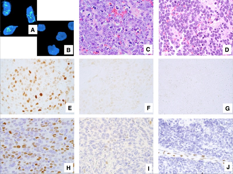
Figure 1. Examples of neuroblastoma, undifferentiated and poorly differentiated subtype with a High Mitosis-Karyorrhexis Index.
(A) Tumor in the MYCN amplified group (FISH, Original × 1,000) Green = 2p23-24 MYCN region probe; Red = CEP 2 region probe. (B) Tumor in the MYCN non-MYCN amplified group (FISH, Original × 1,000) Green = 2p23-24 MYCN region probe; Red = CEP 2 region probe. (C) Tumor with prominent nucleolar formation (H&E, Original x600). (D) Tumor without prominent nucleolar formation (H&E, Original x600). (E) Tumor expressing MYCN protein (+) (Immunostaining without counterstaining, Original × 600). (F) Tumor with MYCN protein (+/−) (Immunostaining without counterstaining, Original × 600). (G) Tumor with MYCN protein (−) (Immunostaining without counterstaining, Original × 600). (H) Tumor expressing MYC protein (+) (Immunostaining with Hematoxylin counterstaining, Original × 600). (I) Tumor with MYC protein (+/−) (Immunostaining with Hematoxylin counterstaining, Original x600). (J) Tumor with MYC protein (−) Note activated endothelial cells positive for MYC protein (Immunostaining with Hematoxylin counterstaining, Original × 600).

