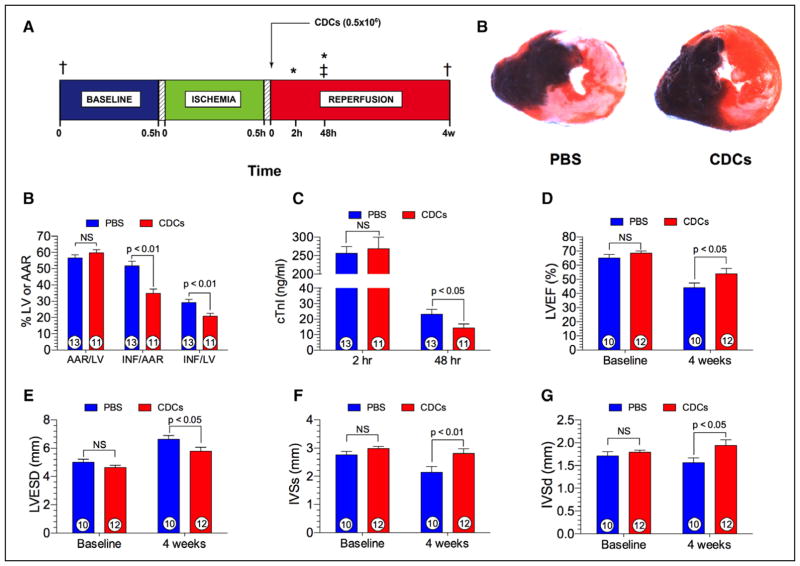
Figure 2. Cellular postconditioning in spontaneously hypertensive rats.
A, Experimental protocol involving male spontaneously hypertensive rats (SHRs) subjected to 30 minutes of left coronary artery ischemia followed by either 48 hours (h) or 4 weeks (w) of reperfusion. Myocardial area-at-risk and infarct size were determined at 48 h postreperfusion. Plasma levels of cardiac troponin I (cTnI) was measured at 2 h and 48 h of reperfusion. At 20 minutes of reperfusion, rat CDCs (0.5×10⁶) or phosphate-buffered saline (PBS) were injected directly into the left ventricular lumen following aortic cross-clamping. B, Representative photomicrographs of SHRs receiving either PBS or CDCs at 20 minutes of reperfusion. Myocardial infarct size is significantly attenuated in the CDC-treated heart. C, Myocardial area-at-risk (AAR) as a percentage of the left ventricle (LV), infarct size (INF) per AAR, and INF as a percent of the LV in rats receiving either PBS or CDCs. Myocardial infarct size per area-at-risk or LV was significantly (P<0.01) reduced in the CDC group. D, Plasma cardiac troponin I (cTnI) levels at 2 and 48 h following reperfusion. cTnI levels are significantly (P<0.05) reduced at 48 h postreperfusion. E, Left ventricular ejection fraction (LVEF) at baseline and at 4 weeks following reperfusion. LVEF is similar at baseline and significantly (P<0.05) greater in animals receiving CDCs. F, Left ventricular end-systolic dimension (LVESD) at baseline and 4 weeks of reperfusion in the PBS and CDC groups. LVESD is significantly (P<0.05) reduced in the CDC group in comparison with PBS. G, Interventricular septal dimension at end-systole (IVSs) at baseline and 4 weeks following reperfusion. IVSs was significantly (P<0.01) greater in hearts treated with CDCs than with PBS. H, Interventricular septal dimension at end-diastole (IVSd) at baseline and 4 weeks postreperfusion. Similar to IVSs, IVSd was significantly (P<0.01) greater in the CDC group than in the PBS group. Numbers inside the bars represent the number of animals in each group. Statistical significance was determined by using the Student t test. CDC indicates cardiosphere-derived cell; and 2,3,5-TTC, 2,3,5-triphenyltetrazolium chloride. *Plasma samples for cTnI. ‡Myocardial infarct size analysis. †2-D Echocardiography, Visual Sonics Vevo 2100.

