Abstract
In the present study, an effort has been made to optimize various culture conditions for enhanced production of anthocyanin. Nutrient content of MS medium (ammonium to potassium nitrate ratio and phosphate concentration) had a profound influence on the cell biomass and anthocyanin accumulation in cell suspension cultures of Daucus carota. Suspension cultures were carried out in shake flasks for 18 days and examined for cell growth, anthocyanin synthesis, anthocyanin yield and development of pigmented cells in relation to the uptake of total sugar, extracellular phosphate, nitrate and ammonia. The addition of NH4NO3 to KNO3 ratio (20.0 mM: 37.6 mM) in the suspension culture media resulted in a 2.85-fold increase in anthocyanin content at day 3. Similarly, a lower concentration of KH2PO4 (0.45 mM) in the MS medium resulted in 1.63-fold increase in anthocyanin content at day 9. The total sugar uptake was closely associated with a significant increase in anthocyanin accumulation. Total sugar and nitrate were consumed until 9–12 days, while ammonia and phosphate were completely consumed within 3 days after inoculation. After 9 days, cell lysis was observed and resulted in the leakage of intracellular substances. These observations suggest that anthocyanin was synthesized only by viable pigmented cells and degraded rapidly after cell death and lysis. This study signifies the utility of D. carota suspension culture for further up-scaling studies of anthocyanin.
Keywords: Ammonium nitrate, Potassium nitrate, Phosphate, Anthocyanin, Nutrient, Suspension culture
Introduction
Production of valuable secondary metabolites in plant cell cultures has received significant attention. Plant cell cultures often produce similar or superior levels of secondary metabolites with diverse profiles when compared with their intact plants (Rajasekaran et al. 2007). Plant in vitro cultures such as cell suspension cultures (Mulabagal and Tsay 2004) and root cultures (Mahendranath et al. 2011) offer attractive alternatives to whole plants for the production of secondary metabolites under controlled, reproducible conditions. Some metabolites in plant in vitro cultures can accumulate at a higher quantity than in parent plants (Akitha Devi and Giridhar 2014), suggesting that the production of plant-specific secondary metabolites in plant cell cultures is industrially feasible instead of using whole plants (Zhong 2001). Secondary metabolite production from wild plants has the problem of seasonal variations and environmental factors, which can be overcome through plant tissue culture. Continuous production of secondary metabolites is possible by manipulating culture medium constituents such as MS salt strength, source of carbohydrates, total nitrogen, phosphate, plant growth regulators and culture conditions such as temperature, illumination, light quality, medium pH, agitation and aeration (Murthy et al. 2014).
Anthocyanins are a class of flavonoids, which impart different colors to plant parts. Plant tissue culture is an alternative for the production of natural colors to replace synthetic dyes. Anthocyanin possesses excellent antioxidant properties (Szymanowska et al. 2015), anti-inflammatory, cardio-protective, antitumor and antidiabetic properties (Ghosh and Konishi 2007; Pallavi et al. 2012). Several investigations have been carried out to enhance the production of anthocyanins in suspension cultures viz., Oxalis linearis (Meyer and Staden 1995), Euphorbia milli (Yamamoto et al. 1989), Vitis sp. (Cai et al. 2012) and Fragaria × ananassa (Edahiro et al. 2005). Commercialization of anthocyanin production has not yet been successful because of insufficient knowledge about the effects of the physicochemical environment on plant cell behavior as well as lack of detailed understanding and control of the physiological state of a cell population. The influence of some physical factors have been studied such as UV irradiation (Takahashi et al. 1991), light (Zhang et al. 2009), nitrogen source (Do and Cormier 1991), type of sugar (Hiratsuka et al. 2001), osmotic stress (Do and Cormier 1990), plant growth regulators (Pasqua et al. 2005) and elicitors (Cai et al. 2012). Cell biomass yield and pigment accumulation of cultures can be affected by the nutrient content of the medium such as, ammonium nitrate to potassium nitrate ratio as well as the phosphate concentration (Sato et al. 1996; Zhang et al. 1998). Experiments were carried out in suspension cultures of D. carota to identify the effect of medium constituents on the cell biomass yield and anthocyanin accumulation because D. carota cell lines are very sensitive to stresses imposed by culture conditions. The cells of D. carota, grown in liquid medium, produced large amounts of cyanidin-3-glucoside and it has application in food, pharmaceutical and cosmetic industries as a natural colorant. Hence, to obtain maximum biomass yield with enhanced production of anthocyanin content, it is necessary to standardize the culture conditions, which would be suitable for cell growth and production of anthocyanins. Therefore, by selecting a high anthocyanin-producing cell line, culture medium or culture conditions optimization may support the continuous production of biomass as well as anthocyanin accumulation. The aim of the present study was to evaluate nutrient uptake and optimization of various physical parameters to achieve higher biomass yield and enriched production of anthocyanins in suspension cultures of D. carota.
Materials and methods
Source of chemicals
Cyanidin 3-glucoside (HPLC grade) were obtained from Sigma Aldrich (Bangalore, India). HPLC grade methanol, formic acid and acetonitrile were obtained from SRL (Mumbai, India). Plant growth regulators (2,4-D, Kin, NAA, BAP and IAA), MS salts, ammonium nitrate (NH4NO3), potassium nitrate (KNO3) and potassium dihydrogen phosphate (KH2PO4) were purchased from Hi-media (Mumbai, India).
Callus induction in Daucus carota
Atomic red (GR004) seeds of carrot (D. carota L.) were obtained from Plantefrø.dk (Denmark) and it was surface sterilized with sodium hypochlorite (4%, [v/v]; Hi-media, Mumbai) for 10 min. The seeds were then rinsed thoroughly (five times) in sterile distilled water. These seeds were inoculated on MS medium (Murashige and Skoog 1962) containing 3% sucrose (w/v, Hi-media, Mumbai), 0.5% CleriGar (w/v, Hi-media, Mumbai) and grown in darkness at 25 ± 2 °C. Germination started within 4–5 days of inoculation. Two-week-old leaves were used to induce callus formation on MS basal medium supplemented with various combinations and concentrations of auxins and cytokinins viz., 4.5 and 9.1 µM 2,4-dichlorophenoxyacetic acid (2,4-D); 5.37 and 10.75 µM 1-Naphthaleneacetic acid (NAA); 4.44 and 8.88 µM 6-Benzylaminopurine (BAP) and 0.46 and 2.32 µM kinetin (Kin). The pH of the media was adjusted to 5.8 with 0.1 N NaOH or 0.1 N HCl before autoclaving at 1.06 kg cm−2 and 121 °C for 15 min. Four-week-old friable and healthy calli were used to induce the pigmentation by placing on medium supplemented with IAA (11.41 µM) and Kin (0.93 µM). These cultures were maintained at 25 ± 2 °C under fluorescent light with a light intensity of 28 μmol s−1 m−2 (16/8 h photoperiod).
Establishment of cell suspension cultures
Cell suspension cultures were established by transferring 1 g (fresh weight) of callus to 25 ml liquid medium in 100 ml Erlenmeyer conical flasks. The basal MS medium supplemented with IAA (11.41 µM) and Kin (0.93 µM) was used as a multiplication medium. The cultures were maintained at 25 ± 2 °C on a rotatory shaker at 90 rpm under fluorescent light with a light intensity of 28 μmol s−1 m−2 (16/8 h photoperiod) and sub-culturing was done every 2 weeks. From 7-day-old sub-cultures, 200 mg cell aggregates was inoculated to 150 ml Erlenmeyer conical flasks containing 40 ml medium of each treatment. The cells were harvested at 3-day intervals from the day of inoculation until 18 days. All experiments were conducted in triplicates.
Manipulation of culture medium constituents
Effects of nitrogen source and phosphate concentration on anthocyanin production
Calli were transferred to MS medium supplemented with 30 g l−1 sucrose, IAA (11.41 µM) and Kin (0.93 µM). To understand the effect of nitrogen on anthocyanin accumulation, ratio of NH4NO3 to KNO3 was varied. The various concentrations of NH4NO3:KNO3 (0:18.8, 10.0:18.8, 40.0:18.8, 20.0:0, 20.0:9.4 or 20.0:37.6 mM and 20.0:18.8 mM as control) were used, with constant amount of KH2PO4 (0.9 mM). In another set of experiments, suspension cultures (5 g l−1) were developed by a medium, which had varied concentrations of KH2PO4 (0, 0.225, 0.45, 1.8 or 2.7 mM and 0.9 mM as control), with constant ratio of NH4NO3:KNO3 (20.0:18.8 mM) as in MS media. The culture conditions were the same as described above and each treatment was repeated three times. After 18 days of growth, the accumulation of anthocyanin was assessed in terms of fresh weight, anthocyanin content and anthocyanin yield was determined. Cell cultures were harvested every 3 days to analyze growth of cells and anthocyanin accumulation.
All the experiments were carried out in 150 ml Erlenmeyer conical flasks containing 40 ml of MS medium. In all the treatments, the culture medium contained 3% (w/v) sucrose IAA (11.41 µM) and Kin (0.93 µM). All cultures were kept under continuous agitation at 90 rpm in an orbital shaker and incubated at 25 °C (16/8 h photoperiod). Culture sampling was done at an interval of 3 days and cell biomass growth and anthocyanin production were measured. Experiments were performed in triplicates.
Determination of callus growth rate
The plant cell biomass is expressed as gram fresh cell weight (FCW) per liter. Cell suspension cultures were filtered and washed with deionized water and then cells were collected and weighed to get fresh weight (FW). For dry weight (DW), the cells were dried in oven at 40 °C until constant weight was achieved.
Determination of anthocyanin content
The anthocyanin content was determined by extracting the weighed calli in 0.1% HCl–methanol at 4 °C overnight (Do and Cormier 1991). The extracts were centrifuged (Hettich Universal 320R, Germany) at 8000×g for 10 min at 4 °C and the supernatant recovered. The absorbance of the supernatant was measured at 520 nm using a UV–Vis spectrophotometer (UV 1800 Shimadzu, Japan). Anthocyanin content is expressed as mg g−1 fresh cell weight. The samples were extracted and quantified in triplicates.
Determination of external phosphate, nitrate, ammonia and total sugar
To establish the kinetics of cell growth and nutrient uptake, cells were harvested from the liquid medium at an interval of 3 days, from day 0 to 18. The cells were washed with distilled water and filtered under vacuum. The residual nutrients (total sugar, phosphate, nitrate and ammonia) of the suspension culture were monitored every 3 days. The culture supernatant was analyzed for phosphate (Singh and Chaturvedi 2012), nitrate (Zhang et al. 1998), ammonia (Weatherburn 1967) and total sugar (Albalasmeh et al. 2013).
Sample preparation for HPLC/DAD
HPLC analysis was conducted following the method of Oh et al. (2008). Calli were extracted overnight using acidic methanol. Crude extracts were loaded on an open glass column packed with XAD-7 (Sigma Aldrich, Mumbai). Sugars and other water-soluble components were removed by eluting double-distilled and purified water (60 ml). After the water washing step, the anthocyanin were collected with an eluting solvent (0.1% HCl in methanol). The extracted solvents were concentrated by rotavapor (Buchi, Switzerland). The anthocyanin extracts were then filtered through a syringe type nylon microfilter (0.22 μm; Nupore Filtration Systems, UP, India). A Shimadzu LC 10-AD high-pressure liquid chromatograph equipped with a dual pump and a UV detector (model SPD-M20A, Shimadzu, Japan) was used to separate, identify and quantify anthocyanins. Separation of anthocyanins was achieved by the column Poroshell 120 SB-C18, 4.6 × 100 mm, 2.7 µm (Agilent Technologies, USA). A temperature-programmable column oven was used to maintain the column temperature at 35 °C during the HPLC analysis. The injection volume of the prepared sample was 20 μl. Solvent A was 1% formic acid in water and solvent B 1% formic acid in acetonitrile. The solvent gradient was 0–1 min, 5% B; 1–3 min, 5% to 8% B; 3–6 min, 8% to 15% B; and 6–14 min, 15% B; and 14–15 min, 15% to 5%. The flow rate of solvent was 1 ml min−1. The wavelength of the UV detector was set at 520 nm.
Statistical analysis
All experiments were performed thrice and the values are represented as mean ± standard deviation of the three analytical replications in the experiment. The investigated parameters were analyzed by two-way ANOVA and significance determined at P < 0.05. The data were analyzed using the SPSS 23 (IBM SPSS Statistics, Armonk) software and significant differences between the means were assessed using the Tukey’s test.
Results
Cell growth and anthocyanin production during a growth cycle
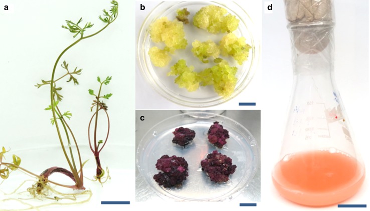
Seedlings obtained after 3–4 weeks were cultured on different combination of hormones to induce callus (Fig. 1a). Among the various combinations of growth regulators tested (2,4-D and Kin; NAA and Kin; BAP and NAA), MS medium supplemented with BAP (8.88 µM) and NAA (5.37 µM) induced maximum callus formation. Three weeks after sub-culturing, light green and friable fresh calli were obtained from leaf explants (Fig. 1b). Calli were multiplied and maintained on fresh medium at 4-week intervals. After 4–5 passages of sub-cultures, fresh and friable calli were transferred to anthocyanin induction medium containing 11.41 µM IAA and 0.93 µM Kin. Pigmented calli masses were obtained after 5–6 subcultures in the same medium (Fig. 1c). Suspension cultures were established by adding 1 g pigmented callus into the medium containing IAA and Kin (Fig. 1d).
Fig. 1.
a Establishment of Daucus carota seedlings on MS medium (bar = 8 cm). b Callus induction in the medium supplemented with BAP and NAA (bar = 5 cm). c Pigment production in the medium with IAA and Kin (bar = 5 cm) and d Suspension culture (bar = 8 cm)
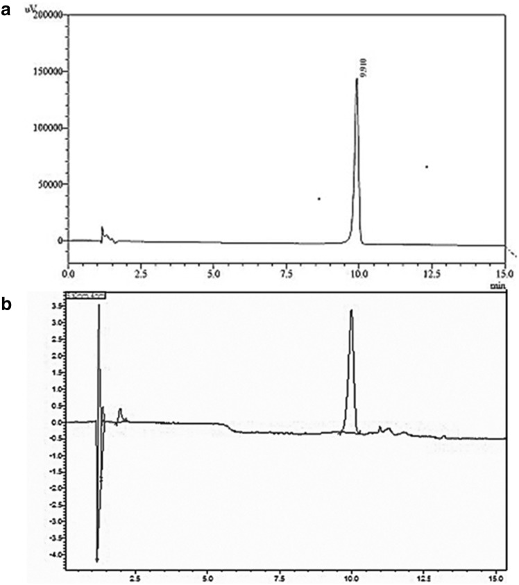
Screening of anthocyanin content in suspension cultures of D. carota was determined using HPLC. A representative HPLC chromatogram of anthocyanin and standards in D. carota is depicted in Fig. 2. Reversed-phase HPLC analyses with the two solvent systems showed one major peak. The major compound was identified as cyanidin 3-Glucoside at 520 nm.
Fig. 2.
HPLC chromatogram of anthocyanin profile a Cyanidin 3-Glucoside b Daucus carota suspension culture
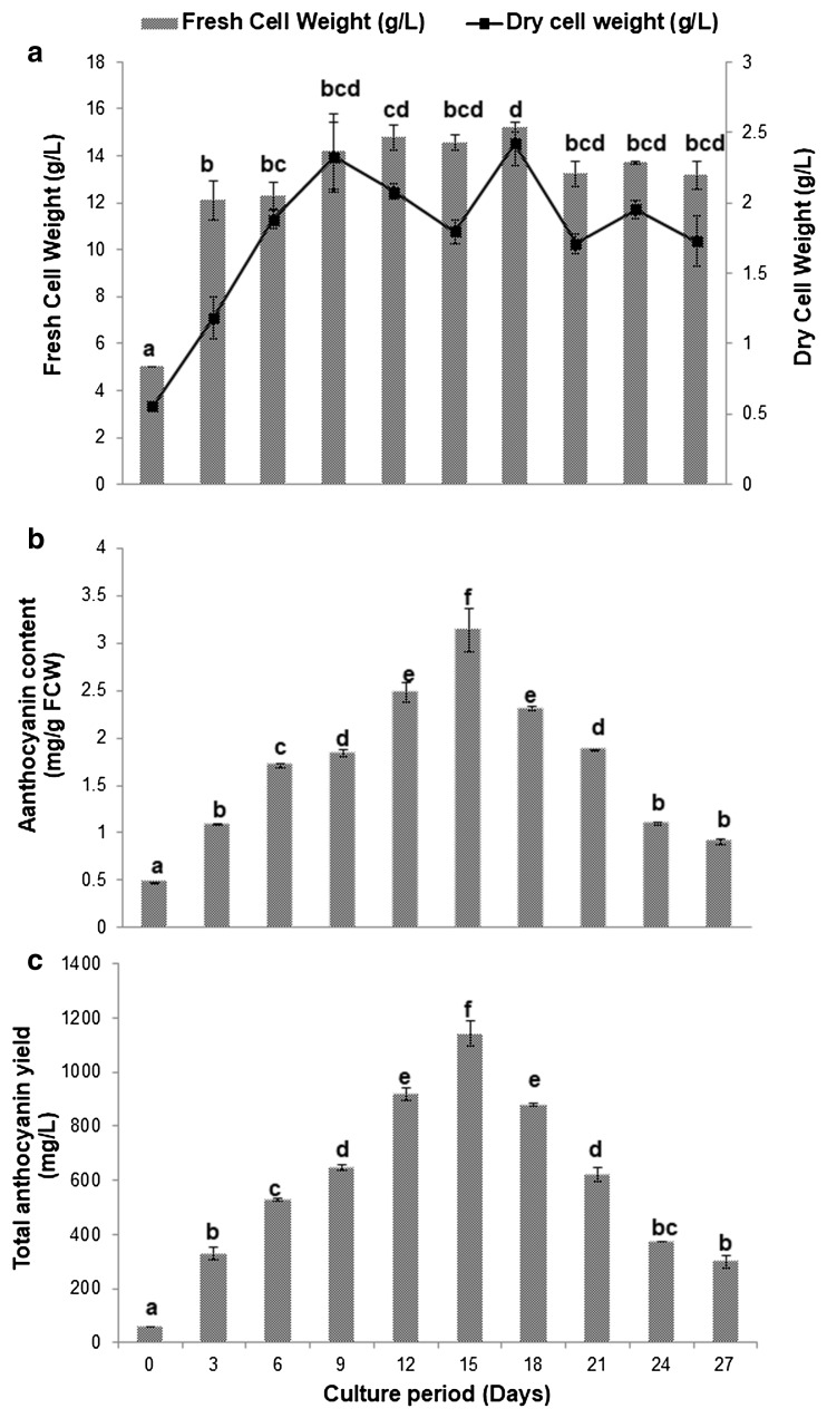
The kinetic profiles of cell growth represented by fresh cell weight, dry cell weight, anthocyanin content and anthocyanin yield in the suspension cultures are presented in Fig. 3. Optimum inoculum density is required for continuous growth and production of secondary metabolites in cell suspension cultures. Too high or too low inoculum levels may either significantly reduce or stop the growth and production of secondary metabolites (Enaksha and Arteca 1994). Cell growth was characterized by a 3-day lag phase, followed by an exponential phase lasting until day 18 of the culture and then attaining a stationary phase (Fig. 3a), which may be due to consumption of nutrients and lack of oxygen in the medium. For anthocyanin estimation, the cells harvested at every 3 days from suspension cultures revealed that the anthocyanin accumulation started at the end of the lag phase (3 days). During the exponential phase, anthocyanin content increased and was found to be growth associated and showed an increase with the increase in biomass until 15 days (Fig. 3b). The initial anthocyanin content was 0.48 ± 0.01 mg g−1 FCW at 0 d and the production peaked by 15 days (3.14 ± 0.23 mg g−1 FCW) and was significantly higher (P < 0.001) with respect to the production observed on previous and successive days (Fig. 3b). Subsequently, anthocyanin yield declined rapidly due to the nutrient depletion and cell death. Anthocyanin yield analyzed at 3-day intervals showed the highest accumulation (1142.15 ± 45.65 mg l−1) after 15 days (Fig. 3c). Anthocyanin accumulation strongly correlated with the growth of cells and anthocyanin content. Triplicate experiments showed a similar trend for anthocyanin accumulation. These results suggest that anthocyanin in suspension cultures were synthesized only by the viable cells and degraded rapidly after cell death and lysis.
Fig. 3.
Growth kinetics of Daucus carota cell suspensions cultured in MS medium with IAA (11.41 µM), Kin (0.93 µM) and 3% sucrose. a Fresh cell weight and dry cell weight. b Total anthocyanin content and c Total anthocyanin yield. The data shown are means of three replications ± standard deviation. Means marked with different letters are significant different according to one-way ANOVA at P < 0.05 (Tukey’s test)
Effect of NH4NO3:KNO3 ratio
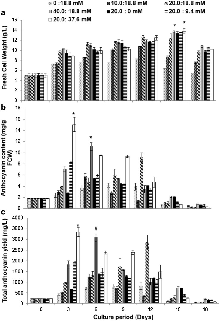
To find out the optimum levels of nitrate in the medium for enhanced production of anthocyanin in suspension culture of D. carota, the influence of nitrogen in the form of potassium nitrate and ammonium nitrate was tested. In MS medium, nitrogen is present as NH4NO3 and KNO3. When the concentration of total nitrogen was varied from 0 to 40 mM, a drastic change was observed both in cell growth and anthocyanin production (Fig. 4). To investigate the effect of different ratios of nitrogen (ammonium and potassium nitrate), inorganic nitrogen sources in the medium were prepared with potassium nitrate or ammonium nitrate (Fig. 4). Various combinations of NH4NO3 to KNO3 were added to the MS medium to provide sufficient concentrations of ammonium and potassium nitrate as a nitrogen source. Different concentration balances of NH4+/NO3− were tested (0:18.8, 10.0:18.8, 40.0:18.8, 20.0:0, 20.0:9.4 or 20.0:37.6 mM) along with 20.0:18.8 mM (NH4NO3:KNO3) as a control. As shown in Fig. 4, cell growth and anthocyanin production were maximal when the ratio of ammonium to nitrate was 1:2 (20.0 mM:37.6 mM). The results with nitrogen treatment suggest that an increased ammonium to nitrate ratio in the medium had a positive effect on anthocyanin production. Typical time course profiles of anthocyanin content and total anthocyanin yield with different ratios of NH4NO3 and KNO3 are presented in Fig. 4. The figure shows that the cell cultures of D. carota incubated in medium supplemented with NH4NO3 and KNO3 at a ratio of 20.0:37.6 mM supported the highest fresh cell weight (13.82 ± 0.08 g l−1) compared to other ratios of NH4NO3 to KNO3. The lowest pigment content (4.2 ± 0.38 mg g−1 FCW) was seen at the 0:18.8 mM NH4NO3 to KNO3 ratio. The culture medium with 20.0:37.6 mM NH4NO3 to KNO3 added to the normal concentration of the basal MS medium showed a reasonably high cell growth and pigment production. Anthocyanin content was enhanced by 2.85-fold in the medium supplemented with a 20.0:37.6 mM NH4NO3:KNO3 ratio compared to the control (Fig. 4a, b). The use of a single nitrogen source in the medium (i.e., either NH4+ or NO3− alone) was not beneficial for either biomass accumulation or anthocyanin production. Hence, a combination of ammonium and nitrate salts (NH4+/NO3−) would be most relevant in achieving higher amounts of anthocyanin in cell cultures. Highest anthocyanin yield was obtained in the medium supplemented with 20.0:37.6 mM (NH4NO3:KNO3 ratio) at day 6 (2379.99 ± 205.75 g l−1). In direct contrast, the 20.0:37.6 mM NH4NO3 to KNO3 ratio not only stimulated anthocyanin accumulation, but also promoted cell growth for D. carota cell cultures, resulting in an increased overall anthocyanin production.
Fig. 4.
Effect of the ratio of ammonium nitrate to potassium nitrate on growth and anthocyanin accumulation in suspension cultures of Daucus carota. a Fresh cell weight. b Total anthocyanin content and c Total anthocyanin yield. The data shown are means of three replications ± standard deviation. Results were analyzed by two-way ANOVA, followed by Tukey’s test. *Denotes P < 0.001 as compared to the other groups, #denotes P < 0.05 as compared to control
Effect of phosphate
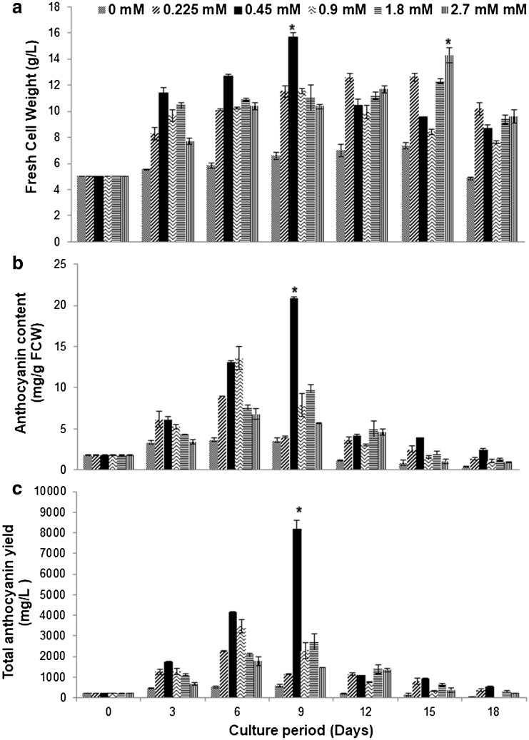
The effect of phosphate concentration on biomass and anthocyanin content is shown in Fig. 5. The cell growth increased significantly with the increase in phosphate concentration, while anthocyanin content tended to decrease at higher concentrations of phosphate (1.8 and 2.7 mM). The cell cultures of D. carota that were incubated in medium without adding KH2PO4 showed no significant increase in anthocyanin content. A phosphate level of 0.45 mM in the medium supported the highest fresh cell weight (15.68 ± 0.36 g l−1) as compared to the culture supplemented with different concentrations of KH2PO4 (0, 0.225, 0.9, 1.8 or 2.7 mM). A reduction in anthocyanin content was seen during phosphate deficiency (0 mM), whereas maximum anthocyanin content was seen for 0.45 mM phosphate, which was 1.63-fold higher than in the control culture. The lowest biomass (7.35 ± 0.24 g l−1) and anthocyanin content (3.66 ± 0.23 mg g−1 FCW) was recorded in the medium without KH2PO4 as compared to the other treatments. There was a significant difference in terms of the cell biomass yield between different concentrations of KH2PO4. The medium supplemented with 0.45 mM KH2PO4 produced the highest anthocyanin content (20.90 ± 0.13 mg g−1 FCW, Fig. 5b). However, the different concentrations of KH2PO4 had a significant effect on the anthocyanin production. Anthocyanin yield was higher in the medium with 0.45 mM KH2PO4 (2.3-fold increase). The addition of 0.45 mM KH2PO4 to the anthocyanin induction medium was seen as the optimum amount.
Fig. 5.
Effect of phosphate concentrations on growth and anthocyanin accumulation in the suspension cultures of Daucus carota. a Fresh cell weight; b Total anthocyanin content and c Total anthocyanin yield. Each treatment represents the mean of three replications ± standard deviation Results were analyzed by two-way ANOVA, followed by Tukey’s test. *Denotes P < 0.001 as compared to the other groups
Nutrient uptake
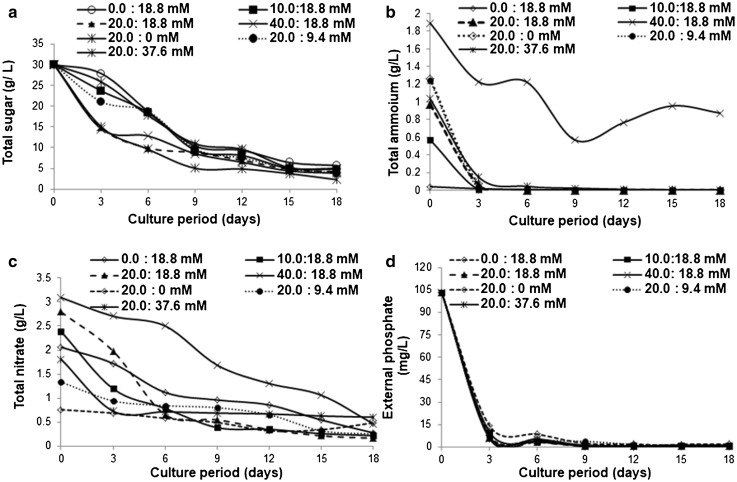
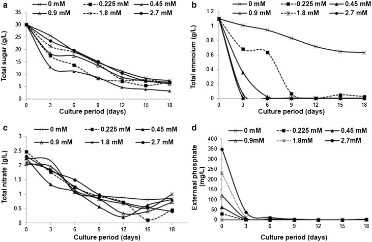
Profiles of nutrient consumption (total sugar, ammonium, nitrate and phosphate) during suspension culture are shown in Figs. 6, 7. Sucrose was utilized until day 18. After 6 days, practically all of the sucrose was absorbed or hydrolysed (Fig. 6a). Cells cultured in medium containing 20.0 mM NH4NO3 and 37.6 mM KNO3 consumed more sucrose compared to the other ratios of NH4NO3 to KNO3 tested. Similar results were obtained in the medium containing different concentrations of phosphate, where total sugar was absorbed by the cells throughout the growth cycle (Fig. 7a). In medium containing 0.45 mM phosphate, total sugar was utilized faster than other concentrations of phosphate in the medium (Fig. 7a). Cell growth represented by cell fresh weight and total anthocyanin content was closely linked to the uptake of the sugar in the medium. The onset of reduction in cell fresh weight coincided with the complete uptake of all the sugar from the medium and anthocyanin content increased with utilization of sugar from the medium. From the above observation, it can be concluded that cell growth and anthocyanin production in D. carota cell suspension cultures are strongly correlated with sugar uptake.
Fig. 6.
Nutrient depletion during a culture period of 18 days of a MS suspension culture of Daucus carota containing different concentration of NH4NO3 and KNO3. a Total sugar. b External ammonium. c External nitrate and d External phosphate
Fig. 7.
Nutrient depletion during a culture period of 18 days of MS suspension culture of Daucus carota containing different concentrations of phosphate. a Total sugar. b External ammonium. c External nitrate and d External phosphate
Figures 6b, 7b show the profile of nitrogen source consumption. Suspension cells completely consumed the ammonium within 3–6 days after inoculation (Figs. 6b, 7b) in the medium containing different ratios of NH4NO3 to KNO3 and phosphate concentration. The combination of 0:18.8 mM NH4NO3 to KNO3 ratio and 0 mM KH2PO4 gave a slower rate of ammonium consumption compared to other concentrations and directly influenced the anthocyanin accumulation. Thus, the slower the consumption, the lower the accumulation of anthocyanin in the medium.
Nitrate uptake occurred at a relatively slower rate in comparison to phosphate and sucrose. The fastest uptake rate was from days 0 to 6, a slower uptake rate was observed from days 6 to 18, and a slightly faster uptake took place during the stationary phase (Figs. 6c, 7c). Practically, the entire amount of nitrate was removed from the medium at the end of the growth cycle. A trend towards a leak of nitrate from the cells into the medium was observed in the final part of the growth cycle in the medium with varying phosphate concentrations (Fig. 7c).
Phosphate is another key nutrient for plant cell growth and metabolite formation. Phosphate is useful for the energy metabolism and biosynthesis. As shown in Figs. 6d, 7d, phosphate concentration showed a sharp drop within the first 3 days and no phosphate was detected during days 12–18. Phosphate uptake was fastest from day 0 to 6 and then occurred at a slower rate up to day 18 in the medium containing different ratios of NH4NO3 to KNO3 and phosphate concentrations (Figs. 6d, 7d). Complete uptake of phosphate was observed on day 3 in both the experimental media.
Discussion
Micro- and macro-elements of MS medium have considerable influence on the cell growth and secondary metabolite production in plant cell cultures. Changing the concentrations of ammonium nitrate, potassium nitrate and phosphate in the MS medium supports anthocyanin accumulation. The presence or absence of these nutrients (below normal level) can lead to growth retardation in plant cells and thereby enhance the secondary metabolite production (Murthy et al. 2014). However, different plants respond differently in response to different nutrient concentrations. In the present work, we established fast growing anthocyanin-producing suspension cultures of D. carota and analyzed the influence of medium composition on anthocyanin production. Alterations of the ammonium nitrate to potassium nitrate ratio and the phosphate concentration in the medium significantly enhanced production of anthocyanins in D. carota cell suspension cultures. To understand uptake of medium constituents, the dynamic profiles of medium sugar, ammonia, nitrate and phosphate levels were also analyzed. The present work demonstrated that changing the ratio of ammonium nitrate to potassium nitrate as well as the phosphate concentration can enhance the accumulation of anthocyanin in plant cell cultures. The ratio of ammonium nitrate to potassium nitrate in the MS medium can be optimized to augment the production of desired secondary metabolites, without adverse effects on plant cell growth. There have been many reports on the effects of nutrient deficiency on secondary metabolite biosynthesis in plants viz. phosphorus deficiency, nitrogen deficiency, or both, was reported to enhance flavonoid content in many plants (Bongue-Bartelsman and Phillips 1995; Morandi and Le Quere 1991). The influence of nitrogen was studied in relation to production of alkaloids in the suspension culture of Holarrhena antidysenterica (Panda et al. 1992), anthocyanin production in Vitis vinifera (Yamakawa et al. 1983; Do and Cormier 1991), shikonin production by Lithospermum erythrorhizon cell cultures (Fujita et al. 1981), diosgenin production in Dioscorea deltoidea cell cultures (Tal et al. 1982), Taxus yunnanensis for taxol production (Chen et al. 2003), camptothecin production in the suspension culture of Nothapodytes nimmoniana (Karwasara and Dixit 2013) and production of ginseng saponins and polysaccharides in suspension cultures of Panax ginseng cells (Zhang et al. 1996). Morard et al. (1998) reported that ammonium uptake acidifies the medium and thus indirectly promotes nitrate uptake, thereby enhancing the anthocyanin production in suspension culture. In the present study, the highest production of anthocyanin was recorded in the suspension culture medium containing 20.0 mM NH4NO3 and 37.6 mM KNO3. Similarly, Kaul and Hoffman (1993), found that ammonium as the nitrogen source inhibited callus growth of Pinus strobus. However, the inhibitory effect could be eliminated by providing 10 mM of KNO3 into the medium.
Secondary metabolite production in plant tissue cultures is also influenced by phosphate concentration in the medium. For growth of suspension cultures, inorganic phosphate is the most efficient nutrient. Cell growth is enhanced by higher concentrations of phosphate, whereas it had a negative influence on anthocyanin accumulation. Lower phosphate concentration stimulates the accumulation of anthocyanin (Jiang et al. 2007). Potassium ions play important biochemical and biophysical roles in plant cells. K+ serves as a major contributor to osmotic potential, a specific requirement for protein synthesis and an activator for particular enzyme systems (Jiang et al. 2007; Zhong 2001). Jiang et al. (2007) and Zhong (2001) showed that a higher concentration of K+ is responsible for the longer cell growth stage and the changes in specific oxygen uptake rate (SOUR) of the cultured cells. With K+ deficiency, more soluble sugars are stored within the cells. Due to deficiency of phosphate, plants have evolved a number of developmental and metabolic responses to adapt both the internal Pi and external Pi availability to sustain growth. These responses resulted in the accumulation of anthocyanins (Jiang et al. 2007). Studies in Arabidopsis showed that Pi deficiency promotes the accumulation of anthocyanins (Lopez-Bucio et al. 2002; Nacry et al. 2013). Due to increases in activities of PAP1, F3H, LDOX and UF3GT, the production of anthocyanins is stimulated in Pi-starved media. A lower concentration of phosphate induced the production of ajmalicine and phenolics in Catharanthus roseus and nicotin in Nicotiana tabacum (Mantell et al. 1983). In contrast, increased phosphate was shown to stimulate synthesis of digitoxin in Digitalis purpurea and betacyanin in Chenopodium rubrum (Berlin et al. 1986). Dedaldechamp et al. (1995) showed that phosphate deficiency leads to a reorientation towards secondary metabolism. Phosphate deprivation increases the anthocyanin production and activity of DFR gens in the suspension culture of grapes. The trend in the present study is similar to the production of camptothecin in the suspension culture of Nothapodytes nimmoniana (Karwasara and Dixit 2013). According to earlier reports, phosphate inhibits both hypericin and hyperforin biosynthesis, even though it stimulated biomass production in shoot cultures of Hypericum hirsutum and Hypericum maculatum (Coste et al. 2011). There have been a number of reports showing that phosphate limitation could improve the production of secondary metabolites. For example, increase in the synthesis of caffeine and anthocyanin was reported to occur alongside phosphate deprivation in cell suspension cultures of Coffea arabica (Bramble et al. 1991) and Vitis vinifera (Dedaldechamp et al. 1995), respectively. However, little is known about the molecular links between the plant P-status, phytohormone signaling, accumulation of metabolites and plant growth. Thus, nitrogen (ammonium to potassium nitrate ratio) and phosphate content are important factors for secondary metabolite production. In this context, the use of culture medium with a low phosphate concentration and a change in the NH4+/NO3− ratio may lead to the intensification of secondary metabolite production. Thus, the outcomes from the present study serve as a background for the future research related to large-scale manufacturing of anthocyanins in bioreactors.
Conclusion
Plant tissue cultures are the most efficient method for production of secondary metabolites under sterile conditions. Cell cultures were successfully established from calli of D. carota. To develop optimized medium conditions, the ammonium nitrate to potassium nitrate ratio as well as the phosphate concentration was manipulated to reduce the time for accumulation of anthocyanins. With optimized medium constitutes, anthocyanin accumulation reached the highest levels approximately at 3–9 days and the cell growth and anthocyanin synthesis correlated to nutrient consumption. Increasing the ammonium nitrate to potassium nitrate ratio from 18.8 to 37.6 mM enhanced cell growth and anthocyanin accumulation, whereas reducing phosphate concentration from 0.9 to 0.45 mM supported higher anthocyanin accumulation. The present work has demonstrated that the combination of changes in the ammonium nitrate to potassium nitrate ratio as well as the phosphate concentration can increase accumulation of anthocyanins in plant cell cultures without harmful effects on plant cell growth. It is evident that medium manipulation is a well-established and major way of enhancing the culture efficiency of plant cells for biomass and secondary metabolite production. Therefore, the present investigation will be of great importance in up-scaling processes, since it has shown the potential of enhanced anthocyanin productivity in D. carota calli. It is also helpful for production of bioactive compound from particular species in plant bioreactors. Further work is in progress to enhance the anthocyanin production from carrot suspension culture for biotechnological exploitation in bioreactors to produce natural food colorants.
Acknowledgements
The authors are thankful to the Council of Scientific and Industrial Research, New Delhi and Central Food Technological Research Institute, Mysuru (India) for financial assistance. Encouragement by Director CSIR-CFTRI, Mysuru, is also gratefully acknowledged.
Abbreviations
- BAP
6-Benzylaminopurine
- 2,4-D
2,4-Dichlorophenoxyacetic acid
- NAA
α-Naphthaleneacetic acid
- IAA
Indole-3-acetic acid
- Kin
Kinetin
- Pi
Inorganic phosphate
Compliance with ethical standards
Conflict of interest
The authors declare that there is no conflict of interest.
References
- Akitha Devi MK, Giridhar P. Isoflavone augmentation in soybean cell cultures is optimized using response surface methodology. J Agric Food Chem. 2014;62:3143–3149. doi: 10.1021/jf500207x. [DOI] [PubMed] [Google Scholar]
- Albalasmeh AA, Berhe AA, Ghezzehei TA. A new method for rapid determination of carbohydrate and total carbon concentrations using UV spectrophotometry. Carbohydr Polym. 2013;97:253–261. doi: 10.1016/j.carbpol.2013.04.072. [DOI] [PubMed] [Google Scholar]
- Berlin J, Sieg S, Strack D, et al. Production of betalains by suspension cultures of Chenopodium rubrum L. Plant Cell Tissue Organ Cult. 1986;5:163–174. doi: 10.1007/BF00040126. [DOI] [Google Scholar]
- Bongue-Bartelsman M, Phillips DA. Nitrogen stress regulates gene expression of enzymes in the flavonoid biosynthetic pathway of tomato. Plant Physiol Biochem. 1995;33:539–546. [Google Scholar]
- Bramble JL, Graves DJ, Brodelius P. Calcium and phosphate effects on growth and alkaloid production in Coffea arabica: experimental results and mathematical model. Biotechnol Bioeng. 1991;37(9):859–868. doi: 10.1002/bit.260370910. [DOI] [PubMed] [Google Scholar]
- Cai Z, Kastell A, Mewis I, et al. Polysaccharide elicitors enhance anthocyanin and phenolic acid accumulation in cell suspension cultures of Vitis vinifera. Plant Cell Tissue Organ Cult. 2012;108:401–409. doi: 10.1007/s11240-011-0051-3. [DOI] [Google Scholar]
- Chen Y-Q, Yi F, Cai M, Luo J-X. Effects of amino acids, nitrate, and ammonium on the growth and taxol production in cell cultures of Taxus yunnanensis. Plant Growth Regul. 2003;41:265–268. doi: 10.1023/B:GROW.0000007502.72108.e3. [DOI] [Google Scholar]
- Coste A, Vlase L, Halmagyi A, et al. Effects of plant growth regulators and elicitors on production of secondary metabolites in shoot cultures of Hypericum hirsutum and Hypericum maculatum. Plant Cell Tissue Organ Cult. 2011;106:279–288. doi: 10.1007/s11240-011-9919-5. [DOI] [Google Scholar]
- Dedaldechamp F, Uhel C, Macheix JJ (1995) Enhancement of anthocyanin synthesis and dihydroflavonol reductase (DFR) activity in response to phosphate deprivation in grape cell suspensions. Phytochemistry 40(5):1357–1360
- Do CB, Cormier F. Accumulation of anthocyanins enhanced by a high osmotic potential in grape (Vitis vinifera L.) cell suspensions. Plant Cell Rep. 1990;9:143–146. doi: 10.1007/BF00232091. [DOI] [PubMed] [Google Scholar]
- Do CB, Cormier F. Effects of low nitrate and high sugar concentrations on anthocyanin content and composition of grape (Vitis vinifera L.) cell suspension. Plant Cell Rep. 1991;9:500–504. doi: 10.1007/BF00232105. [DOI] [PubMed] [Google Scholar]
- Edahiro JI, Nakamura M, Seki M, Furusaki S. Enhanced accumulation of anthocyanin in cultured strawberry cells by repetitive feeding of l-phenylalanine into the medium. J Biosci Bioeng. 2005;99:43–47. doi: 10.1263/jbb.99.43. [DOI] [PubMed] [Google Scholar]
- Enaksha RM, Arteca RN (1994) Taxus cell suspension cultures: optimizing growth and production of taxol. J Plant Physiol 144(2):183–188
- Fujita Y, Hara Y, Suga C, Morimoto T. Production of shikonin derivatives by cell suspension cultures of Lithospermum erythrorhizon. Plant Cell Rep. 1981;1:61–63. doi: 10.1007/BF00269273. [DOI] [PubMed] [Google Scholar]
- Ghosh D, Konishi T. Anthocyanins and anthocyanin-rich extracts: role in diabetes and eye function. Asia Pac J Clin Nutr. 2007;16(2):200–208. [PubMed] [Google Scholar]
- Hiratsuka S, Onodera H, Kawai Y, et al. ABA and sugar effects on anthocyanin formation in grape berry cultured in vitro. Sci Hortic. 2001;90:121–130. doi: 10.1016/S0304-4238(00)00264-8. [DOI] [Google Scholar]
- Jiang C, Gao X, Liao L, Harberd NP, Fu X. Phosphate starvation root architecture and anthocyanin accumulation responses are modulated by the gibberellin-DELLA signaling pathway in Arabidopsis. Plant Physiol. 2007;145(4):1460–1470. doi: 10.1104/pp.107.103788. [DOI] [PMC free article] [PubMed] [Google Scholar]
- Karwasara VS, Dixit VK. Culture medium optimization for camptothecin production in cell suspension cultures of Nothapodytes nimmoniana (J. Grah.) Mabberley. Plant Biotechnol Rep. 2013;7:357–369. doi: 10.1007/s11816-012-0270-z. [DOI] [Google Scholar]
- Kaul K, Hoffman SA. Ammonium ion inhibition of Pinus strobus L. callus growth. Plant Sci. 1993;88(2):169–173. doi: 10.1016/0168-9452(93)90088-H. [DOI] [Google Scholar]
- Lopez-Bucio J, Hernandez-Abreu E, Sanchez-Calderon L, Nieto-Jacobo MF, Simpson J, Herrera-Estrella L. Phosphate availability alters architecture and causes changes in hormone sensitivity in the Arabidopsis root system. Plant Physiol. 2002;129:244–256. doi: 10.1104/pp.010934. [DOI] [PMC free article] [PubMed] [Google Scholar]
- Mahendranath G, Venugopalan A, Parimalan R, et al. Annatto pigment production in root cultures of Achiote (Bixa orellana L.) Plant Cell Tissue Organ Cult. 2011;106:517–522. doi: 10.1007/s11240-011-9931-9. [DOI] [Google Scholar]
- Mantell SH, Pearson DW, Hazell LP, Smith H. The effect of initial phosphate and sucrose levels on nicotine accumulation in batch suspension cultures of Nicotiana tabacum L. Plant Cell Rep. 1983;2:73–77. doi: 10.1007/BF00270169. [DOI] [PubMed] [Google Scholar]
- Meyer HJ, Staden JV. The in vitro production of an anthocyanin from callus cultures of Oxalis linearis. Plant Cell Tissue Organ Cult. 1995;40:55–58. doi: 10.1007/BF00041119. [DOI] [Google Scholar]
- Morandi D, Le Quere JL. Influence of nitrogen on accumulation of isosojagol (a newly detected coumestan in soybean) and associated isoflavonoids in roots and nodules of mycorrhizal and non-mycorrhizal soybean. New Phytol. 1991;117:75–79. doi: 10.1111/j.1469-8137.1991.tb00946.x. [DOI] [Google Scholar]
- Morard P, Fulcheri C, Henry M. Kinetics of mineral nutrient uptake by Saponaria officinalis L. suspension cell cultures in different media. Plant Cell Rep. 1998;18:260–265. doi: 10.1007/s002990050568. [DOI] [PubMed] [Google Scholar]
- Mulabagal V, Tsay HS. Plant cell cultures-an alternative and efficient source for the production of biologically important secondary metabolites. Int J Appl Sci Eng. 2004;2(1):29–48. [Google Scholar]
- Murashige T, Skoog F (1962) A revised medium for rapid growth and bio assays with tobacco tissue cultures. Physiol Plant 15(3):473–497
- Murthy HN, Lee E-J, Paek K-Y. Production of secondary metabolites from cell and organ cultures: strategies and approaches for biomass improvement and metabolite accumulation. Plant Cell Tissue Organ Cult. 2014;118:1–16. doi: 10.1007/s11240-014-0467-7. [DOI] [Google Scholar]
- Nacry P, Bouguyon E, Gojon A. Nitrogen acquisition by roots: physiological and developmental mechanisms ensuring plant adaptation to a fluctuating resource. Plant Soil. 2013;370:1–29. doi: 10.1007/s11104-013-1645-9. [DOI] [Google Scholar]
- Oh YS, Lee JH, Yoon SH, Oh CH, Choi DS, Choe E, Jung MY. Characterization and quantification of anthocyanins in grape juices obtained from the grapes cultivated in Korea by HPLC/DAD, HPLC/MS, and HPLC/MS/MS. J Food Sci. 2008;73(5):C378–C389. doi: 10.1111/j.1750-3841.2008.00756.x. [DOI] [PubMed] [Google Scholar]
- Pallavi R, Elakkiya S, Tennety SSR, Devi PS. Anthocyanin analysis and its anticancer property from sugarcane (Saccharum officinarum L.) peel. IJRPC. 2012;2:338–345. [Google Scholar]
- Panda AK, Mishra S, Bisaria VS. Alkaloid production by plant cell suspension cultures of Holarrhena antidysenterica: I. Effect of major nutrients. Biotechnol Bioeng. 1992;39:1043–1051. doi: 10.1002/bit.260391008. [DOI] [PubMed] [Google Scholar]
- Pasqua G, Monacelli B, Mulinacci N, et al. The effect of growth regulators and sucrose on anthocyanin production in Camptotheca acuminata cell cultures. Plant Physiol Biochem. 2005;43:293–298. doi: 10.1016/j.plaphy.2005.02.009. [DOI] [PubMed] [Google Scholar]
- Rajasekaran T, Giridhar P, Ravishankar G. Production of steviosides in ex vitro and in vitro grown Stevia rebaudiana Bertoni. J Sci Food Agric. 2007;87:420–424. doi: 10.1002/jsfa.2713. [DOI] [Google Scholar]
- Sato K, Nakayama M, Shigeta J. Culturing conditions affecting the production of anthocyanin in suspended cell cultures of strawberry. Plant Sci. 1996;113:91–98. doi: 10.1016/0168-9452(95)05694-7. [DOI] [Google Scholar]
- Singh M, Chaturvedi R. Evaluation of nutrient uptake and physical parameters on cell biomass growth and production of spilanthol in suspension cultures of Spilanthes acmella Murr. Bioprocess Biosyst Eng. 2012;35:943–951. doi: 10.1007/s00449-012-0679-3. [DOI] [PubMed] [Google Scholar]
- Szymanowska U, Złotek U, Karaś M, Baraniak B. Anti-inflammatory and antioxidative activity of anthocyanins from purple basil leaves induced by selected abiotic elicitors. Food Chem. 2015;172:71–77. doi: 10.1016/j.foodchem.2014.09.043. [DOI] [PubMed] [Google Scholar]
- Takahashi A, Takeda K, Ohnishi T. Light-induced anthocyanin reduces the extent of damage to DNA in UV-irradiated Centaurea cyanus cells in culture. Plant Cell Physiol. 1991;32:541–547. [Google Scholar]
- Tal B, Gressel J, Goldberg I. The effect of medium constituents on growth and diosgenin production by Dioscorea deltoidea cells grown in batch cultures. Planta Med. 1982;44:111–115. doi: 10.1055/s-2007-971414. [DOI] [PubMed] [Google Scholar]
- Weatherburn MW. Phenol-hypochlorite reaction for determination of ammonia. Anal Chem. 1967;39(8):971–974. doi: 10.1021/ac60252a045. [DOI] [Google Scholar]
- Yamakawa T, Kato S, Ishida K, Kodama T, Minoda Y. Production of anthocyanins by Vitis cells in suspension culture. Agric Biol Chem. 1983;47(10):2185–2191. [Google Scholar]
- Yamamoto Y, Kinoshita Y, Watanabe S, Yamada Y (1989) Anthocyanin production in suspension cultures of high-producing cells of Euphorbia millii. Agric Biol Chem 53(2):417–423
- Zhang YH, Zhong JJ, Yu JT (1996) Effect of nitrogen source on cell growth and production of ginseng saponin and polysaccharide in suspension cultures of Panax notoginseng. Biotechnol Prog 12(4):567–571
- Zhang W, Seki M, Furusaki S, Middelberg APJ. Anthocyanin synthesis, growth and nutrient uptake in suspension cultures of strawberry cells. J Ferment Bioeng. 1998;86:72–78. doi: 10.1016/S0922-338X(98)80037-8. [DOI] [Google Scholar]
- Zhang T, Li L, Song L, Chen W. Effects of temperature and light on the growth and geosmin production of Lyngbya kuetzingii (Cyanophyta) J Appl Phycol. 2009;21(3):279–285. doi: 10.1007/s10811-008-9363-z. [DOI] [Google Scholar]
- Zhong J-J. Plant cells. Berlin: Springer; 2001. Biochemical engineering of the production of plant-specific secondary metabolites by cell suspension cultures; pp. 1–26. [DOI] [PubMed] [Google Scholar]


