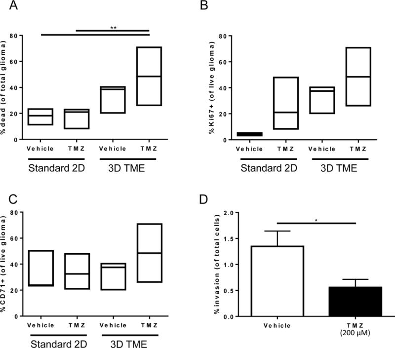
Figure 4. Detection of chemotherapeutic efficacy of temozolomide in a human 3D in vitro system of the brain tumor microenvironment.

A) Viability of U251 glioma cells after treatment in standard culture compared to tissue engineered model with and without temozolomide treatment represented as % dead cells of total populations of tumor cells. B) Percentage of live U251 glioma cells that are proliferating as assessed by Ki67 positivity. C) Percentage of live U251 glioma cells those are stem-like as assessed by CD71 positivity. Box plots show minimum, median, and maximum values assessed for n≥3 biological replicates. D) Invasion of U251 glioma cells across the tissue culture insert membrane from the tumor microenvironment model after treatment with temozolomide. Analyzed by two-way ANOVA, followed by posthoc t-tests. *p<0.05, **p<0.01.
