

Abstract
OBJECTIVES
To measure microleakage around zirconia crown margins cemented with self-adhesive resin or resin modified glass ionomer (RMGI) cement after ultrasonic scaling.
METHODS
16 molars were prepared for crowns (margin 0.5mm coronal of cementum-enamel junction). Preparations were digitally scanned and zirconia crowns milled. Specimens were divided into two groups (n=8): self-adhesive resin (RelyX Unicem 2) or resin modified glass ionomer (RMGI) (RelyX Luting Plus) cements. After cementation, specimens were ultrasonic scaled with a piezoelectric device (60sec, hand pressure). After thermocycling (20,000 cycles /5-55°C), specimens were immersed in 5wt% fuchsine dye before sectioning bucco-lingually. Microleakage was examined under 40× light magnification. Statistical comparisons were made using a paired t-test and a two-sample t-test (α=0.05).
RESULTS
Ultrasonic scaling did not alter microleakage at the margins of crowns (p=0.31). There was no significant difference in microleakage of scaled and untreated margins with the use of different cements (p=0.21). The amount of microleakage around margins that were scaled was not significantly different between cements (p=0.14). Untreated margins of crowns cemented with RelyX Luting Plus showed a significantly higher microleakage than those cemented with RelyX Unicem 2 (p=0.005).
CONCLUSIONS
Piezoelectric ultrasonic scaling did not increase microleakage at the margin of zirconia crowns cemented with self-adhesive resin or RMGI cements.
CLINICAL SIGNIFICANCE
Piezoelectric ultrasonic scaling around zirconia crowns did not impact marginal microleakage cemented with self-adhesive resin or RMGI cements.
Introduction
Introduction of computer-aided design and computer-assisted manufacturing (CAD/CAM) as a method of fabrication of ceramic fixed restorations has increased their use to surpass that of metal-ceramic crowns.1, 2 A recent survey from the National Practice-Based Research Network reported that monolithic zirconia is the most prescribed material for posterior single unit crowns.3 An important criteria for the long term success of a zirconia crown is the maintenance of a marginal seal. Microleakage at marginal interfaces may cause restoration failure due to the dynamic passage of bacteria, oral fluids, molecules, and ions between the interface of the restoration and tooth, causing secondary caries and discoloration of margins as well as tooth hypersensitivity.4–12 A recent systemic review reported that the estimated annual complication rate for single unit zirconia crowns was 0.09% for secondary caries and 0% for marginal staining.13 No information was provided regarding the incidence of hypersensitivity.
The selection of dental cement is one factor that may affect marginal seal. Different types of cement may affect the marginal seal as they differ based on their polymerization shrinkage,14 hygroscopic expansion,14 coefficient of thermal expansion,15 bond with tooth structure,16 and bond with zirconia.17 One of the clinical advantages of zirconia crowns is that they may be conventionally cemented with resin-modified glass ionomer (RMGI) cement or adhesively bonded with resin cement without affecting the strength of the restoration.18 A 2013 survey by Clinician’s Report found that 55% of dentists use RMGI cement and 39% of dentists use a resin cement for zirconia crowns.1 Several in vitro studies have shown increased microleakage, as measured by dye penetration, around margins of crowns cemented with RMGI cements than resin cements.7,19 In contrast, an in vitro study reported less bacterial microleakage in crowns cemented with RMGI cement than resin cement.20
The marginal fit of the crown may also affect its marginal seal, as this fit will influence the amount of cement exposed to the oral environment.21 Previous studies indicate cement gaps of 1-161 μm in conventionally fabricated all-ceramic crowns and 17-118 μm in CAD/CAM fabricated all-ceramic crowns.21 The exposed cement may undergo dissolution caused by oral fluids which may lead to microleakage.21,22 Additionally, some cement may be susceptible to mechanical removal or roughening when cleaned using manual or ultrasonic scalers. Hygiene instrumentation may cause roughening of the marginal interface or increase in marginal gaps which may lead to plaque accumulation, microleakage and secondary decay.4, 23,24
Sonic and ultrasonic scalers are common tools used for removing plaque and calculus from tooth and root surface. Ultrasonic scalers are divided into two common units: magnetostrictive (elliptical vibration pattern active on all sides of the tip) and piezoelectric (linear vibration pattern with only two active sides of the tip). Sonic scalers vibrate between 3,000 to 8,000 cycles per second (Cps) whereas magnetostrictive and piezoelectric units vibrate between 18,000 to 45,000 Cps and 25,000 to 50,000 Cps respectively.23 An in vitro study found more adverse effects on surface roughness of resin-based restorative materials using magnetostrictive ultrasonic scalers than sonic scalers.25 Piezoelectric ultrasonic scalers are clinically favorable due to their quieter operation, smaller tips and handpieces, and ease of use.4, 19 It is possible that the vibrational forces produced by piezoelectric ultrasonic scalers may disrupt the bond formed at the crown margin.
A previous study demonstrated that ultrasonic scaling with a piezoelectric unit caused microleakage at the cementum margin of Class V restorations.4 It follows that ultrasonic scaling margins of a crown may cause disruption of the cement bond leading to microleakage. The null hypothesis for this study is that there is no difference between microleakage observed in zirconia crowns with and without ultrasonic scaling. The second null hypothesis is that there is no difference between the microleakage caused by ultrasonic scaling with zirconia crowns cemented with self-adhesive resin and RMGI cements.
Materials and methods
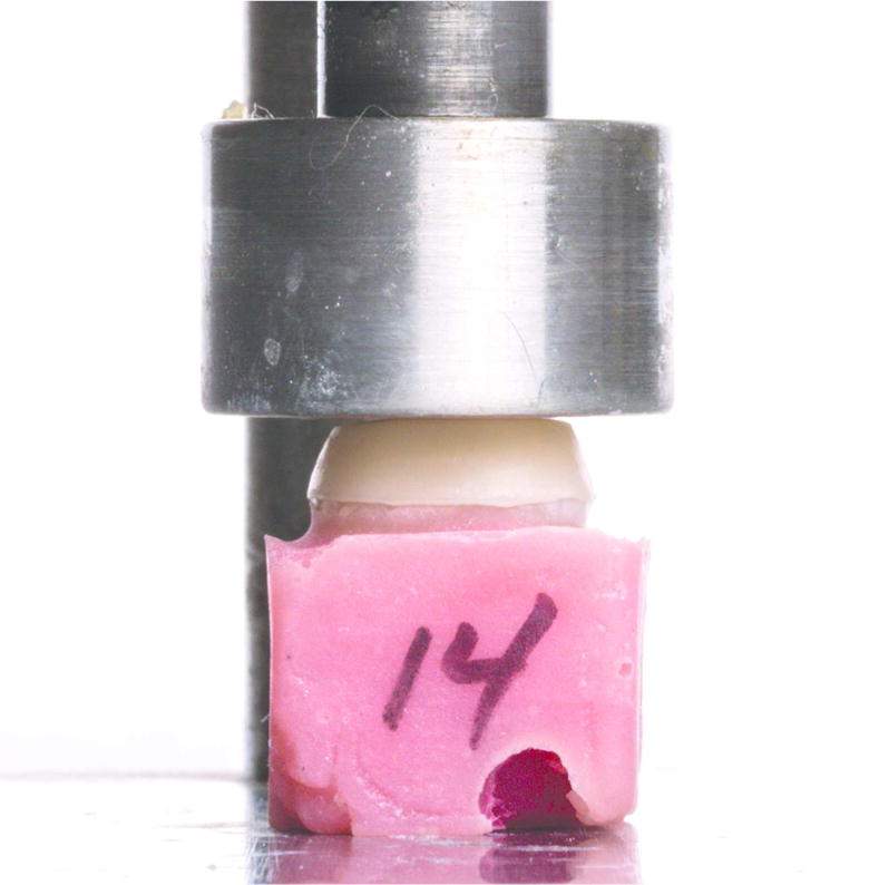
Following Institutional Review Board non-human research subjects approval, sixteen human molars were collected from the University of Alabama at Birmingham Oral and Maxillofacial Surgery department. All teeth were examined under 20× magnification (VHX 600, Keyence, Osaka, Japan) for any cracks and fracture lines. All teeth were screened for caries using an explorer. Any teeth presenting with cracks, fracture lines, or caries were excluded from this study. Teeth were notched at the roots and embedded into an acrylic resin base for ease of sectioning and blocking open apices. Crowns were prepared in a standardized crown preparation device (Figure 1) with all walls and occlusal table in dentin. The finish line was placed within 0.5 mm coronal of the cementum-enamel junction. The finish line was designed as a 1 mm chamfer.
Figure 1.

Specimen preparation (from top left to bottom right): Crown preparation with 60° taper and chamfer margin, standardized 2 kg weight to cement zirconia crown, piezoelectric ultrasonic scaling crown margin for 60 seconds, sectioning specimen bucco-lingually using a diamond disc
The tooth preparations were captured with an intraoral scanner (True Definition Scanner, 3M ESPE, St. Paul, MN, USA). The .stl files were exported to a commercial laboratory (Custom Milling Center, Golden, CO, USA) which fabricated zirconia (Bruxzir Glidewell, Newport Beach, CA, USA) crowns. The crowns were fabricated with a 0.6 mm wall thickness and a 100 μm cement gap.
Teeth were divided into two test groups (n=8): self-adhesive resin cement (RelyX Unicem 2, 3M ESPE, St. Paul, MN, USA) or a resin modified glass ionomer (RMGI) cement (RelyX Luting Plus, 3M ESPE, St. Paul, MN, USA). Prior to cement application, the zirconia crowns were airborne particle abraded with 50 μm alumina at 2 bar pressure and cleaned for 5 minutes in an ultrasonic water bath. The crowns were seated with finger pressure to verify complete seating and excess cement was removed around the marginal interface with a microbrush. The cement was then allowed to self-polymerize for 6 minutes under a 2 kg lead weight at room temperature (Figure 1). Specimens were stored in distilled water at 37°C for 24 hours.
Ultrasonic scaling with a piezoelectric device (Varios 750, NSK-Nakanishi Inc., Kaunda, Japan) with a scaling tip (model G6, NSK-Nakanishi Inc., Kaunda, Japan) was used at full power with distilled water. The lateral side of the tip was used to trace the crown-dentin interface for 60 seconds under moderate hand pressure on one side of the crown (Figure 1).
Specimens underwent 20,000 thermo cycles of 5°C and 55°C in water baths with a 15-second dwell time. After thermocycling, specimens were immersed in freshly mixed 5wt% fuchsine solution (Fischer Scientific Company, Fairlawn, NJ, USA) for 24 hours. All specimens were sectioned bucco-lingually through the center of the crown with a dental sectioning disc (Figure 1; Abrasive Discs, Zermatt, Buehler, Lake Bluff, IL, USA) and examined under light microscopy (VHX 600, Keyence, Itasca, IL, USA) at 25× and 40× magnification (Figure 2). Distance (μm) of dye penetration from the external crown surface to the point where no dark purple dye could be seen was measured with the built-in image analysis software (Measurement tool, Keyence) at 40× magnification. Percentage microleakage was measured by dividing the linear distance of dye penetration by the linear distance from the external margin to the axial-occlusal line angle (Figure 3). Using 100× micrographs, the cement gap was measured as the distance from the external crown margin to the external tooth preparation finish line. Using 100× micrographs, the diameter of the scaler tip was measured based on the external outline of the tip curvature.
Figure 2.


Representative sectioned specimens under light microscopy (40X) to examine microleakage represented by dark purple 5wt% Fuchsine stains (from top left to bottom right): RelyX Unicem 2 Control, RelyX Luting Plus Control, RelyX Unicem 2 Scaled, RelyX Luting Plus Scaled
Figure 3.

Diagram of method used to measure microleakage
Descriptive statistics (mean and standard deviation) were used to summarize the amount of microleakage (%) around dental crowns cemented using two different types of cement (RelyX Unicem 2 and RelyX Luting Plus) with and without ultrasonic scaler treatment. The mean and SD were computed for cement groups altogether and individually. A paired t-test was used to assess whether the use of ultrasonic scaler affects the amount of microleakage from the treated and untreated surface within each specimen. The change in microleakage was computed by taking the difference of microleakage of treated and untreated surfaces. Then a two-sample t-test was used to compare the change between the two types of cement. The microleakage for each surface (treated and untreated) of the specimen between two cement types was evaluated using the two-sample t-test. The normality assumption for the data was verified using Shapiro-Wilk test. A p-value of < 0.05 was considered statistically significant in two-tailed statistical tests. To ensure the adequacy of our sample size for the reported significant finding (Fig 4c), a post-hoc power analysis was conducted using nQuery Advisor v7, based on a sample size of 8 per group, a two-sided statistical test and a type I error rate of 0.05. All analyses were conducted using SAS 9.4 software (SAS Institute, Cary, NC, USA). The box and whisker plots were created using R 3.3.0.
Figure 4.

c. Box and whisker plot showing the microleakage (%) of untreated surface between two types of cement.
Results
Descriptive statistics for the 16 cemented dental crown specimens were presented in Table 1. The mean microleakage with the use of the ultrasonic scaler was 24.5±18.9% and without was 28.9±19.4%. In specimen cemented with RelyX Unicem 2, the mean microleakage for the treated and untreated surface was 17.3±14.4% and 16.3±11%, respectively. In RelyX Luting Plus specimen, the mean microleakage for the treated surface was 31.6 ±21.1% and 41.4±18.1% for the untreated surface. All variables in comparison met the assumption of normality (p > 0.05, Table 2).
Table 1.
Percent microleakage at zirconia-dentin interface
| Treatment Group | Cement | n | % Microleakage
|
|
|---|---|---|---|---|
| Mean ± SD | Range | |||
| Ultrasonic Scaled | RelyX Unicem 2 + RelyX Luting Plus |
16 | 24.5±18.9% | 2–72% |
| RelyX Unicem 2 | 8 | 17.3±14.4% | 18–65% | |
| RelyX Luting Plus | 8 | 31.6±21.1% | 8–72% | |
| Control | RelyX Unicem 2 + RelyX Luting Plus |
16 | 28.9±19.4% | 5–65% |
| RelyX Unicem 2 | 8 | 16.3±11.0% | 5–33% | |
| RelyX Luting Plus | 8 | 41.4±18.1% | 2–41% | |
Table 2.
Result of Shapiro-Wilk test for normality
| Treatment Group | Cement | n | Shapiro-Wilk Test Statistics (W) | p |
|---|---|---|---|---|
| Ultrasonic Scaled | RelyX Unicem 2 + RelyX Luting Plus | 16 | 0.889 | 0.053 |
| Control | RelyX Unicem 2 + RelyX Luting Plus | 16 | 0.901 | 0.08 |
| Control | RelyX Unicem 2 | 8 | 0.859 | 0.12 |
| RelyX Luting Plus | 8 | 0.88 | 0.19 | |
| Ultrasonic Scaled | RelyX Unicem 2 | 8 | 0.837 | 0.07 |
| RelyX Luting Plus | 8 | 0.87 | 0.15 |
Ultrasonic scaling did not alter microleakage at the margins of dental crowns (p=0.31). There was no significant difference in microleakage of scaled and untreated margins with the use of RelyX Unicem 2 and RelyX Luting Plus (p=0.21, Figure 4a). The amount of microleakage around margins that were scaled was not significantly different between cements (p=0.14, Figure 4b). Untreated margins of crowns cemented with RelyX Luting Plus showed a significantly higher microleakage than those cemented with RelyX Unicem 2 (p=0.005, Figure 4c). Even with this small sample of 8 per group, the two-sample t-test had 87% power to detect the 25±15% microleakage difference of the untreated surface between the two types of cement at a type I error rate of 0.05.
Figure 4.

a. Box and whisker plot showing the difference in microleakage (%) of treated and untreated surface between two types of cement.
The two-sample t-test p values are displayed for cement comparisons. The asterisk denotes the mean; the line inside the box depicts the median; the upper and lower hinges represent 25th and 75th percentile, respectively.
Figure 4.

b. Box and whisker plot showing themicroleakage (%) of treated surface between two types of cement.
The cement gaps were measured as 153.8±86 μm (control) and 135.2±66.1 μm (scaled) for the RMGI cement and 97.4±44.6 μm (control) and 115.5±62.2 μm (scaled) for the resin cement. The diameter of the ultrasonic scaling tip was 270.6 μm (Figure 5).
Figure 5.

NSK Model G6 scaler tip under 100× light microscopy
Discussion
Based on the results of this study, there is no difference between microleakage of zirconia crowns with and without piezoelectric ultrasonic scaling of the crown margins; and therefore, the first null hypothesis is accepted. Additionally, there was no difference in microleakage of scaled and untreated margins with the use of RelyX Unicem 2 and RelyX Luting Plus; and therefore, the second null hypothesis is accepted. However, higher microleakage was found using an RMGI cement (RelyX Luting Plus) than using a self-adhesive resin cement (RelyX Unicem 2) when no ultrasonic scaling was performed. The clinical implication of this study is that piezoelectric ultrasonic scaling may be used around the margins of zirconia crowns cemented with RMGI or self-adhesive resin cements.
Ultrasonic scaling was predicted to create marginal microleakage due to increased mechanical stimulation of the cement margin. In a previous study from Goldstein et al.4, increased microleakage was observed at composite-cementum interfaces that were subjected to ultrasonic scaling; however, composite-enamel margins were unaffected by ultrasonic scaling. All margins in the current study were located on enamel. Perhaps if the margins would have been located apical to the cementum-enamel junction, increase microleakage may have occurred. A study by Angerame et al.24 examined composite crowns placed on enamel margins that were subjected to mechanical instrumentation with hand instruments. The crowns subjected to manual instrumentation showed an increased marginal gap but a decrease in marginal microleakage. The authors explained this observation by suggesting debris from instrumentation filled the marginal gaps which protected against microleakage. A recent study by Rohani et al.26 found that there was no significant differences in microleakage between ultrasonic scaling and hand instrumentation when used on cervical glass ionomer, resin composite, and amalgam restorations, either at the cementum or enamel margins. The results of these studies suggest that ultrasonic or manual cleaning of crown margins are safe treatment options, particularly when crown margins are located on enamel.
Ultrasonic scaling was also hypothesized to create marginal microleakage from cement roughening and removal. The cement gaps were measured as 153.76±85.96 μm (control) and 135.2±66.1 μm (scaled) for the RMGI cement and 97.40±44.6 μm (control) and 115.51±62.23 μm (scaled) for the resin cement. These values are similar to previously reported cement gaps of 17-118 μm for CAD/CAM fabricated all-ceramic crowns.21 This amount of marginal opening is smaller than the 270.63 μm diameter of the ultrasonic scaler tip and therefore it would not have been able to ditch out cement from the cement gap. If a crown were to have a marginal opening larger than 270 μm, however, the scaler tip may have been able to remove cement to create a marginal gap as well as create a ledge from which vibrational forces could be applied. Therefore, it is important to realize that the results of this study may not apply to clinical situations in which an open margin is present.
In this study, there was more microleakage observed in the untreated teeth cemented with RMGI than self-adhesive resin cement. Previous studies have also reported that RMGI cements show more microleakage than resin cements.7,19,27 A study by Albert and El-Mowafy reported that both ceramic and metal ceramic crowns demonstrated less microleakage when cemented with resin cement than RMGI cement.7 In their study, the superior performance of the resin cement was credited to the higher bond between dentin and the resin cement. A study by Rossetti et al.19 compared microleakage of metal cast crowns with resin and RMGI cement. Slightly more microleakage was reported with the RMGI cement; however, there was no statistical difference. Lyons et al.27 detected microleakage around metal crowns cemented with glass-ionomer cement but not resin-cemented crowns.
Several factors may differentiate resin cements and RMGI regarding their ability to seal a crown margin, including their shrinkage/expansion,14 thermal coefficient,15 bond to tooth structure,16 and bond to zirconia.17 The stresses applied to the RMGI cement bond during water storage and thermocycling may have been greater than those applied to the resin cement bond. A recent study determined that a glass ionomer material demonstrated significantly less post-gel shrinkage than a resin-based material.14 Following storage in water, however, the glass ionomer material underwent significantly more hygroscopic expansion that more than compensated for post-gel shrinkage. The coefficient of dimensional change of both RMGI materials (−85 ×10−6 C°−1) and resin materials (64.5 ×10⁻⁶ C°−1) are both greater than that of tooth structure (11 ×10−6 C°−1).25 However, RMGI materials contract at elevated temperatures unlike resin materials and tooth structure which expand.15 If the RMGI cement was contracting while tooth structure was expanding, increased stress would be experience at the cement-tooth interface of the crown margin.
Additionally, the resin cement may have experienced less microleakage due to its superior bond. A study evaluating the bond between bovine tooth and zirconia showed a greater bond using two self-adhesive resin cements than the reference RMGI cement.16 A study by Turker et al.17 demonstrated that a resin cement containing 10-methacryloyloxydecyl dihydrogen phosphate (MDP) produced a significantly greater bond to zirconia than an RMGI cement. The self-adhesive resin cement used in this study (RelyX Unicem 2) also contains MDP unlike the RMGI cement (RelyX Luting Plus). Examination of the sectioned crowns reveals that the dye stain is more intense at the interface between the cement and the tooth than the cement and the crown. This observation suggests that the bond between the cement and the tooth is most susceptible to microleakage. A similar observation was reported in a previous study.7
Thermocycling was performed following ultrasonic scaling as it has been shown to reproduce the effects of temperature changes that occur intraorally.28 Extreme temperature changes cause mechanical thermal stresses on the restoration margins due to the differences in coefficient of thermal expansion between restorative materials, cements, and natural tooth structure. Furthermore, constant gap dimensional changes allow oral fluids including pathogenic bacteria to flow in and out of microscopic gaps found in the restorative margins.6 Based on an average of 20 to 50 thermocycles in the oral cavity per day, several 10,000 thermocycles in vitro may represent a year of in vivo function.6, 29 This current study attempted to mimic short-term clinical outcomes by thermocycling specimens for 20,000 cycles at 15 seconds dwell time, equivalent to about one to two years of in vivo function.
The use of a microleakage test based on dye penetration should not be viewed as the definitive measure of the clinical performance of a dental cement. Several reviews have reported that dye penetration does not correlate directly to the incidence of clinical outcomes, such as hypersensitivity, marginal discoloration, and secondary caries.5,6 Additionally, the test methodology used in this study did not accurately reproduce all the variables found in the oral cavity such as occlusal forces, presence of varying amounts of saliva with varying pH, and Streptococcus mutans and lactobacillus.6 There is no single laboratory test that can predict the clinical outcome of a restoration but laboratory tests allow researchers and clinicians to obtain some insight into factors that may influence future clinical outcomes in patients.5
Several other clinical variables were not included in this study which may have underrepresented the amount of microleakage observed in this study. All specimens were prepared, scanned and bonded outside of the oral cavity, allowing for complete visualization and circumferential access to the margins for each step. These conditions allow for nearly perfect preparation of margins, ideal scanning of the tooth margins and emergence profile, and complete access for cleaning excess cement. Therefore, most of the crowns in this study allowed a nearly perfect fit with all cement flushed with the tooth and crown. In clinical situations, there may be areas with open margins, ditched cement gaps, or non-ideal bonding. Perhaps these areas are more susceptible to further degradation from ultrasonic scaling. Future studies should be performed on crowns with open margins and margins located on cementum to determine if there is a detrimental effect of ultrasonic scaling along their margin.
Conclusion
Within the limitations of this study, piezoelectric ultrasonic scaling could be performed around zirconia crown margins cemented with self-adhesive resin or RMGI cements without increasing microleakage at the margin of the restoration.
Figure 1.
Specimen preparation (from top left to bottom right): Crown preparation with 60° taper and chamfer margin, standardized 2 kg weight to cement zirconia crown, piezoelectric ultrasonic scaling crown margin for 60 seconds, sectioning specimen bucco-lingually using a diamond disc
Figure 1.

Specimen preparation (from top left to bottom right): Crown preparation with 60° taper and chamfer margin, standardized 2 kg weight to cement zirconia crown, piezoelectric ultrasonic scaling crown margin for 60 seconds, sectioning specimen bucco-lingually using a diamond disc
Figure 1.

Specimen preparation (from top left to bottom right): Crown preparation with 60° taper and chamfer margin, standardized 2 kg weight to cement zirconia crown, piezoelectric ultrasonic scaling crown margin for 60 seconds, sectioning specimen bucco-lingually using a diamond disc
Acknowledgments
The authors would like to acknowledge the Research Grant from the Alpha Omega Dental Foundation for the support of this research project. Statistical analysis of research reported in this publication was partially supported by National Center for Advancing Translational Sciences of the National Institutes of Health under award number UL1TR00165.
Footnotes
Disclosure
The authors do not have any financial interest in the companies whose materials are included in this article.
References
- 1.Christensen G. How to Prepare Zirconia and IPS e.max Restorations for Cementation. Clinicians Report. 2013;6:1–3. [Google Scholar]
- 2.Christensen GJ. Is the rush to all-ceramic crowns justified? J Am Dent Assoc. 2014;145:192–4. doi: 10.14219/jada.2013.19. [DOI] [PubMed] [Google Scholar]
- 3.Makhija SK, Lawson NC, Gilbert GH, et al. Dentist material selection for single-unit crowns: Findings from the National Dental Practice-Based Research Network. J Dent. 2016;55:40–47. doi: 10.1016/j.jdent.2016.09.010. [DOI] [PMC free article] [PubMed] [Google Scholar]
- 4.Goldstein RE, Lamba S, Lawson NC, et al. Microleakage around Class V Composite Restorations after Ultrasonic Scaling and Sonic Toothbrushing around their Margin. J Esthet Restor Dent. 2016;29:41–48. doi: 10.1111/jerd.12262. [DOI] [PMC free article] [PubMed] [Google Scholar]
- 5.Heintze SD. Systematic Reviews: I. The Correlation Between Laboratory Tests on Marginal Quality and Bond Strength. II. The Correlation Between Marginal Quality and Clinical Outcome. Journal of Adhesive Dentistry. 2007;9:77–106. [PubMed] [Google Scholar]
- 6.Schmid-Schwap M, Graf A, Preinerstorfer A, et al. Microleakage after thermocycling of cemented crowns—A meta-analysis. Dental Materials. 2011;27:855–69. doi: 10.1016/j.dental.2011.05.002. [DOI] [PubMed] [Google Scholar]
- 7.Albert FE, El-Mowafy OM. Marginal adaptation and microleakage of Procera AllCeram crowns with four cements. Int J Prosthodont. 2004;17:529–35. [PubMed] [Google Scholar]
- 8.Chazine M, Casucci A, Mazzoni A, et al. Interfacial nanoleakage and internal cement thickness of three esthetic crown systems. Dent Mater. 2012;28:1105–11. doi: 10.1016/j.dental.2012.07.005. [DOI] [PubMed] [Google Scholar]
- 9.Ng J, Ruse D, Wyatt C. A comparison of the marginal fit of crowns fabricated with digital and conventional methods. J Prosthet Dent. 2014;112:555–60. doi: 10.1016/j.prosdent.2013.12.002. [DOI] [PubMed] [Google Scholar]
- 10.Ortega R, Gonzalo E, Gomez-Polo M, et al. SEM evaluation of the precision of fit of CAD/CAM zirconia and metal-ceramic posterior crowns. Dent Mater J. 2017;36:387–393. doi: 10.4012/dmj.2016-305. [DOI] [PubMed] [Google Scholar]
- 11.Freire Y, Gonzalo E, Lopez-Suarez C, et al. The Marginal Fit of CAD/CAM Monolithic Ceramic and Metal-Ceramic Crowns. J Prosthodont. 2017;00:1–6. doi: 10.1111/jopr.12590. [DOI] [PubMed] [Google Scholar]
- 12.Yuksel E, Zaimoglu A. Influence of marginal fit and cement types on microleakage of all-ceramic crown systems. Braz Oral Res. 2011;25:261–6. doi: 10.1590/s1806-83242011000300012. [DOI] [PubMed] [Google Scholar]
- 13.Sailer I, Makarov NA, Thoma DS, et al. All-ceramic or metal-ceramic tooth-supported fixed dental prostheses (FDPs)? A systematic review of the survival and complication rates. Part I: Single crowns (SCs) Dent Mater. 2015;31:603–23. doi: 10.1016/j.dental.2015.02.011. [DOI] [PubMed] [Google Scholar]
- 14.Suiter EA, Watson LE, Tantbirojn D, et al. Effective Expansion: Balance between Shrinkage and Hygroscopic Expansion. J Dent Res. 2016;95:543–9. doi: 10.1177/0022034516633450. [DOI] [PubMed] [Google Scholar]
- 15.Sidhu SK, Carrick TE, McCabe JF. Temperature mediated coefficient of dimensional change of dental tooth-colored restorative materials. Dent Mater. 2004;20:435–40. doi: 10.1016/j.dental.2003.02.001. [DOI] [PubMed] [Google Scholar]
- 16.Prylinska-Czyzewska A, Piotrowski P, Prylinski M, et al. Various cements and their effects on bond strength of zirconia ceramic to enamel and dentin. Int J Prosthodont. 2015;28:279–81. doi: 10.11607/ijp.4261. [DOI] [PubMed] [Google Scholar]
- 17.Turker SB, Ozcan M, Mandali G, et al. Bond strength and stability of 3 luting systems on a zirconia-dentin complex. Gen Dent. 2013;61:e10–3. [PubMed] [Google Scholar]
- 18.Nakamura K, Mouhat M, Nergård JM, et al. Effect of cements on fracture resistance of monolithic zirconia crowns. Acta Biomater Odontol Scand. 2016;2:12–19. doi: 10.3109/23337931.2015.1129908. [DOI] [PMC free article] [PubMed] [Google Scholar]
- 19.Rossetti PH, do Valle AL, de Carvalho RM, et al. Correlation between margin fit and microleakage in complete crowns cemented with three luting agents. J Appl Oral Sci. 2008;16:64–9. doi: 10.1590/S1678-77572008000100013. [DOI] [PMC free article] [PubMed] [Google Scholar]
- 20.Zmener O, Pameijer CH, Hernández S. Resistance against bacterial leakage of four luting agents used for cementation of complete cast crowns. Am J Dent. 2014;27:51–5. [PubMed] [Google Scholar]
- 21.Martinez-Rus F, Suarez MJ, Rivera B, et al. Influence of CAD/CAM systems and cement selection on marginal discrepancy of zirconia-based ceramic crowns. Am J Dent. 2012;25:67–72. [PubMed] [Google Scholar]
- 22.Yuksel E, Zaimoglu A. Influence of marginal fit and cement types on microleakage of all-ceramic crown systems. Braz Oral Res. 2011;25:261–6. doi: 10.1590/s1806-83242011000300012. [DOI] [PubMed] [Google Scholar]
- 23.Arabaci T, Cicek Y, Canakci CF. Sonic and ultrasonic scalers in periodontal treatment: a review. Int J Dent Hyg. 2007;5(1):2–12. doi: 10.1111/j.1601-5037.2007.00217.x. [DOI] [PubMed] [Google Scholar]
- 24.Angerame D, Sorrentino R, Cettolin D, et al. The effects of scaling and root planing on the marginal gap and microleakage of indirect composite crowns prepared with different finish lines: an in vitro study. Oper Dent. 2012;37:650–9. doi: 10.2341/11-347-L. [DOI] [PubMed] [Google Scholar]
- 25.Lai YL, Lin YC, Chang CS, et al. Effects of sonic and ultrasonic scaling on the surface roughness of tooth-colored restorative materials for cervical lesions. Oper Dent. 2007;32:273–8. doi: 10.2341/06-77. [DOI] [PubMed] [Google Scholar]
- 26.Rohani B, Barekatain M, Farhad SZ, et al. Influence of Hand Instrumentation and Ultrasonic Scaling on the Microleakage of various Cervical Restorations: An in vitro Study. J Contemp Dent Pract. 2017;18:437–42. doi: 10.5005/jp-journals-10024-2061. [DOI] [PubMed] [Google Scholar]
- 27.Lyons KM, Rodda JC, Hood JA. Use of a pressure chamber to compare microleakage of three luting agents. Int J Prosthodont. 1997;10:426–33. [PubMed] [Google Scholar]
- 28.White SN, Yu Z, Tom JF, Sangsurasak S. In vivo microleakage of luting cements for cast crowns. J Prosthet Dent. 1994;71:333–8. doi: 10.1016/0022-3913(94)90090-6. [DOI] [PubMed] [Google Scholar]
- 29.Gale MS, Darvell BW. Thermal cycling procedures for laboratory testing of dental restorations. J Dent. 1999;27:89–99. doi: 10.1016/s0300-5712(98)00037-2. [DOI] [PubMed] [Google Scholar]