

Abstract
BACKGROUND:
Pigmented lesions represent a broad spectrum of clinical conditions, both benign and malignant. The precise diagnosis is often a challenge, while the clinical diagnostic criteria could be misleading, as a result of the frequently atypical presentation of otherwise completely benign in nature lesions. The variety of therapeutic options for benign pigmented lesions including shave curettage, local laser destruction, electrocoagulation removal could sound enticingly both for the physician and patient, but they destroy the possibility for histological examination and provide a deceptively feeling of calm, that the problem is solved. If there is even a minimum chance for misdiagnosis, the risk could be a human life. Furthermore, a simple surgical excision could provide total resolution of the problem, with correct histological verification and further therapeutic measurements, if needed.
CASE REPORT:
We present a case of a patient, with advanced pretibial melanoma with multiple lung metastases, misdiagnosed as a seborrheic keratosis, treated with shave-curettage 6 months earlier, as we want to emphasize the importance of the correct therapeutic method in all cases with pigmented lesions with unknown origin, in order to minimize the risk of dramatic consequences of misdiagnosis of melanoma. So, we want to ask you- is this risk justified?
CONCLUSION:
So, we want to ask you - is this risk justified?
Keywords: melanoma, seborrheic keratosis, imitation, surgery, risk behaviour, outcome
Introduction
Pigmented lesions represent a broad spectrum of clinical conditions, both benign and malignant [1]. The precise diagnosis is often a challenge, while the clinical diagnostic criteria could be misleading, as a result of the frequently atypical presentation of otherwise completely benign in nature lesions [1]. In contrast, numerous malignant keratinocyte cutaneous tumours could also be atypically presented, especially in their pigmented variants, where the precise clinical diagnosis is almost impossible [2]. Furthermore, while the treatment of pigmented lesions includes several approaches, from local destruction to incision biopsy, and the clinician’s choice depends on the nature of the cutaneous lesion, mistakes could be quite dangerous, as in the presented case. The implication of dermoscopy in routine dermatologic practice achieve an improvement of the malignant/benign diagnostic ratio in excised lesions, leading to a more appropriate selection of pigmented lesions referred to surgery [2]. In another hand, is this risk justified, if the bet is a human life?
Case report
A 71-year-old Caucasian female patient, presented with a 6-months history of rapidly increasing in size pigmented lesion, located on the medium front part of her right lower leg. The lesion has been presented for many years, occurring as small pigmented maculae, gradually increasing in size. Two years ago, the lesion had been removed by shave curettage, with the clinical observation of pigmented seborrheic keratosis. No, follow up had been recommended to the patient. A couple of months after the procedure, the lesion occurred again, but rapidly increased its size from a small coin to a palm. Clinical examination revealed well-demarcated, oval-shaped, dark-brown to the black uneven coloured pigmented lesion, measuring approximately 15/5 cm, irregularly bordered, covered with yellow crusts and partially ulcerated surface, located on the frontal medium part of the right lower leg (Fig. 1a,b).
Figure 1.
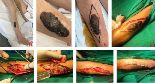
a,b – a Clinical manifestation of advanced pretibial melanoma, 6 months after shave curettage of the so-called “seborrheic keratosis”; 1c – Preoperative marking of the surgical safety margins; 1d, e, f, g – Intraoperative findings: Elliptic surgical excision under local anesthesia; 1h- Postoperative findings. Closing of the wound with single stitches
Burning sensations and formications of the same leg were presented as subjective complaints. Arterial hypertension, controlled with medicine and status post thrombosis of v. popliteal dextra, with patch plastic autogenous, performed two years ago, were reported from the medical history. Family history was negative for cutaneous diseases. Total surgical excision of the lesion was performed under local anaesthesia; with the field of safety margins 1cm. Histological examination verified the diagnosis of melanoma, with a tumor thickness of 4 mm.
Laboratory blood tests established elevated level of glucose (6.5 mmol/l), direct bilirubin (4.4 mmol/l), alcal phosphatase (185 UI/l), LDH (2381 UI/L) and hsCRP (101.8 mg/l). The performed screening vie X-ray revealed multiple metastases in both of the lungs, with maximal diameter 40 mm. The diagnosis of melanoma staged IV was made.
One metastasis was referred for BRAF testing, as therapy with BRAF and MEK inhibitors was planned, while pembrolizumab was considered in case of negative BRAF status.
Discussion
Although most often benign, small pigmented lesions remain the most significant diagnostic challenge, both in naked-eye and in dermatoscopic examinations, particularly in the diagnosis of small melanoma [3]. When considering the diagnosis of pigmented lesions, a balance between therapeutic aspects and cosmetic concerns have to be taken into account, with attention for the differential diagnosis between benign and malignant lesions [4]. The variety of therapeutic options for benign pigmented lesions including shave curettage, local laser destruction, electrocoagulation removal could sound enticingly both for the physician and patient, but they destroy the possibility for histological examination and provide a deceptively feeling of calm, that the problem is solved [2].
Nevertheless, the recurrence rate is the smallest problem that should be taken into consideration. The most severe challenge in such cases is the correct differentiation between the benign and potentially malignant nature of the lesion that must be treated [4]. As it was already mentioned, the diagnosis is not often correct even with dermoscopy or confocal microscopy, although these diagnostic weapons could be high sensitive and precise [5][6]. When considering a pigmented lesion with unknown origin, once should always keep in mind that the incidence of primary cutaneous melanoma has been increasing dramatically for several decades, as this kind of tumour is responsible for the majority of skin cancer-related deaths. Furthermore, the most important diagnostic point is the early diagnosis and treatment with simple excision, because if the 5 year- survival rate for patient in stage I is 95% AND 70-90% for stage II, these percents decreased dramatically up to 15% in patients with stage IV [7]. If there is even a minimum chance for misdiagnosis, the risk could be a human life. Furthermore, a simple surgical excision could provide total resolution of the problem, with correct histological verification and further therapeutic measurements, if needed [7].
We present a case of a patient, with advanced pretibial melanoma with multiple lung metastasis, misdiagnosed as a seborrheic keratosis and treated with shave-curettage 6 months earlier. We want to emphasize the importance of the correct therapeutic method in all cases with pigmented lesions with unknown origin, in order to minimize the risk of dramatic consequences of misdiagnosis of melanoma. So, we want to ask you- is this risk justified?
Footnotes
Funding: This research did not receive any financial support
Competing Interests: The authors have declared that no competing interests exist
References
- 1.Blum A, Kreusch J, Stolz W, Haenssle H, Braun R, Hofmann-Wellenhof R, Tschandl P, Zalaudek I, Kittler H. Dermoscopy for malignant and benign skin tumours:Indication and standardized terminology. Hautarzt. 2017;68(8):653–73. doi: 10.1007/s00105-017-4013-5. https://doi.org/10.1007/s00105-017-4013-5 PMid:28721529. [DOI] [PubMed] [Google Scholar]
- 2.Carli P, De Giorgi V, Crocetti E, Mannone F, Massi D, Chiarugi A, Giannotti B. Improvement of malignant/benign ratio in excised melanocytic lesions in the ‘dermoscopy era’:a retrospective study 1997-2001. Br J Dermatol. 2004;150(4):687–92. doi: 10.1111/j.0007-0963.2004.05860.x. https://doi.org/10.1111/j.0007-0963.2004.05860.x PMid:15099364. [DOI] [PubMed] [Google Scholar]
- 3.Marino ML, Carrera C, Marchetti MA, Marghoob AA. Practice Gaps in Dermatology:Melanocytic Lesions and Melanoma. Dermatol Clin. 2016;34(3):353–62. doi: 10.1016/j.det.2016.03.003. https://doi.org/10.1016/j.det.2016.03.003 PMid:27363893. [DOI] [PubMed] [Google Scholar]
- 4.de Giorgi V, Savarese I, Rossari S, Gori A, Grazzini M, Crocetti E, Sara Longo A, Oranges T, Massi D. Features of small melanocytic lesions:does small mean benign? A clinical-dermoscopic study. Melanoma Res. 2012;22(3):252–6. doi: 10.1097/CMR.0b013e3283527430. https://doi.org/10.1097/CMR.0b013e3283527430 PMid:22430838. [DOI] [PubMed] [Google Scholar]
- 5.Schein O, Westreich M, Shalom A. Effect of dermoscopy on diagnostic accuracy of pigmented skin lesions emphasizing malignant melanoma. Harefuah. 2009;148(12):820–3, 855. PMid:20088434. [PubMed] [Google Scholar]
- 6.Figueroa-Silva O, Cinotti E, de Almeida Silva T, Moscarella E, Lallas A, Ciardo S, Argenziano G, Pellacani G, Piana S, Longo C. Diagnostic accuracy of reflectance confocal microscopy for lesions typified by dermoscopic island. J Eur Acad Dermatol Venereol. 2016;30(9):1594–8. doi: 10.1111/jdv.13632. https://doi.org/10.1111/jdv.13632 PMid:27109574. [DOI] [PubMed] [Google Scholar]
- 7.Bichakjian CK, Halpern AC, Johnson TM, Foote Hood A, Grichnik JM, Swetter SM, Tsao H, Barbosa VH, Chuang TY, Duvic M, Ho VC, Sober AJ, Beutner KR, Bhushan R, Smith Begolka W. Guidelines of care for the management of primary cutaneous melanoma. American Academy of Dermatology. J Am Acad Dermatol. 2011;65(5):1032–47. doi: 10.1016/j.jaad.2011.04.031. https://doi.org/10.1016/j.jaad.2011.04.031 PMid:21868127. [DOI] [PubMed] [Google Scholar]