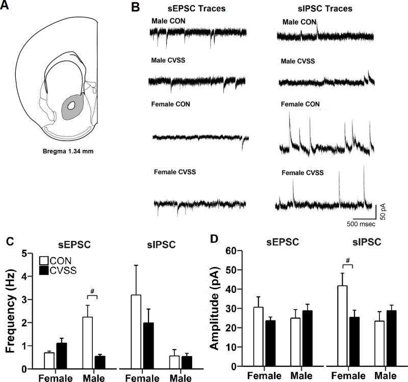
Figure 7.
Exposure to adolescent CVSS altered synaptic transmission nucleus accumbens (NAC) neurons. (A) Illustration of coronal half section of NAC; gray area depicts the region where recordings were performed. (B) Representative traces of sEPSC and sIPSC from CVSS and CON mice. Exposure to CVSS decreased (C) sEPSC frequency in males, but not females, and (D) sIPSC amplitude in females, but not males, relative to CON mice (p < 0.001 and p < 0.05, respectively). Data are presented as mean ± SEM (n = 5–8/group). #Significant ‘Sex × Stress Condition’ interaction

