Abstract
Chemical composition and porosity characteristics of calcium silicate-based endodontic cements are important determinants of their clinical performance. Therefore, the aim of this study was to investigate the chemical composition and porosity characteristics of various calcium silicate-based endodontic cements: MTA-angelus, Bioaggregate, Biodentine, Micromega MTA, Ortho MTA, and ProRoot MTA. The specific surface area, pore volume, and pore diameter were measured by the porosimetry analysis of N2 adsorption/desorption isotherms. Chemical composition and powder analysis by scanning electron microscope (SEM) and energy dispersive spectroscopy (EDS) were also carried out on these endodontic cements. Biodentine and MTA-angelus showed the smallest pore volume and pore diameter, respectively. Specific surface area was the largest in MTA-angelus. SEM and EDS analysis showed that Bioaggregate and Biodentine contained homogenous, round and small particles, which did not contain bismuth oxide.
1. Introduction
Mineral trioxide aggregate (MTA) was introduced in endodontic field as root end filling material and perforation repair material in early 1990s [1]. Due to its superior biocompatibility [2] and sealing ability [3], MTA has been widely used for perforation repair [4], root end filling [5], pulp capping [6], one-visit apexification [7], and pulpal revascularization [8]. However, MTA has been described to have drawbacks such as long setting time [9], tooth discoloration potential [10], and handling difficulty [11]. To overcome these drawbacks, many calcium silicate-based cements such as MTA-angelus [12], Bioaggregate [13], Biodentine [12], Micromega MTA (MM-MTA) [6], and Ortho MTA [14] have been introduced in market and showed good clinical and experimental results.
There are many reports that proved superior sealing ability of MTA in the MTA-tooth interface [15, 16]. However, the porosity existing in MTA itself has not been studied extensively [17–19]. Considering that the porosity of MTA is related to its ability to resist microbial penetration and leakage [20], there is relative lack of knowledge on this issue currently.
Thus, the aim of this study was to investigate the pore volume, pore diameter, and the specific surface area of various commercial calcium silicate-based endodontic cements. The surface morphology and chemical compositions of these cements were also investigated.
2. Materials and Methods
2.1. Materials Used
The materials used in this study were MTA-angelus, Bioaggregate, Biodentine, MM-MTA, Ortho MTA, and ProRoot MTA. The compositions of these materials are listed in Table 1.
Table 1.
Names and compositions of calcium silicate-based endodontic cements which were used in this study.
| Products | Compositions |
|---|---|
| MTA-angelus (Londrina, PR, Brazil) | Tricalcium silicate, dicalcium silicate, tricalcium aluminate, tetracalcium aluminoferrite, bismuth oxide (MSDS) |
| Bioaggregate (Diadent, Burnaby, Canada) | Tricalcium silicate, dicalcium silicate, tantalum pentoxide, calcium phosphate monobasic, amorphous silicon oxide (MSDS) |
| Biodentine (Septodont, St. Maur-des-Fossés, France) | Tricalcium silicate, dicalcium silicate, calcium carbonate and oxide, iron oxide, zirconium oxide [21] |
| MM-MTA (MicroMega, Besançon, France) | Mixture of several mineral oxides and bismuth oxides (MSDS) |
| Ortho MTA (BioMTA, Seoul, Korea) | Calcium carbonate, silicon dioxide, aluminum oxide, dibismuth trioxide (MSDS) |
| ProRoot MTA (Dentsply, Tulsa, OK, USA) | Portland cement, bismuth oxide (MSDS) |
2.2. BET Surface Area and Porosimetry Analyzer
Surface area and pore structure were measured by N2 adsorption/desorption isotherms (ASAP 2020 series) at 77 and 273 K for nitrogen and carbon dioxide within relative pressures from 0 to 1.0 and from 0 to 0.03, respectively. Before analysis, the samples were degassed in the degas port of the adsorption analyzer at 423 K for 10 hours. The surface area, pore volume, and pore diameter were analyzed using ASAP 2020 v3.00 software (Micromeritics, Norcross, GA, USA).
2.3. Scanning Electron Microscope (SEM) and Energy Dispersive Spectroscopy (EDS) Analysis
The morphology of the powders and chemical constitutions was measured on JEOL JSM-6700 scanning electron microscope. Prior to SEM measurement, the samples were coated with platinum using sputter for 45 seconds.
3. Results
3.1. BET Surface Area and Porosimetry Analysis
The specific surface area (m2/g), pore volume (cm3/g), and pore diameter (nm) values of all the samples are listed in Table 2. Specific surface area was the largest in MTA-angelus and the smallest in ProRoot MTA. Pore volume was the largest in MTA-angelus and the smallest in Biodentine. Pore diameter was the largest in MM-MTA and the smallest in MTA-angelus.
Table 2.
Porosity in tested calcium silicate-based endodontic cements.
| MTA-angelus | Bioaggregate | Biodentine | MM-MTA | Ortho MTA | ProRoot MTA | |
|---|---|---|---|---|---|---|
| S A | 6.2 | 5.5 | 4.0 | 3.5 | 4.5 | 3.2 |
| V pore | 0.016 | 0.014 | 0.0080 | 0.0086 | 0.014 | 0.0097 |
| d pore | 9.3 | 11.9 | 13.5 | 21.5 | 11.1 | 17.3 |
S A: specific surface area (m2/g) calculated by BET equation; Vpore: pore volume (cm3/g) calculated by BJH equation; dpore: pore diameter (nm) calculated by BJH equation.
3.2. Scanning Electron Microscope (SEM) and Energy Dispersive Spectroscopy (EDS) Analysis
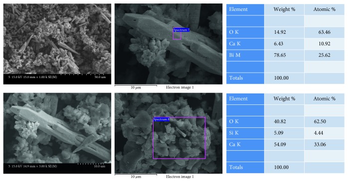
MTA-angelus (Figure 1) showed multiple aggregates of round particles. EDS analysis showed that these round particles are mainly composed of calcium and silica. Among these round particles, long spindle-shaped particles were shown. EDS analysis showed that these long spindle-shaped particles were mainly composed of bismuth.
Figure 1.
SEM and EDS analysis results of MTA-angelus.
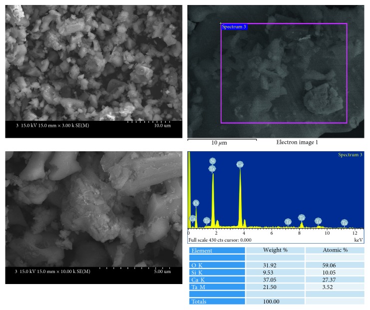
Bioaggregate (Figure 2) showed relatively homogenous aggregates of small round particles. EDS analysis showed that these particles were mainly composed of calcium, silicon, and tantalum. Bioaggregate did not contain bismuth.
Figure 2.
SEM and EDS analysis results of Bioaggregate.
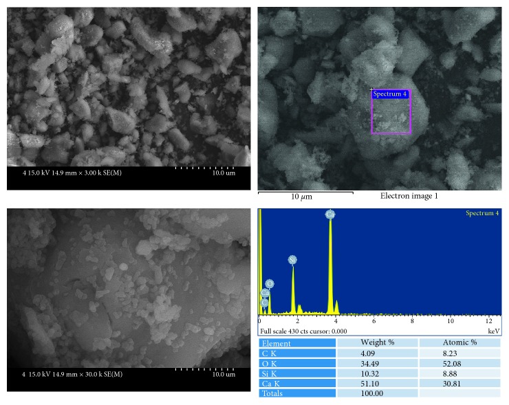
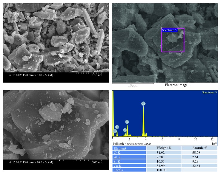
Biodentine (Figure 3) showed that relatively large particles were covered with small particles. EDS analysis showed that these particles were mainly composed of calcium and silicon.
Figure 3.
SEM and EDS analysis results of Biodentine.
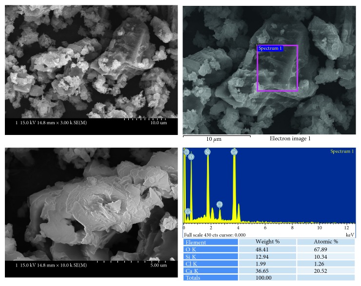
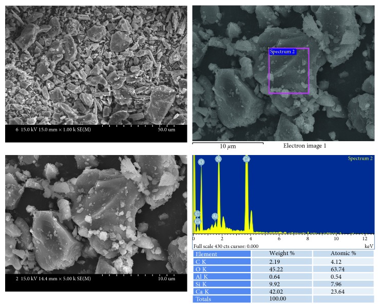
MM-MTA (Figure 4) also showed the mixtures of relatively larger particles and smaller particles. EDS analysis showed that these particles were mainly composed of calcium and silicon.
Figure 4.
SEM and EDS analysis results of MM-MTA.
Ortho MTA (Figure 5) showed large particles, small particles, and long spindle-shaped particles at the same time. All these particles were shown to be mainly composed of calcium and silicon.
Figure 5.
SEM and EDS analysis results of Ortho MTA.
ProRoot MTA (Figure 6) showed relatively homogenous particles which are mainly composed of calcium and silicon.
Figure 6.
SEM and EDS analysis results of ProRoot MTA.
4. Discussion
Porosity of mineral trioxide aggregate is important in that it is related to bacterial leakage [20]. However, there are few studies which investigated the porosity of MTA [17–19]. Regarding the porosity characteristics, one previous study [17] reported that the apparent porosity of ProRoot MTA was 29.36% while that of Dycal was 9.04%. However, this study used Archimedes' principle to calculate the porosity of MTA samples. In this reason, this study had a limitation that it could not give information regarding the characteristics such as pore diameter and specific surface area of MTA.
Porosity-related properties of a certain material are specific surface area (m2/g), pore volume (cm3/g), and pore diameter [22]. Most previous studies which investigated MTA porosity used mercury intrusion porosimetry [18, 19]. It was reported that the detection range of mercury intrusion porosimetry is from 3 nm to 200 μm, whereas that of N2 adsorption/desorption isotherms is from 0.3 nm to 300 nm [22]. According to this report [22], N2 adsorption/desorption isotherms can detect the small pores which could not be detected by mercury intrusion porosimetry. In this reason, the study of porosity of MTA using N2 adsorption/desorption isotherms as well as mercury intrusion porosimetry could be regarded as ideal methods.
The previous studies reported that the pore volume for ProRoot MTA was 0.1025 cm3/g at pH 7.4 [19]. The pore volume for ProRoot MTA was 0.0097 cm3/g in this study. This difference could be attributed to the experimental conditions such as time elapsed for MTA setting and the environment around the MTA setting.
The pore volume inside the specimen was the largest in MTA-angelus group (0.016 cm3/g). The pore volume inside Bioaggregate and Ortho MTA was the same and was 0.014 cm3/g. The pore volume of MM-MTA was 0.0086 cm3/g. The pore volume of Biodentine was the smallest of all the tested groups. (0.0080 cm3/g).
In addition to the total pore volume, the size of pore diameter is important [19]. Unfortunately, there has been no study which evaluated pore diameters of mineral trioxide aggregate. In the present study, pore diameter was the largest in MM-MTA (21.5 nm) and decreased in the order of ProRoot MTA, Biodentine, Bioaggregate, Ortho MTA, and MTA-angelus. MTA-angelus has the smallest pore diameter, and it was 9.3 nm. Considering that the average size of Enterococcus faecalis (representative endodontic bacterium) is 0.6–2.5 μm [23], it is quite unlikely that bacteria could penetrate well-condensed and hydrated MTA. Another characteristic investigated in this study was specific surface area. Specific surface area could affect the adhesion of contacting cells [24]. The larger surface area is considered to be the more favorable condition to cellular adhesion [24]. In the present study, the specific surface area was the largest in MTA-angelus and decreased in the order of Bioaggregate, Ortho MTA, Biodentine, MM-MTA, and ProRoot MTA. ProRoot MTA has the smallest specific surface area, and it was 3.2 m2/g. The effect of these different specific surface areas should be investigated further in future study.
5. Conclusion
In conclusion, this study showed that Biodentine and MTA-angelus showed the smallest pore volume and pore diameter, respectively, which could be regarded as superior physicochemical properties from the perspective of clinical endodontics.
Conflicts of Interest
The authors declare that they have no conflicts of interest related to this study.
References
- 1.Torabinejad M., Rastegar A. F., Kettering J. D., Pitt Ford T. R. Bacterial leakage of mineral trioxide aggregate as a root-end filling material. Journal of Endodontics. 1995;21(3):109–112. doi: 10.1016/s0099-2399(06)80433-4. [DOI] [PubMed] [Google Scholar]
- 2.Torabinejad M., Hong C. U., Pitt Ford T. R., Kettering J. D. Cytotoxicity of four root end filling materials. Journal of Endodontics. 1995;21(10):489–492. doi: 10.1016/s0099-2399(06)80518-2. [DOI] [PubMed] [Google Scholar]
- 3.Torabinejad M., Smith P. W., Kettering J. D., Pitt Ford T. R. Comparative investigation of marginal adaptation of mineral trioxide aggregate and other commonly used root-end filling materials. Journal of Endodontics. 1995;21(6):295–299. doi: 10.1016/s0099-2399(06)81004-6. [DOI] [PubMed] [Google Scholar]
- 4.Main C., Mirzayan N., Shabahang S., Torabinejad M. Repair of root perforations using mineral trioxide aggregate: a long-term study. Journal of Endodontics. 2004;30(2):80–83. doi: 10.1097/00004770-200402000-00004. [DOI] [PubMed] [Google Scholar]
- 5.Torabinejad M., Pitt Ford T. R., McKendry D. J., Abedi H. R., Miller D. A., Kariyawasam S. P. Histologic assessment of mineral trioxide aggregate as a root-end filling in monkeys. International Endodontic Journal. 1997;42(5):408–411. doi: 10.1111/j.1365-2591.2009.01556.x. [DOI] [PubMed] [Google Scholar]
- 6.Chang S. W., Bae W. J., Yi J. K., et al. Odontoblastic differentiation, inflammatory response, and angiogenic potential of 4 calcium silicate-based cements: Micromega MTA, ProRoot MTA, RetroMTA, and experimental calcium silicate cement. Journal of Endodontics. 2015;41(9):1524–1529. doi: 10.1016/j.joen.2015.04.018. [DOI] [PubMed] [Google Scholar]
- 7.Simon S., Rilliard F., Berdal A., Machtou P. The use of mineral trioxide aggregate in one-visit apexification treatment: a prospective study. International Endodontic Journal. 2007;40(3):186–197. doi: 10.1111/j.1365-2591.2007.01214.x. [DOI] [PubMed] [Google Scholar]
- 8.Corbella S., Ferrara G., El Kabbaney A., Taschieri S. Apexification, apexogenesis and regenerative endodontic procedures: a review of the literature. Minerva Stomatologica. 2014;63(11-12):375–389. [PubMed] [Google Scholar]
- 9.Chung C. J., Kim E., Song M., Park J. W., Shin S. J. Effects of two fast-setting calcium-silicate cements on cell viability and angiogenic factor release in human pulp-derived cells. Odontology. 2016;104(2):143–151. doi: 10.1007/s10266-015-0194-5. [DOI] [PubMed] [Google Scholar]
- 10.Ramos J. C., Palma P. J., Nascimento R., et al. 1-year in vitro evaluation of tooth discoloration induced by 2 calcium silicate-based cements. Journal of Endodontics. 2016;42(9):1403–1407. doi: 10.1016/j.joen.2016.06.012. [DOI] [PubMed] [Google Scholar]
- 11.Cianconi L., Palopoli P., Campanella V., Mancini M. Composition and microstructure of MTA and Aureoseal Plus: XRF, EDS, XRD and FESEM evaluation. European Journal of Paediatric Dentistry. 2016;17(4):281–285. [PubMed] [Google Scholar]
- 12.Chang S. W., Lee S. Y., Ann H. J., Kum K. Y., Kim E. C. Effects of calcium silicate endodontic cements on biocompatibility and mineralization-inducing potentials in human dental pulp cells. Journal of Endodontics. 2014;40(8):1194–1200. doi: 10.1016/j.joen.2014.01.001. [DOI] [PubMed] [Google Scholar]
- 13.Chang S. W., Lee S. Y., Kum K. Y., Kim E. C. Effects of ProRoot MTA, Bioaggregate, and Micromega MTA on odontoblastic differentiation in human dental pulp cells. Journal of Endodontics. 2014;40(1):113–118. doi: 10.1016/j.joen.2013.09.036. [DOI] [PubMed] [Google Scholar]
- 14.Chang S. W., Baek S. H., Yang H. C., et al. Heavy metal analysis of ortho MTA and ProRoot MTA. Journal of Endodontics. 2011;37(12):1673–1676. doi: 10.1016/j.joen.2011.08.020. [DOI] [PubMed] [Google Scholar]
- 15.Bird D. C., Komabayashi T., Guo L., Opperman L. A., Spears R. In vitro evaluation of dentinal tubule penetration and biomineralization ability of a new root-end filling material. Journal of Endodontics. 2012;38(8):1093–1096. doi: 10.1016/j.joen.2012.04.017. [DOI] [PMC free article] [PubMed] [Google Scholar]
- 16.Parirokh M., Torabinejad M. Mineral trioxide aggregate: a comprehensive literature review–Part III: clinical applications, drawbacks, and mechanism of action. Journal of Endodontics. 2010;36(3):400–413. doi: 10.1016/j.joen.2009.09.009. [DOI] [PubMed] [Google Scholar]
- 17.Gandolfi M. G., Siboni F., Primus C. M., Prati C. Ion release, porosity, solubility, and bioactivity of MTA Plus tricalcium silicate. Journal of Endodontics. 2014;40(10):1632–1637. doi: 10.1016/j.joen.2014.03.025. [DOI] [PubMed] [Google Scholar]
- 18.Oliveira I. R., Pandolfelli V. C., Jacobovitz M. Chemical, physical and mechanical properties of a novel calcium aluminate endodontic cement. International Endodontic Journal. 2010;43(12):1069–1076. doi: 10.1111/j.1365-2591.2010.01770.x. [DOI] [PubMed] [Google Scholar]
- 19.Saghiri M. A., Asgar K., Lotfi M., Karamifar K., Neelakantan P., Ricci J. L. Application of mercury intrusion porosimetry for studying the porosity of mineral trioxide aggregate at two different pH. Acta Odontologica Scandinavica. 2012;70(1):78–82. doi: 10.3109/00016357.2011.597777. [DOI] [PubMed] [Google Scholar]
- 20.Saghiri M. A., Lotfi M., Saghiri A. M., et al. Effect of pH on sealing ability of white mineral trioxide aggregate as a root-end filling material. Journal of Endodontics. 2008;34(10):1226–1229. doi: 10.1016/j.joen.2008.07.017. [DOI] [PubMed] [Google Scholar]
- 21.About I. Biodentine: from biochemical and bioactive properties to clinical applications. Giornale Italiano di Endodonzia. 2016;30(2):81–88. doi: 10.1016/j.gien.2016.09.002. [DOI] [Google Scholar]
- 22.Westermarck S. Use of Mercury Porosimetry and Nitrogen Adsorption in Characterisation of the Pore Structure of Mannitol and Microcrystalline Cellulose Powders, Granules, and Tablets. Helsinki, Finland: Department of Pharmacy, University of Helsinki; 2000. Academic dissertation. [Google Scholar]
- 23.2017. Available from: https://catalog.hardydiagnostics.com/cp_prod/Content/hugo/Enterococcus.htm.
- 24.Chen M., Patra P. K., Lovett M. L., Kaplan D. L., Bhowmick S. Role of electrospun fibre diameter and corresponding specific surface area (SSA) on cell attachment. Journal of Tissue Engineering and Regenerative Medicine. 2009;3(4):269–279. doi: 10.1002/term.163. [DOI] [PubMed] [Google Scholar]


