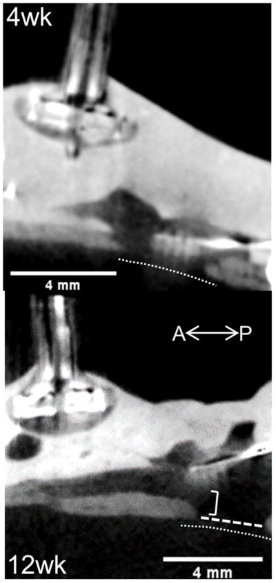
Figure 2.

Sagittal microCT imaging of uncoated implants following perfusion, but preceding dissection (intact head). Top: 4 weeks post-implantation Bottom: 12 weeks. Implant bed and shanks visible from right side of image near craniotomy opening in skull (bottom right area of each image). Thin dotted line indicates brain tissue surface, wide dotted line spans craniotomy, bracket highlights implant displacement.
